Pacemakers use low-energy pulses to help regulate heart rhythm, while defibrillators deliver a stronger charge to stop certain dangerous rhythms or respond to cardiac arrest. Some implanted defibrillators also provide pacing.
A digital design becomes a physical device through successive material layers, but the freedom to shape complex forms brings practical questions about material behavior, fit, and manufacturing quality.
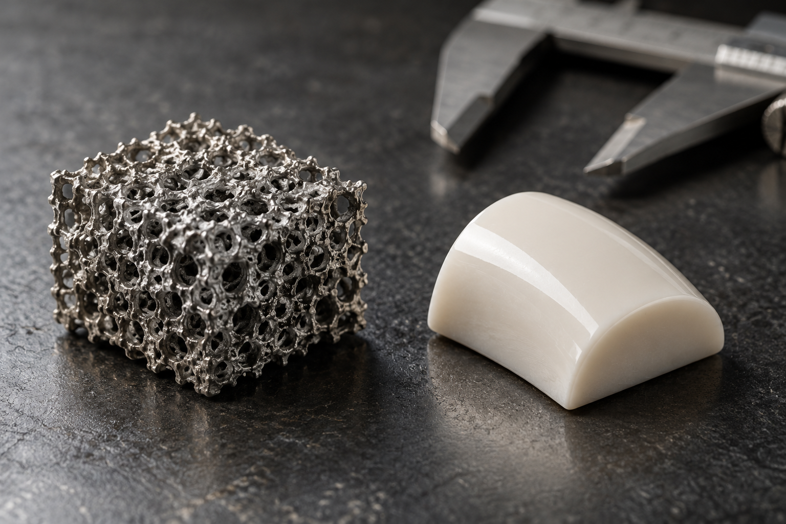
A porous scaffold can look like a solid object while behaving more like a landscape of channels. The dimensions and connections of those spaces influence how fluids, cells, and materials move through it.
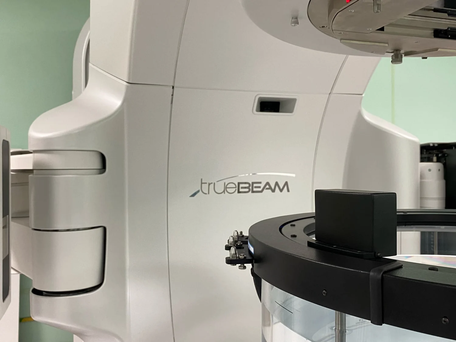
Stereotactic treatments concentrate radiation where beams meet, using careful imaging and immobilization to manage small targets. Their precision has limits, especially when organs move or nearby healthy tissue is radiation-sensitive.
A traveling wave moves through the cochlea’s fluid and excites different regions of its basilar membrane. This spatial arrangement lets the inner ear sort sound frequencies before signals reach the brain.
At small scales, surface interactions and short travel distances can dominate a device’s behavior. Understanding those effects helps explain the opportunities and limits of compact diagnostics and biomaterials.
A vivid optical image can reveal patterns without telling us how much tissue property changed. Moving from appearance to measurement requires calibration, uncertainty estimates, and an account of how light interacted with the sample.
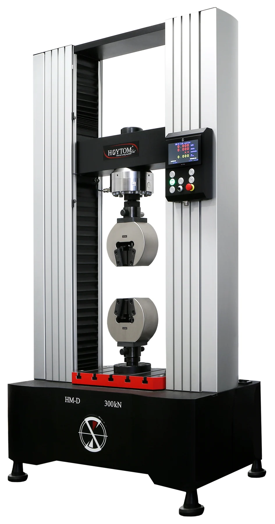
A material can resist bending yet fail at a modest load, or bend readily without breaking. Understanding stiffness and strength helps explain why medical devices must be matched to their specific jobs and environments.
Ultrasound can do more than form images: converging sound waves can concentrate energy at a selected location. The physical mechanism is established, while specific therapeutic applications differ in evidence and clinical availability.
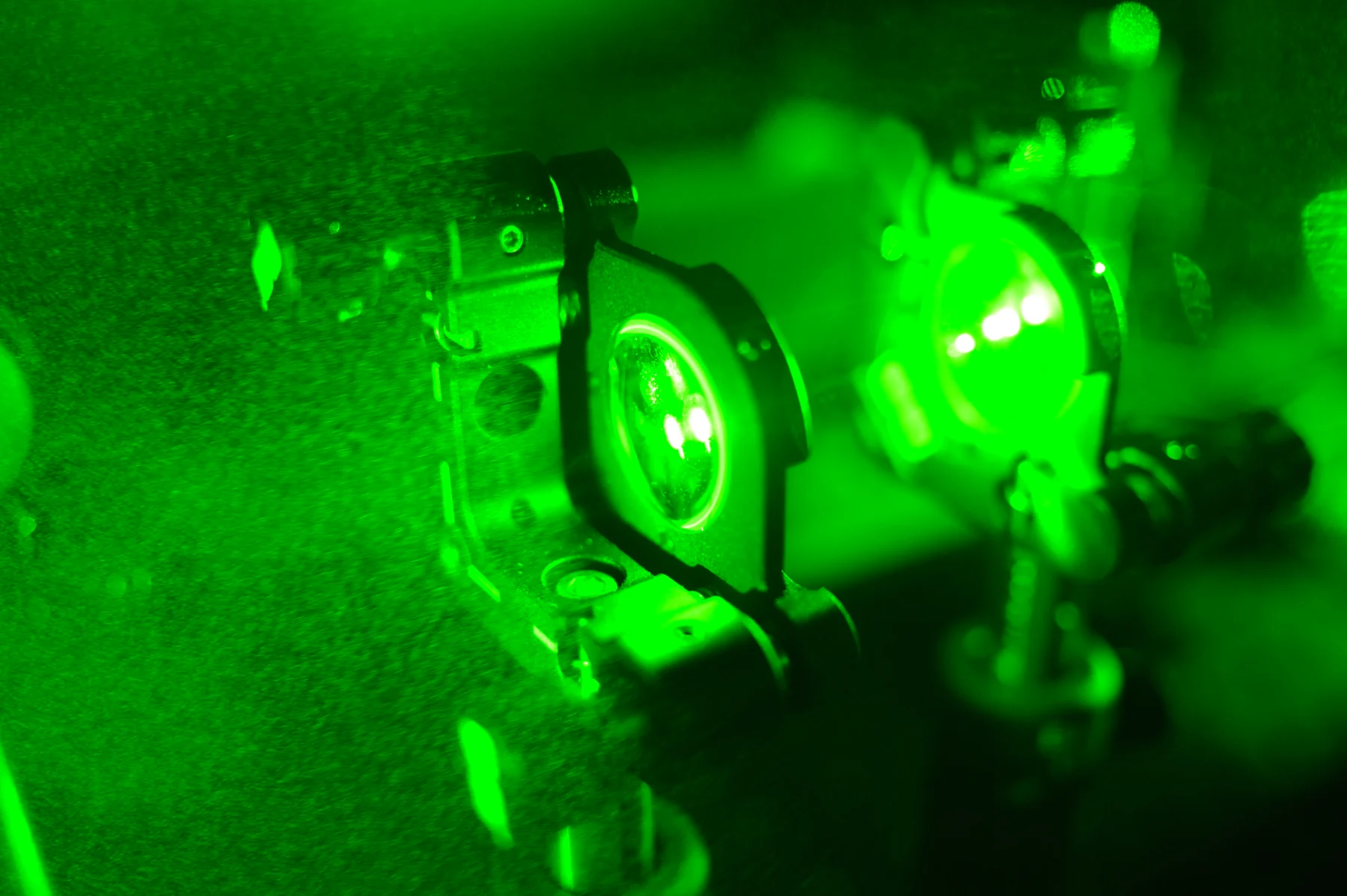
Photodynamic therapy combines a light-activated drug with oxygen to damage selected cells. Its local action depends on where the sensitizer accumulates, where light can reach, and whether oxygen is available nearby.
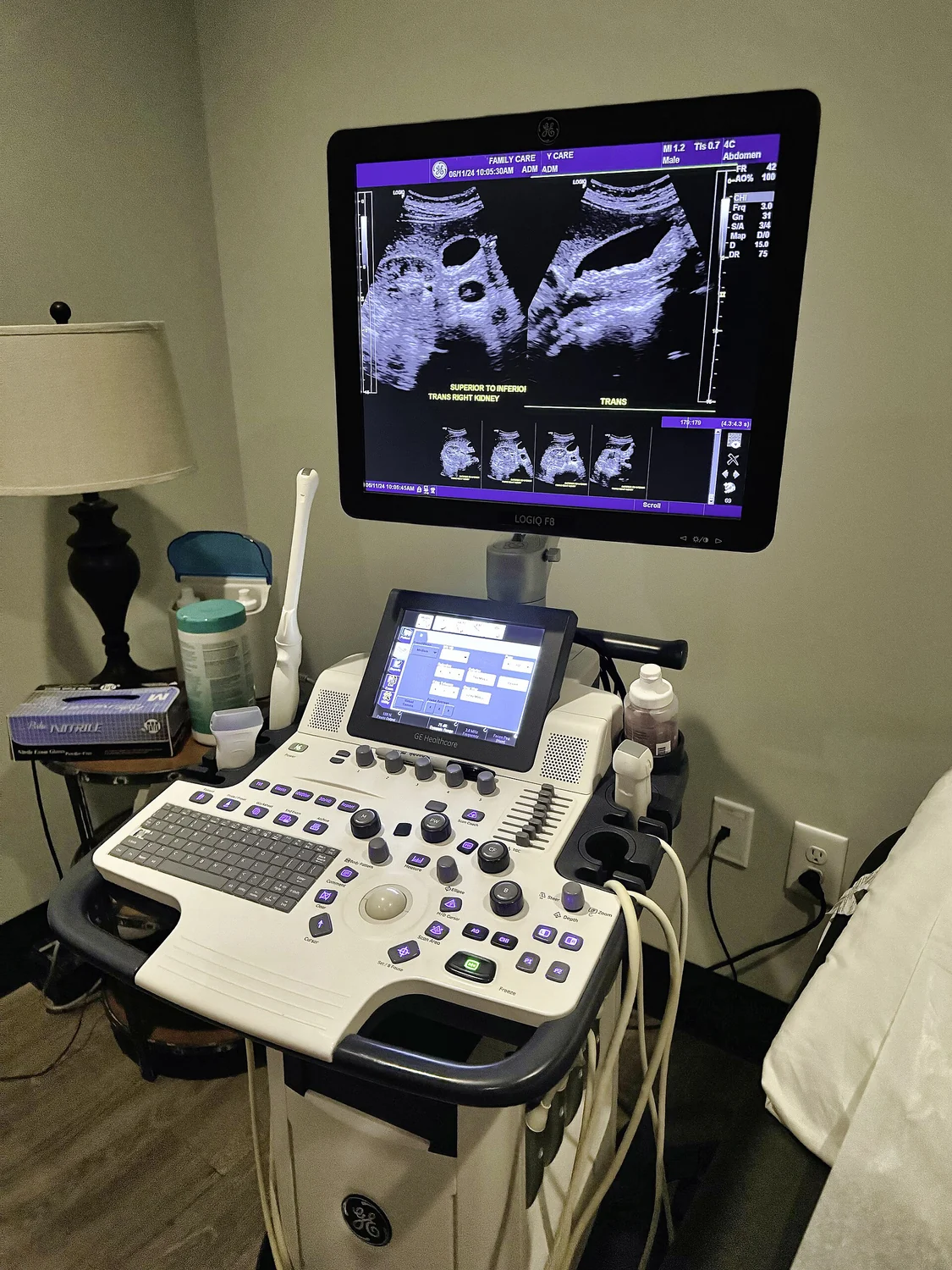
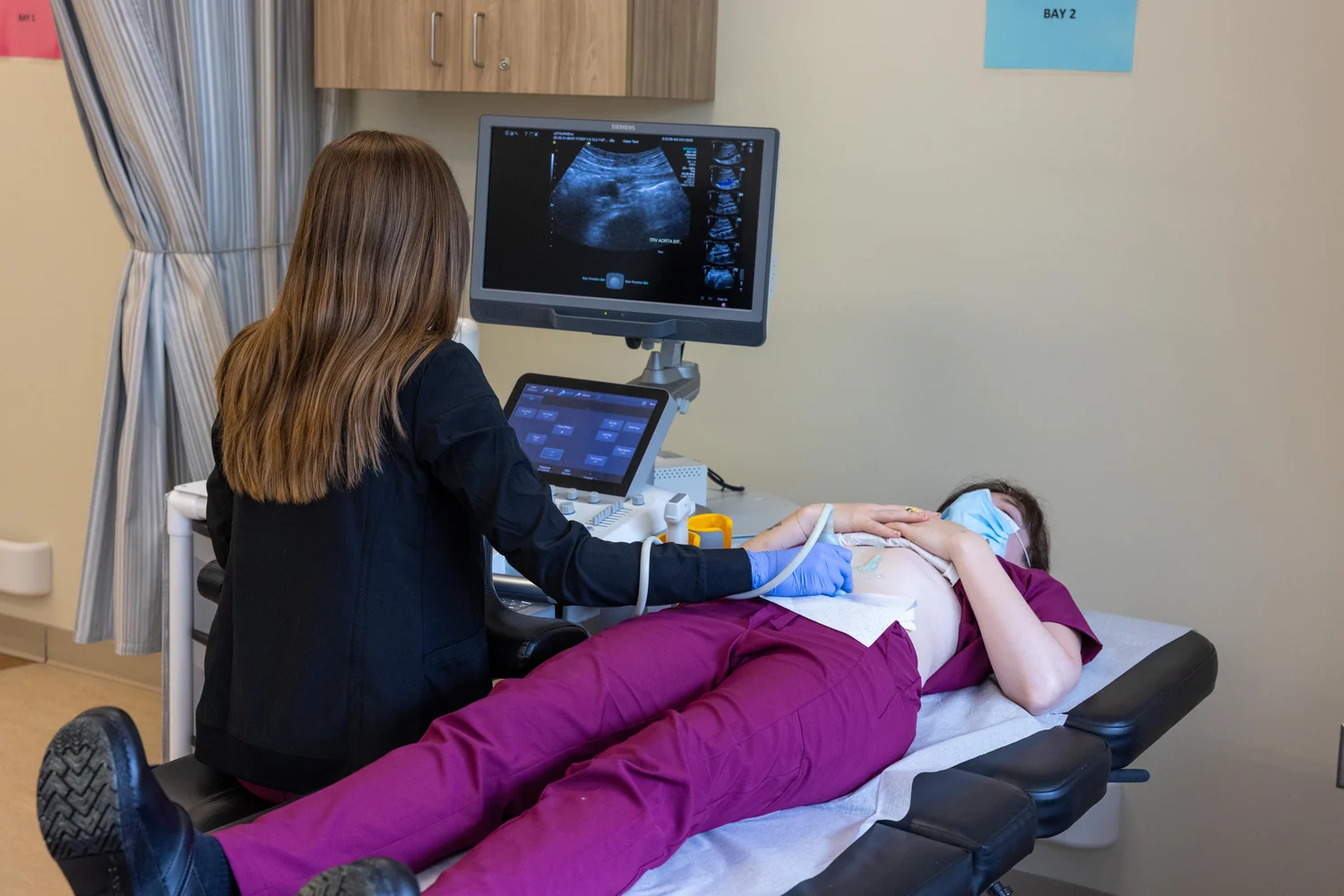
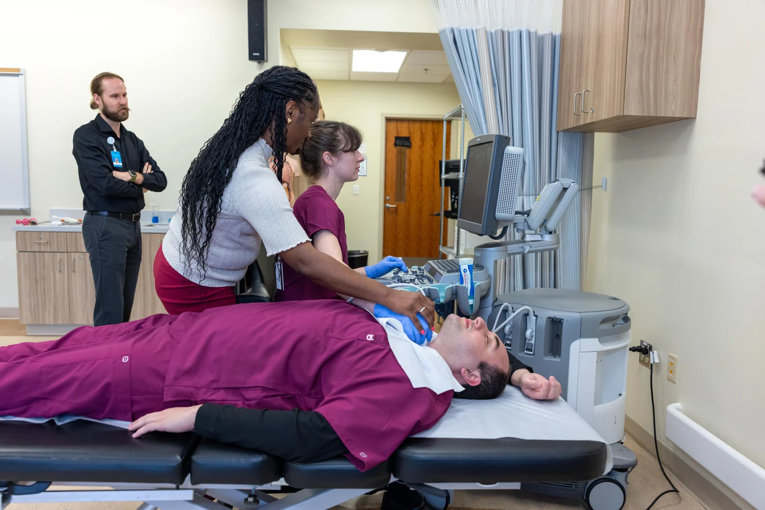
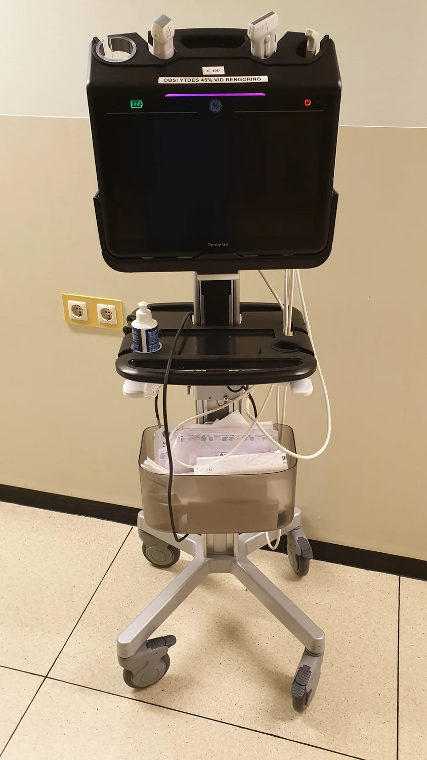
Ultrasound images depend on sound waves entering tissue and returning as echoes. Bone and air create strong boundaries that reflect or disrupt those waves, limiting what lies beyond them even as the same echoes create useful contrast.
A wound’s measured area turns a changing, three-dimensional injury into a number that can be compared over time. Photography, tracing, and careful definitions help researchers interpret that number while recognizing its limits.
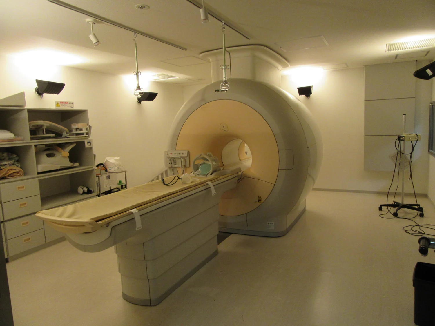
A sequence of medical images can reveal how anatomy or biological activity changes over time. MRI and nuclear medicine create those images through different physical signals, so their comparisons answer different questions.
Amplitude describes the size of a wave’s changing signal; intensity describes how much wave energy passes through an area. Keeping those quantities distinct clarifies how ultrasound scanners interpret echoes and manage exposure.
OCT and ultrasound both build cross-sectional views from returning waves, but one sends infrared light and the other sound. Their shared logic makes a useful comparison; their different physics sets distinct limits and uses.
SPECT turns emissions from a radiotracer inside the body into a three-dimensional map. Its rotating detectors gather projections from many directions, allowing clinicians to see patterns of function as well as anatomy.
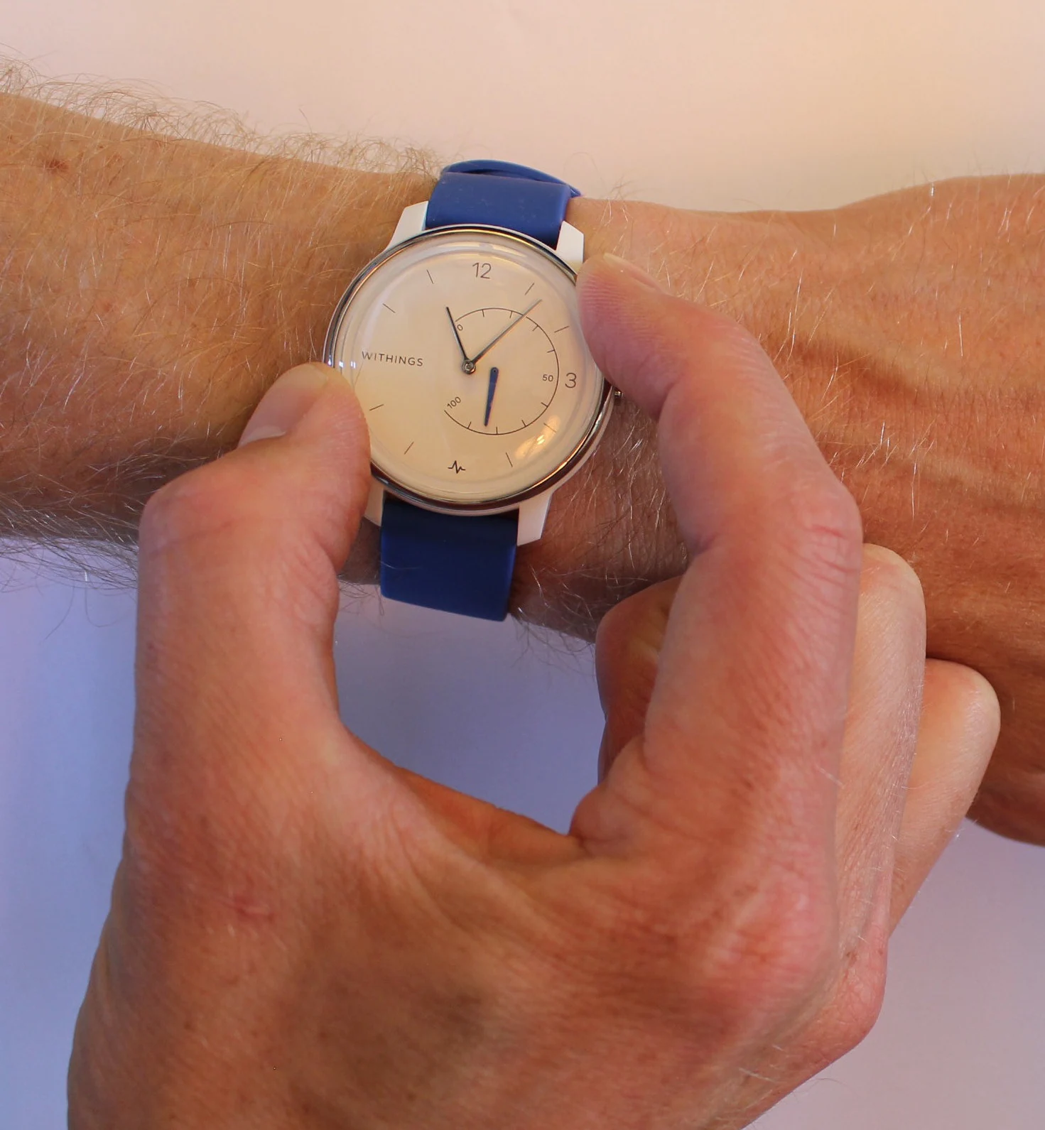
A fingertip pulse sensor detects changing light absorption as blood volume pulses through tissue; an ECG records electrical activity that coordinates heart contractions. Their signals answer related but distinct questions.
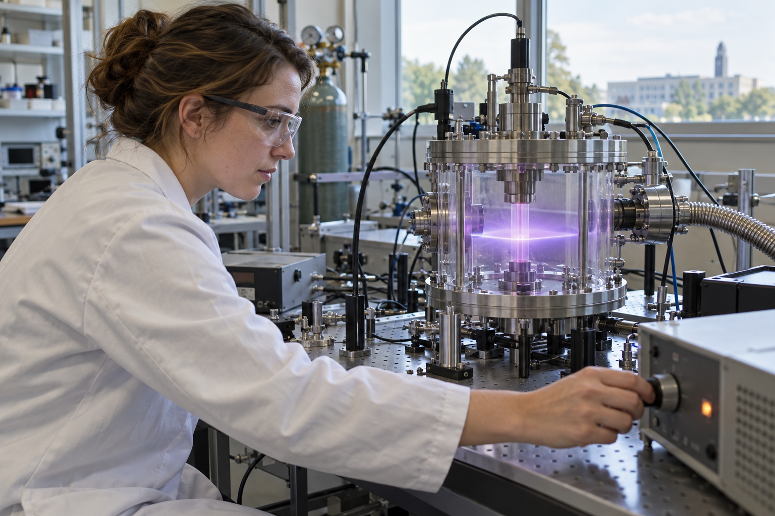
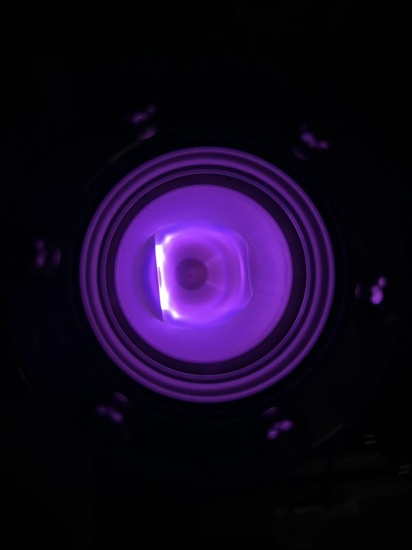
Plasma processes can operate in open air near tissue or inside an evacuated sterilization chamber. Pressure, gas chemistry, energy delivery, and intended target distinguish these technologies and their evidence.