Why can an ultrasound probe show a moving heart but struggle to see through a rib or a pocket of lung air? The answer begins with a simple requirement: sound must travel into the body and return as echoes. When waves meet a sharp change between materials, much of their energy can bounce away or scatter. Bone and air create especially difficult boundaries, so the view behind them may be obscured.
An image built from returning echoes
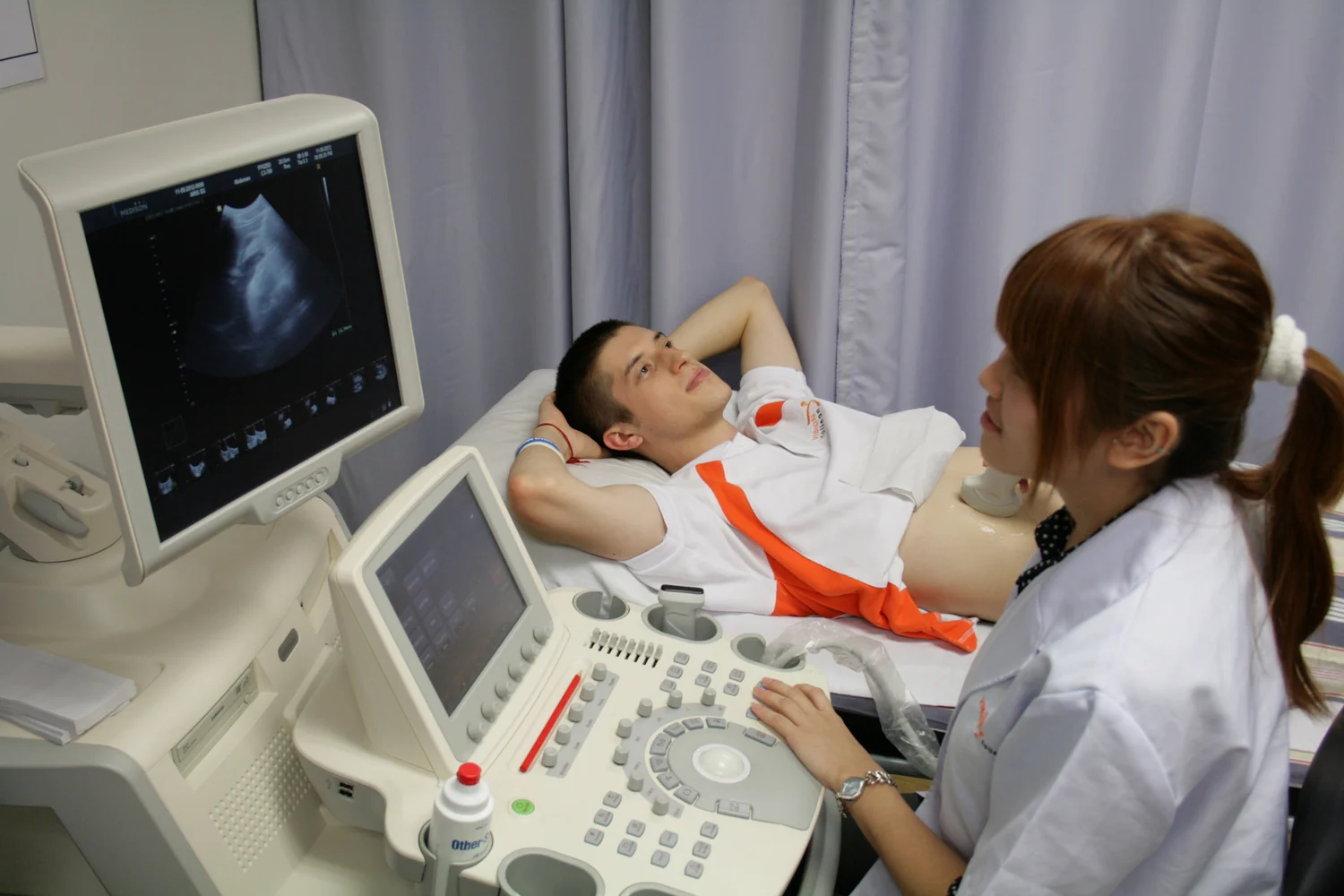
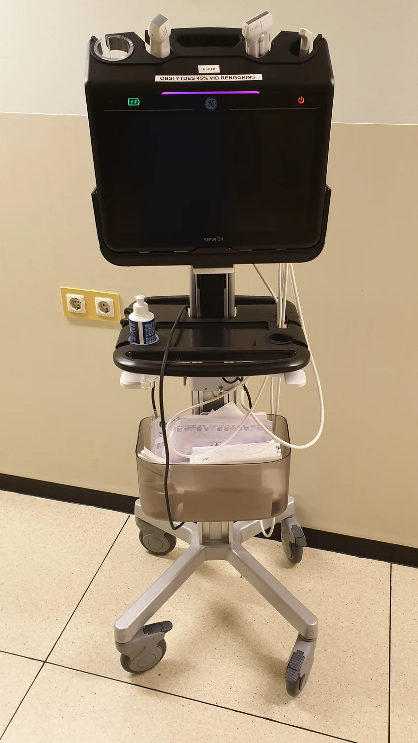
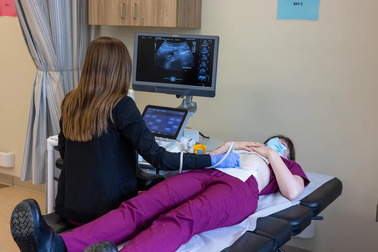
An ultrasound probe, or transducer, sends high-frequency sound into the body and listens for echoes. The time each echo takes to return helps the scanner estimate how far away a tissue boundary lies; echo strength contributes information about the boundary. A computer converts many such measurements into an image [1] [2].
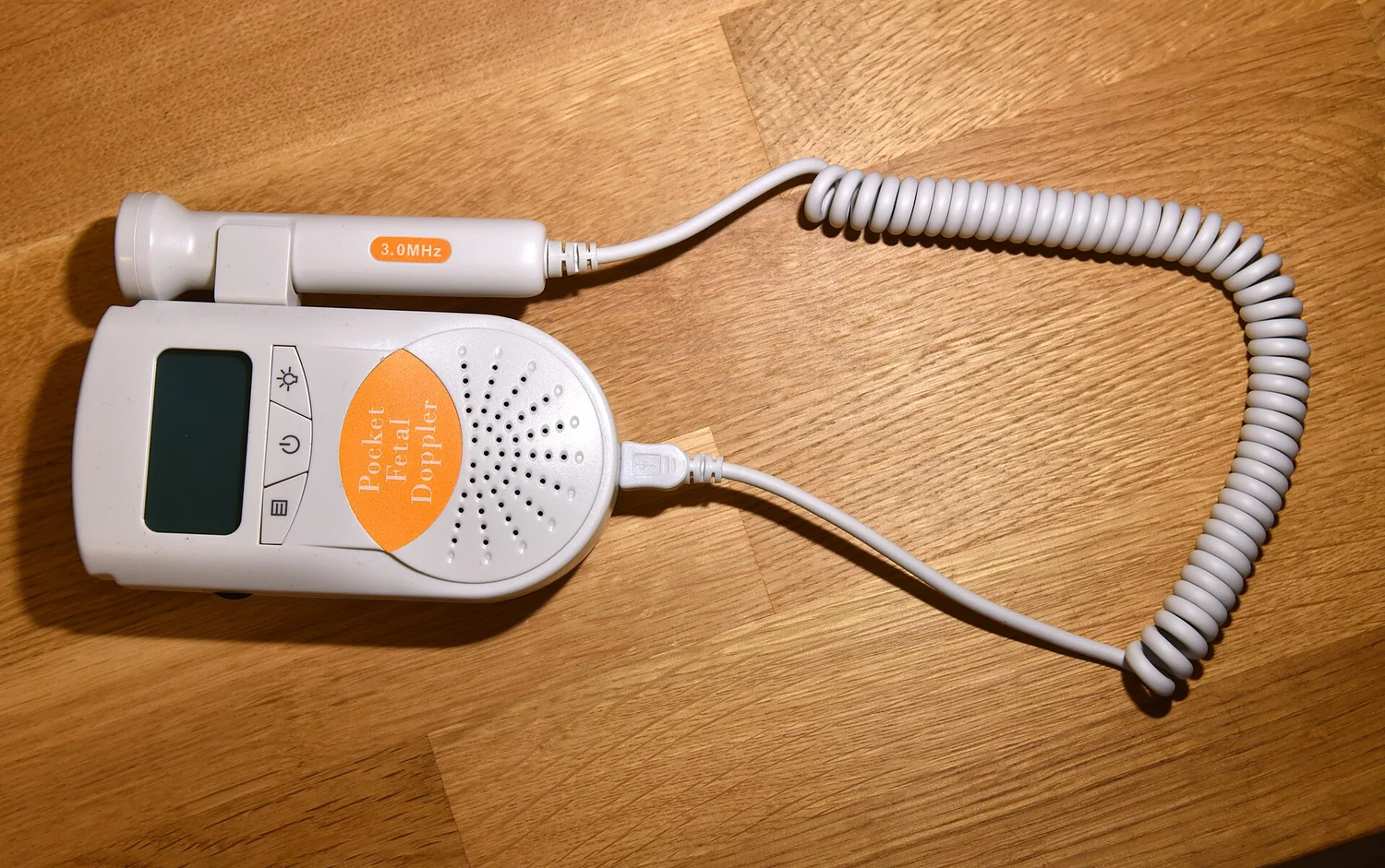
Many probes use piezoelectric materials: applying an electric field makes them vibrate and produce sound, while returning sound makes them generate electrical signals. The same device can therefore act as speaker and microphone. A thin gel layer helps sound pass from the probe into skin by preventing air pockets at that interface [1]. The gel solves a small version of the larger problem: sound transmission depends on what materials touch.
Why boundaries matter so much
A sound wave carries energy through material as a pressure disturbance. At a boundary, the amount reflected depends partly on how different the materials are in their acoustic properties. A modest change between soft tissues can return enough echo to mark an organ edge. A very large mismatch can send much more of the wave back toward the probe, leaving less energy to continue onward.
Picture shouting down a hallway: a doorway may return a clear echo, while a barrier can stop much of the sound from reaching rooms beyond it. The analogy is imperfect because ultrasound involves waves traveling through tissue, but it captures the trade-off. Reflections are not merely obstacles; they are the signal used to locate boundaries. Yet a strong reflection can hide deeper structures by reducing the wave energy that reaches them.
Bone and air make difficult windows
Bone is dense and has acoustic properties that differ sharply from soft tissue. It reflects and absorbs ultrasound, making it difficult to obtain a clear image through it. Air creates an even more challenging interface with tissue: the mismatch causes strong reflection and scattering. As a result, ultrasound is generally poor for imaging bones and air-containing structures such as lungs [1].
This explains why a rib can block a view of the heart from one angle, while a nearby opening between ribs may provide a useful acoustic window. It also explains why the lungs, which contain air, cannot usually be imaged like a fluid-filled organ. Under some conditions, ultrasound can examine the lung surface or surrounding lining when fluid is present, and it can image some bones in fetuses or small babies [1]. The physical obstacle is not an absolute ban; it depends on the route and what lies at the boundary.
The limits shape where ultrasound excels
When waves can travel through soft tissue and return, ultrasound can display internal anatomy in real time and show movement, including blood flow. Doppler techniques use changes in the returning signal to visualize flow direction and speed, while elastography estimates relative tissue stiffness [1]. These capabilities are valuable precisely because echoes respond to tissue boundaries and motion.
Ultrasound does not use ionizing x-rays, but it does introduce energy into the body. Diagnostic examinations are generally considered safe when used prudently by trained providers; energy can slightly heat tissue and, under some conditions, produce cavitation, or small gas pockets [2]. The same physics that limits the view through bone and air also explains why probe placement, angle, and the choice of imaging method matter. Ultrasound is powerful when there is a workable sound path, and constrained when the path is interrupted.
Explore the physics: an explanatory diagram

Sources & further reading
- Ultrasound NIBIB
- Ultrasound Imaging FDA
Prepared for the independent PSI Center reading library. About edition dates and source checking.
This explainer describes scientific principles. It does not assess individual treatment choices.