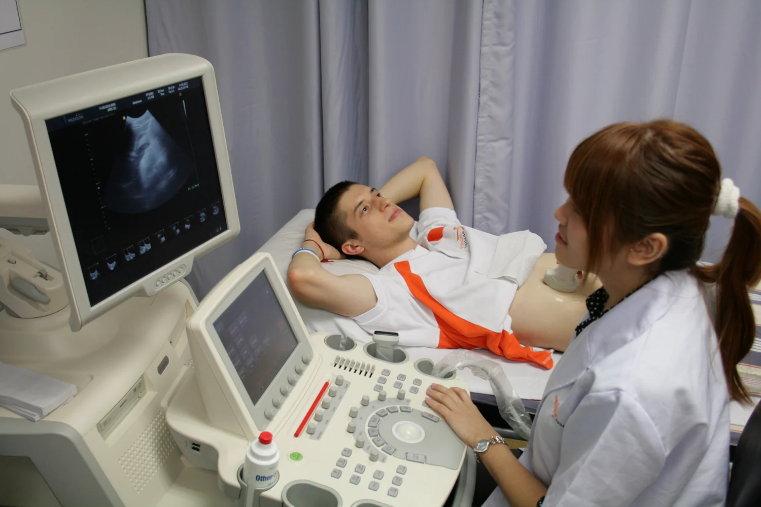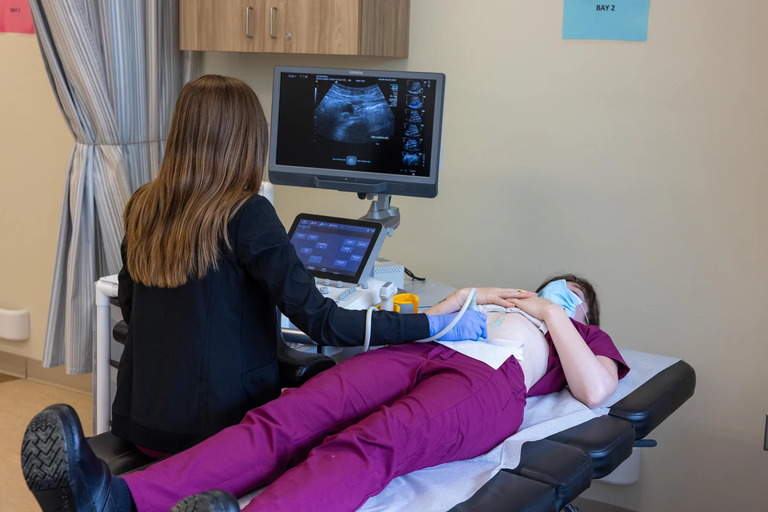
Why does a sonographer spread cool gel before placing an ultrasound probe on the skin? The gel is not a medicine and does not create the image. It solves a physical problem at the surface: trapped air reflects sound strongly, so even a thin air gap can prevent useful waves from entering the body. Gel provides a continuous acoustic path from probe to skin.
The troublesome boundary at the skin
An ultrasound probe sends high-frequency sound waves into the body and detects echoes that return. In the body, differences between tissues reflect some sound at boundaries; those echoes provide the raw information for an image [1]. At the probe-skin boundary, however, air is a poor neighbor for transmitting sound into tissue. A pocket of air reflects much of the incoming wave instead of letting it travel onward.
The problem is similar to trying to pass a vibration from one surface into another when a gap separates them. The wave encounters a sharp change in acoustic properties at the air interface, and the mismatch sends energy back. Even though the gap may look tiny, it interrupts the pathway the scanner depends on.
Gel makes a better acoustic bridge
Gel fills the uneven spaces between the probe and skin, helping prevent air pockets from forming. That allows ultrasound waves to pass from the transducer through the gel and into the body, as described by both NIBIB and the FDA [1] [2]. The gel’s job is therefore practical and local: improve transmission at the contact surface.
It does not need to carry the echoes all the way through the body or “show” organs. Once waves enter the tissue, they travel, reflect at boundaries, and return to the probe. The scanner uses the time between sending a pulse and receiving an echo, along with the speed of sound, to estimate how far away a reflecting boundary lies [1].
From echo timing to a moving picture
The transducer both emits pulses and detects echoes. Many probes use piezoelectric materials, which convert electrical energy into sound and sound back into electrical signals. As returning echoes arrive, the scanner interprets their timing and strength to build an image of tissue structures [1]. Repeated pulses make it possible to display movement in real time, including organ motion or blood flow.
Consider a short pulse that reflects from a boundary close to the probe and another that returns from a deeper boundary. The deeper echo takes longer to arrive, so the scanner assigns it a greater distance. Echo strength also matters: a stronger reflection produces a different image signal than a weaker one. Many such measurements combine into a two-dimensional view, and specialized methods can map motion or tissue stiffness [1].
Useful imaging, with limits
Ultrasound is used to examine many structures, guide procedures, and visualize blood flow. It does not image all materials equally well: bone and air-containing tissue can obstruct or complicate the path, though there are particular situations in which nearby fluid makes some structures more accessible [1]. Gel fixes the surface coupling problem; it cannot remove every limitation deeper in the body.
Diagnostic ultrasound uses non-ionizing sound rather than x-rays, but sound energy can still produce biological effects under some settings. The FDA notes that ultrasound can heat tissue slightly and, in some cases, cause cavitation, so prudent use and trained operation matter [2]. The gel’s simple role sits inside a broader system: sound must enter, echoes must return, and the scanner must interpret them to form an image.
Explore the physics: an explanatory diagram

Sources & further reading
- Ultrasound NIBIB
- Ultrasound Imaging FDA
Prepared for the independent PSI Center reading library. About edition dates and source checking.
This explainer describes scientific principles. It does not assess individual treatment choices.