Ultrasound depends on sound traveling out from a probe and echoes returning. A microscopic air gap at the probe–skin boundary can disrupt that trip because air and soft tissue respond very differently to sound. Coupling gel fills the gap, creating a continuous path between the transducer and skin. Its job is physical: help transmit acoustic energy so the scanner can receive echoes. It does not itself diagnose or treat anything. [1] [4]
The boundary is part of the instrument
A probe face may look flat against skin, but ordinary surfaces have tiny ridges and hollows. If those spaces contain air, the sound encounters a sequence of materials rather than a close acoustic contact. Ultrasound pulses are mechanical pressure waves: particles in one material push on neighboring particles, passing the disturbance along. At a boundary, some energy continues onward and some returns as reflection. A strong reflection at the very first boundary can leave less of the pulse available to enter the body. [1] [4]
The coupling layer is not merely lubricant. Gel displaces air and contacts both probe and skin. FDA describes this as the route by which waves pass from the transducer into the body. The path works in reverse too: returning echoes cross the interface and reach the transducer. A break in acoustic contact can weaken transmission and reception. [1]
Acoustic impedance predicts the size of a mismatch
Acoustic impedance, written Z, is the product of a material’s density and the speed of sound in it: Z = ρc. It describes how pressure and particle motion relate as a wave travels through a medium. At a flat boundary between two ideal materials, the fraction of incident sound intensity reflected at normal incidence is R = ((Z₂ − Z₁)/(Z₂ + Z₁))². A greater mismatch means a larger reflected fraction; closer impedances allow more energy across. [4] [3]
For a simple worked comparison, suppose one side has impedance 1.5 MRayl and the other 1.6 MRayl. The ideal intensity reflection is ((0.1)/(3.1))², or about 0.001, roughly one tenth of one percent. This is a model for one boundary under simplifying assumptions—not the total energy loss through a real probe, gel layer, skin, and heterogeneous tissue. A small mismatch can reflect less than a large one. Real systems also depend on angle, surface shape, frequency, attenuation, and multiple layers. [3]
Why even a tiny air gap matters
Air is unusual beside soft tissue because its acoustic impedance is extremely low. This makes the air–tissue boundary a severe mismatch. A thin pocket can behave like a reflector even though it contains very little material: the wave meets the boundary before it reaches the region being examined. Gel fills those small voids, replacing the air interface with better acoustic contact. In a useful analogy, gel is closer to a bridge between two unlike surfaces than to a lens that focuses the image. [4] [3]
The necessary function is a continuous coupling path without intervening air. Thickness, probe shape, frequency, tissue path, and signal processing also influence the final display.
Matching is useful, but not perfect transmission
Gel is designed as a practical acoustic couplant, but an impedance that resembles soft tissue does not make every pulse pass through unchanged. Some sound can still reflect at boundaries, and energy can be absorbed or scattered as it travels. The FDA-hosted K221999 submission summary reports bench measurements for one gel formulation: sound velocity around 1,538 m/s, acoustic impedance in a specified 1.56–1.63 MRayl range, and attenuation of 0.01 dB/(cm·MHz) in its reported testing. Those are measurements for that submitted product and test context, not universal values for all gels or proof of a clinical outcome. The same summary states that no clinical study was included. [2]
A 2022 experimental paper measured polymer gel against commercial gel using a 2 MHz probe and pulse-echo setup. Such bench measurements characterize material behavior; they do not show improved diagnosis or patient outcomes. [3]
From transmitted pulse to displayed image
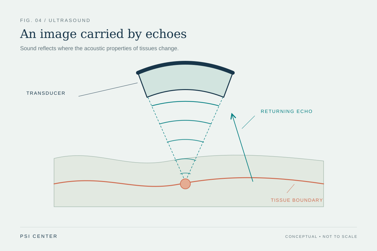
Once the pulse enters tissue, boundaries with differing acoustic properties generate echoes. Their arrival times help estimate depth, while echo amplitudes contribute to image brightness after amplification and other processing. The gel mainly addresses the first contact between probe and body; it does not determine what a deeper echo means. Bone, fluid, soft tissue, beam angle, and attenuation shape the returning signal, and scanner settings affect how that signal is displayed. [1] [4]
This distinction prevents a common category error: a component that improves transmission is not itself evidence of diagnostic accuracy. The coupling layer helps the physical measurement chain begin and end at the probe. Interpretation still depends on image acquisition, modality, anatomy, and clinical expertise. A bright or dark feature cannot be read from gel physics alone, and no material property cited here establishes a treatment benefit.
A small layer with a specific role
The causal chain is straightforward: trapped air creates a large impedance contrast; a large contrast reflects a substantial portion of incident sound; gel displaces air and establishes contact; a more suitable interface allows more sound to pass into tissue and echoes to return. The equations make the mechanism quantitative, while regulatory summaries and laboratory studies show how gel properties can be measured. Together they explain why coupling is built into routine ultrasound imaging. [1] [2] [3]
The limits are just as important. The ideal reflection equation describes a simplified boundary, not an entire scan. One product’s bench results are not a class-wide specification, and material measurements do not establish clinical benefit. Gel is a coupling medium in an imaging system. Its physics supports transmission; diagnosis comes from the complete measurement and interpretation process.
Explore the physics: an explanatory diagram
Sources & further reading
- Ultrasound Imaging U.S. Food and Drug Administration
- 510(k) Summary: Ultrasound Transmission Gels (K221999) U.S. Food and Drug Administration (2022 submission summary)
- Preparation and Evaluation of Polymer-Based Ultrasound Gel and Its Application in Ultrasonography Gels (2022)
- Ultrasound Physics and Instrumentation StatPearls Publishing / NCBI Bookshelf (2023)
Sources checked: 20 November 2026. The calculations illustrate physical models; they are not clinical settings.
Prepared for the independent PSI Center reading library. About edition dates and source checking.
This explainer describes scientific principles. It does not assess individual treatment choices.