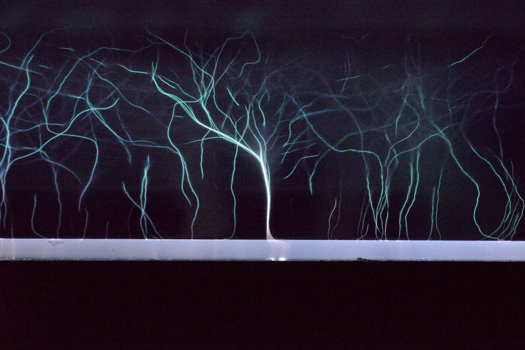
Cold Plasma: Chemistry, Dose, and Wound Healing
A close examination of how a cool electrical discharge could influence wound repair, what randomized trials actually measured, and why seconds of exposure do not define a transferable biological dose.
A closer look at the science
behind better healthcare.
Follow a question from first principles to the clinic. Browse the science of imaging, treatment, materials, and measurement.
343 articles
Deep dives first, then reading editionsA close examination of how a cool electrical discharge could influence wound repair, what randomized trials actually measured, and why seconds of exposure do not define a transferable biological dose.
Follow the signal from a small imbalance in nuclear spins to spatial encoding and reconstruction, with worked examples showing why frequency, resolution, field of view, noise, and scan time are different quantities.
A better dose distribution is a powerful starting point. Range uncertainty, tissue biology, and contrasting randomized trials explain why it is only the beginning of the clinical argument.
How focused sound deposits heat or drives mechanical bubble activity, why imaging and motion control matter, and how indication-specific FDA authorizations differ from evidence of clinical benefit.
A thin gel layer helps ultrasound cross the probe–skin boundary by replacing trapped air and reducing abrupt acoustic changes. The physics explains why contact matters without making gel a diagnostic treatment.
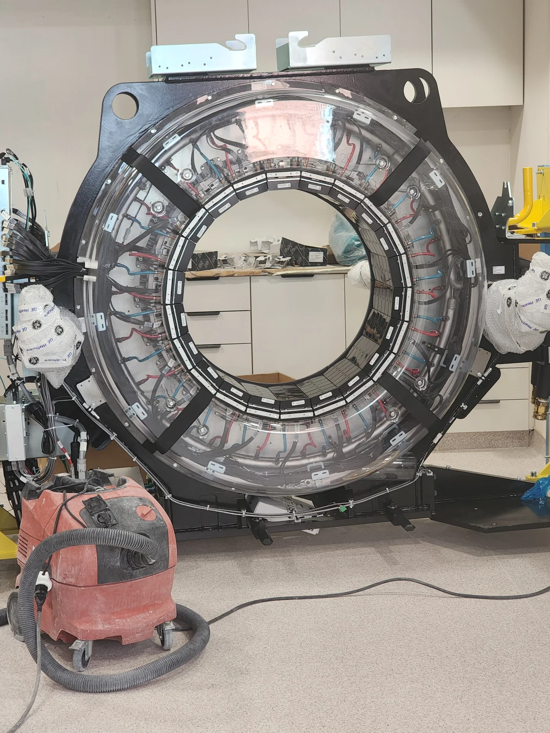
A June 2026 study used physics-constrained machine learning to design an MRI metasurface. Here is what the simulated field gains mean, how RF exposure constrains the design, and what remains untested.
Ultrasound scanners combine timed signals from many probe elements. Delay-and-sum beamforming explains how arrays focus and steer, and why finite geometry and image-quality tradeoffs matter.
An ECG uses voltage differences between electrodes to form leads: calculated views of the heart’s electrical activity. The lead geometry helps explain both the value of a 12-lead tracing and why moving electrodes can change it.
Photon-counting CT converts individual X-ray interactions into electrical pulses and sorts those pulses with energy thresholds. That preserves spectral information, while charge sharing, pulse overlap and calibration still shape the measurement.
A thermal camera converts detected infrared radiation into an estimated surface temperature. Emissivity, reflections, calibration, and image resolution determine how well that estimate represents the object.
An October 2026 study separates cross-channel interference from slower signal drift in ring-array photoacoustic tomography. Its finger and zebrafish tests suggest a useful signal-processing strategy, while leaving clinical performance untested.
A 27 October 2026 study reconstructed richly sampled arterial spin labeling MRI from data undersampled across several dimensions. Its eight-volunteer results preserve research maps, but do not yet establish a faster clinical protocol.
MRI receiver arrays do more than collect signal: their different spatial sensitivities can help reconstruct an image from fewer measurements. The speed gain depends on coil geometry, noise, and how aggressively data are undersampled.
A study published 16 October 2026 compared four photon-counting CT acquisition modes for coronary stenosis assessment. Ultra-high resolution improved image ratings but carried a higher reported dose; a 41-examination retrospective comparison cannot establish a universal best protocol.
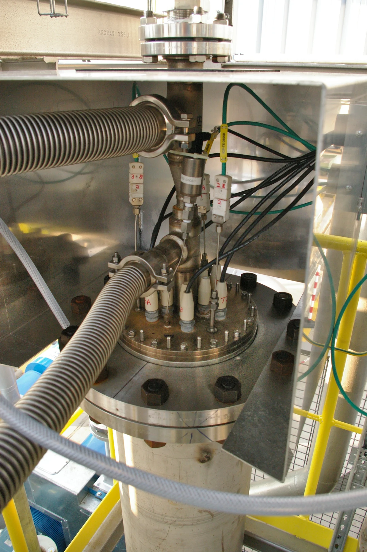
A multijet cold-plasma device study published on 7 November 2026 combined temperature, electrical, coverage and cell tests. Each checks a different part of performance; none alone establishes clinical safety or benefit.
A 2026 pilot study tested iodine-sensitive photon-counting CT measurements in tissue near a resected pancreas. Its signal is promising, but a small retrospective comparison cannot establish a diagnostic cutoff for routine care.
A standardized uptake value (SUV) is a normalized measure of PET tracer concentration, not a count of tumor cells. Its meaning depends on tracer, calculation method, scan protocol, and the question being asked.
Pulsed Doppler estimates motion from returning echoes sampled at intervals. If the frequency shift outruns that sampling rate, the displayed signal folds back into the available range—a measurement limit, not a sudden reversal of flow.