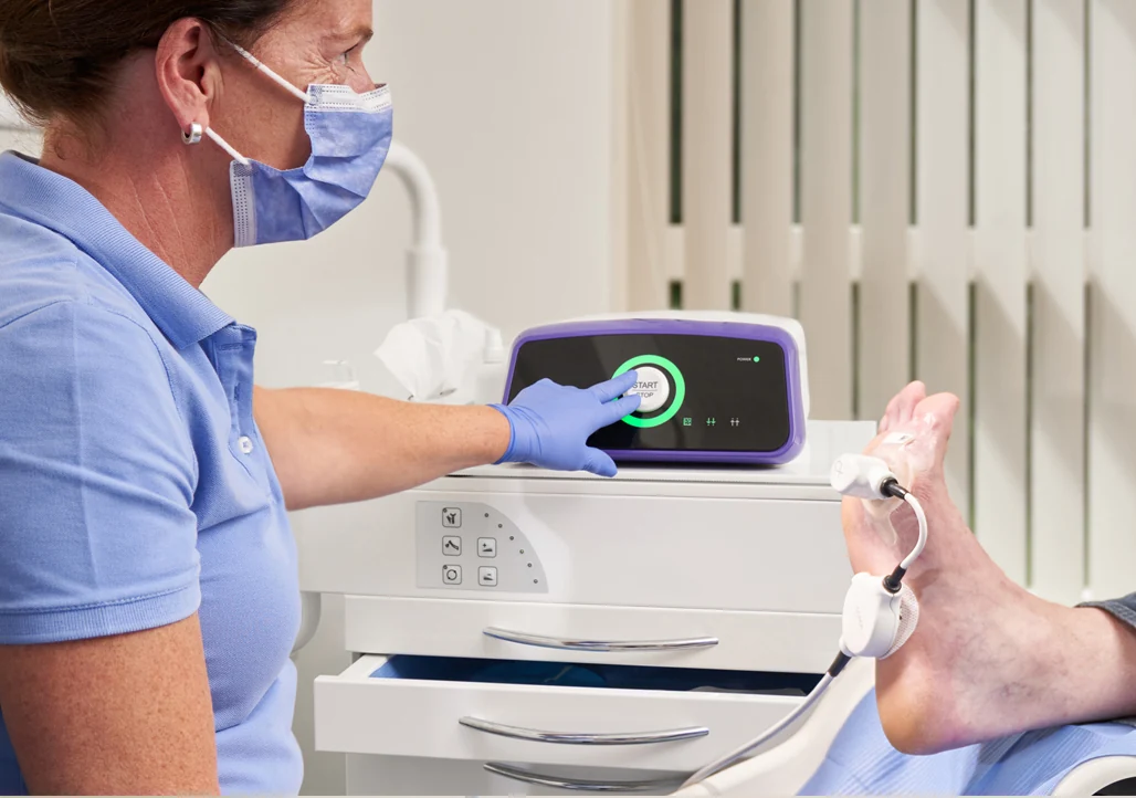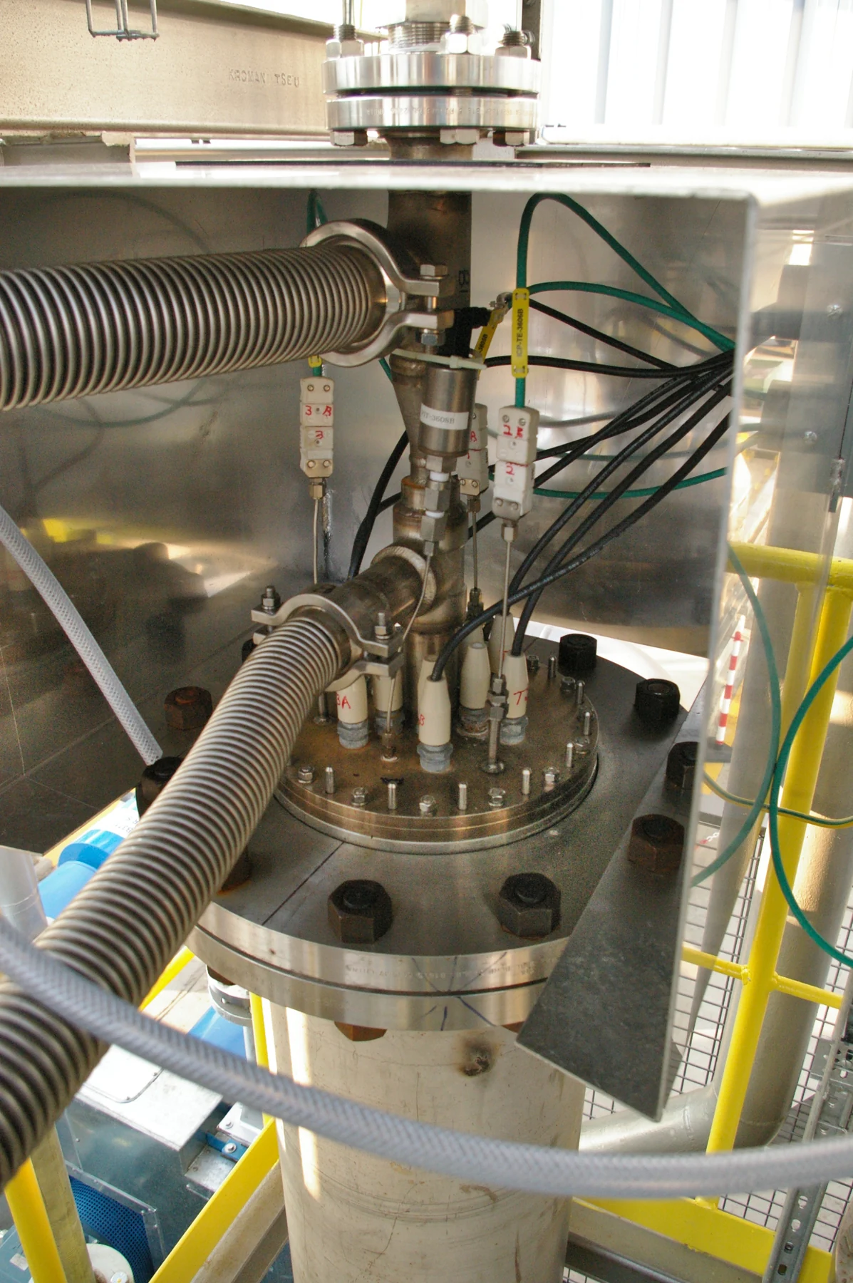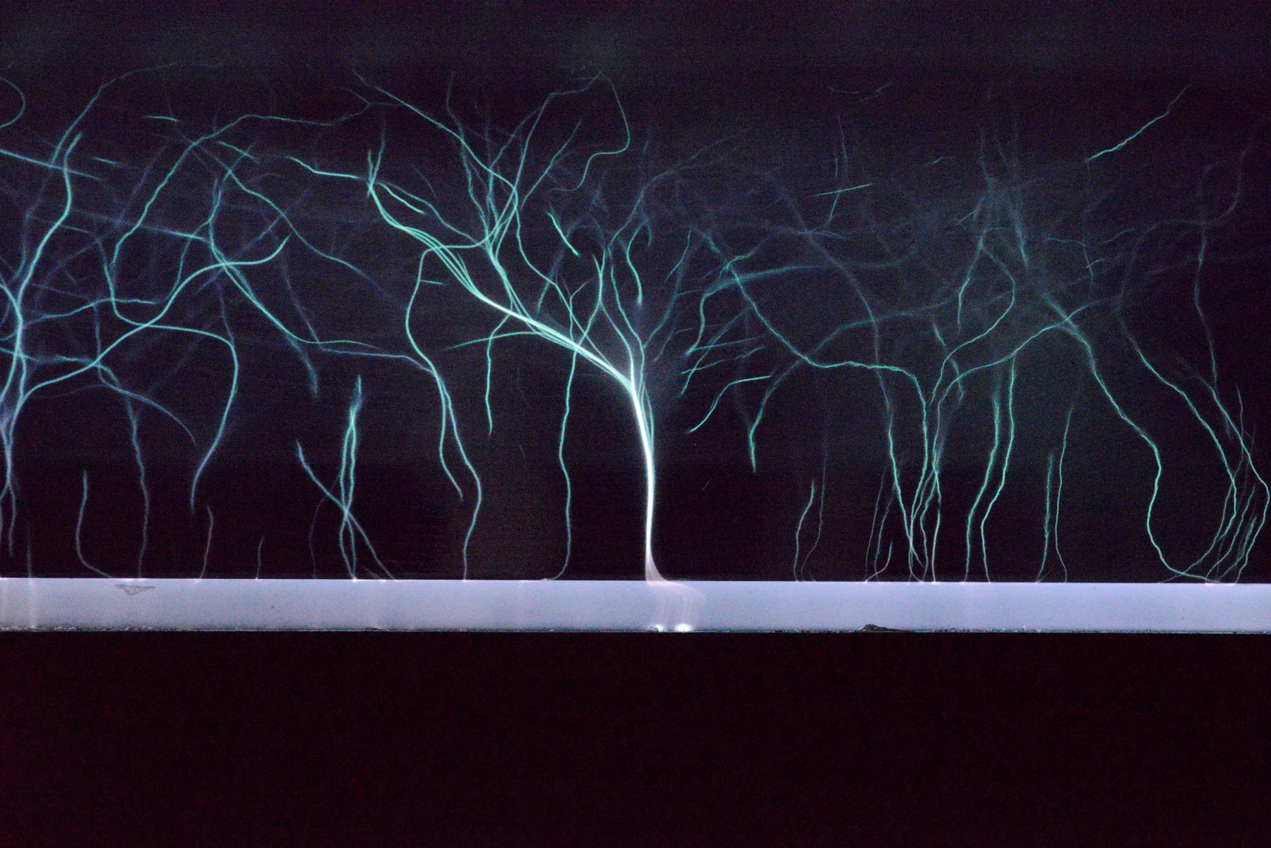
A glowing gas can damage a bacterial membrane and activate a human cell’s defenses. Those effects can coexist, but neither automatically means that a chronic wound will close. Cold atmospheric plasma sits at this difficult junction between electrical engineering, reactive chemistry, and clinical evidence. Several randomized studies report promising results; important questions about devices, comparators, durable healing, and safety remain. Understanding the field requires following energy all the way from an electrode to a wet wound—and then following a study beyond its headline.
1. Cold describes the gas, not the electrons
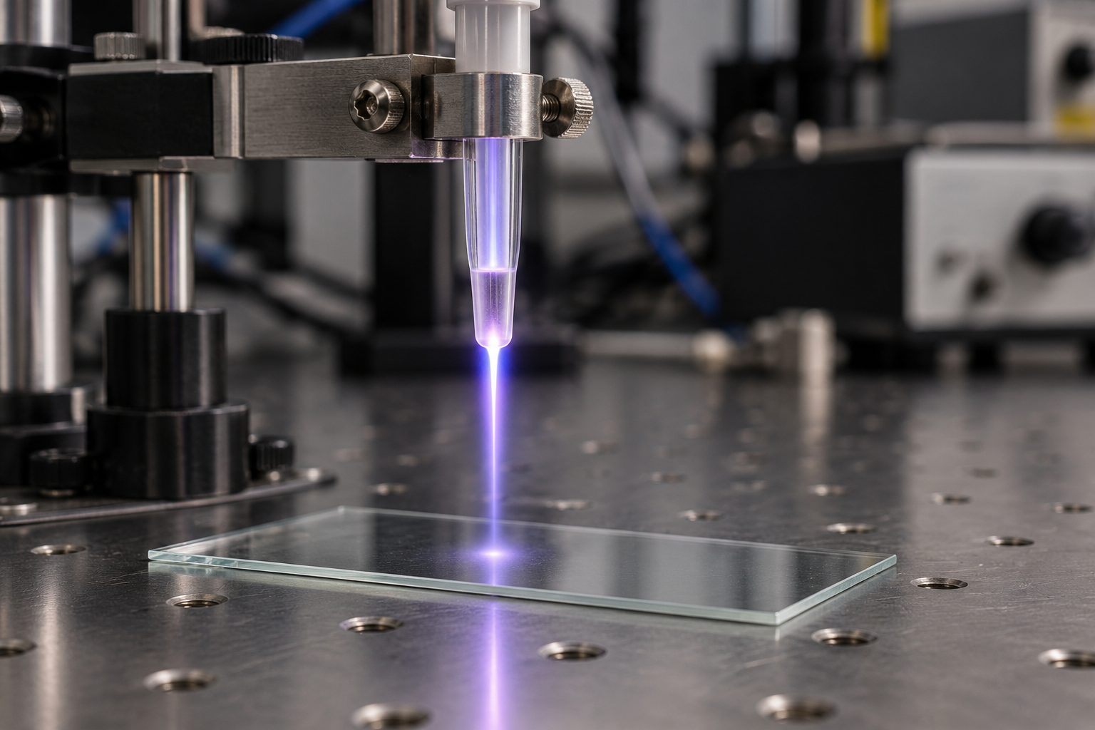
Plasma is a gas containing charged particles alongside neutral atoms and molecules. In a medical cold plasma, an electric field accelerates a small electron population. Electrons can drive excitation, ionization, and molecular dissociation while the much heavier gas particles remain comparatively cool. The electron energy distribution and the gas temperature describe different parts of a system that is far from thermal equilibrium. A photograph of a purple plume conveys little about either quantity.
This separation explains the attraction for fragile tissue: chemistry can be generated without first heating the whole gas to a destructive temperature. It does not establish that an exposure is harmless. Electrical currents, ultraviolet emission, reactive gases, local heating, and the products formed after contact with liquid all require characterization. The current German technical specification addresses medical plasma sources; its existence should not be read as a clinical recommendation for every device or indication. [1]
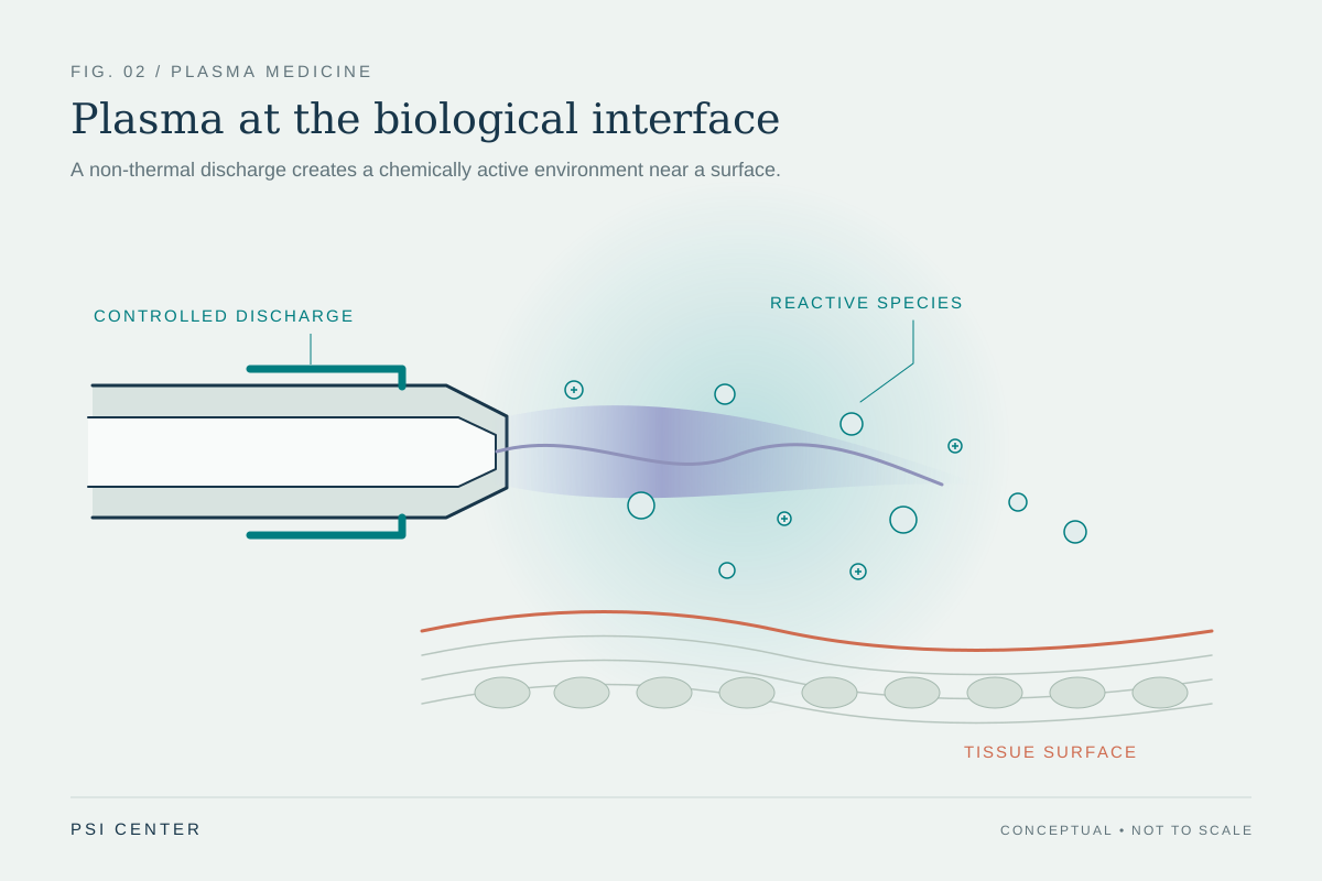
A useful engineering description therefore begins with a chain: electrical input changes electron energies; collisions create reactive species; transport carries some products toward a surface; the surface changes those products again. The final biological response depends on that entire chain. Treating the glow as a single active ingredient hides the very variables that researchers need to control.
2. A jet, a pad, and a chamber deliver different environments
A plasma jet carries a localized discharge or its reactive effluent toward a target in a flowing gas. A dielectric barrier discharge limits current through an insulating barrier; some arrangements incorporate the target into the discharge geometry. Surface microdischarges produce plasma along a dielectric surface and expose a larger region to the resulting environment. These geometries change the footprint, transport path, electric coupling, and importance of gas motion. The construction is part of the intervention.
For a jet, the same total exposure time can be concentrated over a small patch or distributed across a wider area. A larger-area applicator has a different distribution of local exposure even if its nominal electrical power matches the jet’s. Distance matters through both the electrical geometry and the time available for reactions before products reach tissue. A gas-only comparator can also reveal drying or mechanical effects that would otherwise be attributed to plasma chemistry. [2]
Feed gas does not define the complete chemistry. An experimental argon-jet study changed the surrounding gas and observed different reactive chemistry and keratinocyte responses. Ambient air mixes into an effluent, while water vapor supplies additional reaction partners. A device operating in helium, argon, or room air is consequently not just another brand of the same exposure. [3]
| Geometry | Key comparison question |
|---|---|
| Localized flowing jet | How uniformly was the surface covered, and what did gas flow contribute? |
| Discharge coupled to a target | How did target position and electrical properties change the discharge? |
| Surface discharge with a broader footprint | What reached each part of the exposed area? |
3. The wet wound is a chemical reactor
The gas-phase recipe is only the beginning. A wound contains water, salts, proteins, lipids, cells, and extracellular material. A species arriving at this interface may dissolve, react immediately, or be converted into something longer lived. Hydrogen peroxide, nitrite, and nitrate are relatively accessible measurements, but a measurement of one accumulated product is not a census of the reactions that occurred on the way.
Lackmann and colleagues tested this problem with cysteine, a sulfur-containing amino acid. Using spectroscopy, mass spectrometry, and simulations, they found distinguishable product patterns from different plasma sources and gas mixtures. Some less abundant products depended on the source or operating condition. This chemical fingerprinting approach captures information that a single peroxide concentration would miss, although a cysteine solution is much simpler than a wound. [4]
Mixtures also matter. In a helium-plasma cell experiment, hydrogen peroxide and nitrite acted synergistically: the combination had a cell-killing effect that was not explained by considering each component separately. This was a particular liquid-and-cell model, not a universal wound-healing mechanism. It illustrates why equating plasma with a dilute bottle of peroxide is chemically incomplete. [5]
Imagine two wounds exposed to an identical incoming flux. One has a thin liquid layer; another has abundant protein-rich exudate. Their dilution, scavenging capacity, and access to underlying cells differ. Equal delivery at the gas boundary can therefore produce unequal exposure at a cell membrane. This is a transport-and-reaction problem, not simply an uncertainty in the machine’s timer.
4. Oxidative signaling and oxidative injury share ingredients
Reactive oxygen and nitrogen chemistry is not inherently restorative or destructive. Cells use oxidation-sensitive molecular switches, but excessive or poorly located oxidation can damage the same cellular machinery. The relevant distinction includes concentration, location, timing, and the cell’s ability to adapt. A short pulse that initiates a response is biologically different from persistent oxidative stress, even when some chemical species overlap.
In human keratinocyte experiments, Schmidt and colleagues observed activation of NRF2, a transcriptional regulator of antioxidant and detoxification responses. Plasma-treated cells changed the expression of targets including heme oxygenase 1 and NQO1; gene-silencing experiments helped test the pathway’s role. These findings support an adaptive redox response in cultured skin cells. They do not establish a patient-level therapeutic window. [6]
A later study combined diabetic mouse wounds with human wound samples. Plasma-associated healing effects were lost in NRF2-deficient mice, strengthening the case that this pathway mattered in that model. Human wound-exudate measurements also showed changes in NRF2-related and inflammatory signals. The human sampling supports biological plausibility, but does not turn a mouse knockout result into proof that the same pathway explains every clinical benefit. [7]
The tension is fundamental: bacteria and repair cells are exposed in neighboring compartments. An intervention must achieve a useful balance across both, while the balance may shift as a wound changes. “More reactive species” is consequently not a sensible universal optimization target. A good mechanistic study asks which cells were affected, how their function changed, and whether that change helped tissue repair.
5. Why seconds and watts are incomplete dose descriptions
Electrical power measures energy supplied per unit time. It does not specify how much energy reaches tissue as heat, photons, charged particles, or chemical reactivity. Multiplying source power by time gives input energy; dividing by an exposed area gives a nominal energy-per-area value. Neither operation identifies the mixture of biologically active products. Two devices can have the same value while delivering different chemistry and spatial distributions.
A more informative description would track several quantities together: local temperature, electrical coupling, optical emission, reactive-species fluxes, exposed area, and the receiving liquid’s composition. Flux means the amount crossing a unit area per unit time. Its time integral estimates cumulative arrival, but subsequent reactions determine how much remains available to a particular biological target. There is no reason to assume that one scalar will summarize all these processes across devices.
A 2026 comparative laboratory study makes the difficulty concrete. Altering a jet’s circular movement changed cell metabolic responses at identical exposure times. In indirect exposure experiments, changing liquid volume and treatment time did not produce a simple proportional change in biological effect. Transferring treated liquid also omits short-lived species and nonchemical components of direct exposure. These are in-vitro assay findings, not rankings of clinical effectiveness. [2]
For interpreting a trial, the implication is practical: identify the actual device, geometry, application pattern, and accompanying wound care. Evidence for one combination cannot establish an interchangeable dose for another. Reporting only “cold plasma for several minutes” leaves out information needed to understand, reproduce, or compare the exposure.
6. Educational calculation: lifetime sets a transport scale
Consider an idealized molecule diffusing in a uniform liquid without flow. Its characteristic one-dimensional diffusion distance is approximately ℓ = √(2Dτ), where D is the diffusion coefficient and τ is its lifetime before removal. This is an illustrative physical model, not a measured penetration depth for a plasma treatment. Real wounds have binding, barriers, uneven production, and coupled reactions.
Assume D = 2 × 10⁻⁹ m²/s for both of two imaginary species. Give the first a lifetime of one microsecond: ℓ = √(2 × 2 × 10⁻⁹ × 10⁻⁶) m, approximately 6.3 × 10⁻⁸ m, or 63 nanometers. Give the second a lifetime of one second: ℓ is approximately 6.3 × 10⁻⁵ m, or 63 micrometers. A millionfold difference in lifetime creates a thousandfold difference in this distance scale.
The same model gives a diffusion time of roughly 2.5 seconds across a 100-micrometer liquid layer: t ≈ L²/(2D). A species removed much sooner would be unlikely to traverse that layer intact by diffusion alone. Yet it could react near the surface and create a longer-lived product. Biological signaling can also relay a local perturbation without the original molecule reaching every responding cell.
This calculation explains why “penetration” needs a precise subject. The visible plume, an electric field, a short-lived radical, a stable reaction product, and a downstream cellular response have different spatial ranges. An image showing a plume touching skin does not measure any of those ranges. The assumed numbers here illustrate transport scaling; they provide no treatment settings or clinical dose.
7. Fewer bacteria and a healed wound are different endpoints
The early clinical literature often asked an antimicrobial question. Isbary and colleagues’ 2010 randomized study examined 38 chronic infected wounds in 36 people, with each person serving as their own control. The reported bacterial-load reduction was 34%, based on repeated treatment observations. That is evidence about a microbiological endpoint under that study’s conditions, not a 34% increase in the probability of complete healing. [8]
A bacterial culture samples what can be recovered and grown under the assay conditions. It does not directly quantify every organism in a wound, prove that an infection has resolved, or show that tissue has regenerated. Sampling location, timing, recent cleaning, and the organisms targeted can change the result. Repeated observations also need analysis that recognizes their shared patient and wound.
Clinical outcomes form a ladder. At one level are changes in recoverable bacteria. At another are infection signs and symptoms. Wound-area reduction measures tissue progress, but complete epithelial closure is a different endpoint; sustained closure and recurrence address whether repair lasts. Pain, function, admissions, amputations, and quality of life answer further questions. A positive finding on one rung does not establish all the others.
This distinction also separates mechanisms. Improved area reduction without a measurable antimicrobial difference could reflect repair signaling, an insensitive sampling method, or other factors. Conversely, a bacterial decrease without closure could leave the main barrier to healing untouched. Trials need endpoints that match their claims.
8. What the 2020 diabetic-foot sham trial actually showed
Stratmann and colleagues randomized 62 wounds in 43 people with infected diabetic foot ulcers classified as Wagner–Armstrong 1B or 2B. Standard care plus an argon plasma jet was compared with standard care plus a sham, with eight applications over 14 days. At the final assessment, median remaining wound area was 30.5% of baseline in the plasma group versus 55.2% with sham (p = .03). The reported 95% confidence intervals were 12.3–53.5% and 25.2–72.0%, respectively. Infection and microbial-load differences were not significant. Some participants contributed wounds to both groups; the analysis used a patient random effect. No treatment-related adverse events were reported during the observed therapy. [9]
The area result supports a short-term healing signal for this device and study population. It does not mean that 69.5% of patients healed: subtracting remaining area from 100% produces an area reduction, not a person-level closure rate. Nor does the finding demonstrate that bacterial killing caused the improvement. The study’s outcomes must be read separately.
Randomizing wounds can be efficient, but wounds from one person share circulation, metabolism, and care. Treating them as wholly independent would exaggerate the information available. A statistical model can account for that clustering; it cannot transform a small, short study into evidence about long-term recurrence or limb preservation. The distinction matters when a compelling number is reused in a much broader claim.
9. Newer trials add evidence—and different uncertainties
A Dutch venous-leg-ulcer trial randomized 46 people among standard care and two frequencies of direct plasma. Its 12-week complete-healing endpoint favored plasma numerically, but comparisons were inconclusive. Table 3 reported 3/12 healed with control, 8/15 with once-weekly plasma, and 8/13 with twice-weekly plasma; comparison p values were .1560 and .0746. These outcome denominators differ from the randomized group sizes. Recruitment stopped well below the planned 150. The open-label, manufacturer-funded study is informative but cannot settle the effect size. [10]
Strohal and colleagues’ 2025 sham-controlled study enrolled 70 people with chronic wounds of varied origins, predominantly venous or mixed. By day 42, complete closure was reported in 19/35 plasma-treated wounds versus 2/35 controls. Interpretation needs the surrounding care: antiseptic interventions were prohibited in the plasma arm, while infected control wounds could receive antiseptics and antimicrobial dressings. The device manufacturer funded the study and one author was its employee. The result concerns those treatment packages and population, rather than every plasma device or diabetic ulcer. [11]
A July 2026 open-label trial added 50 people with Wagner grade 2 diabetic foot ulcers, comparing standard care with or without a helium-based plasma device over three weeks. It reported favorable wound-size and targeted bacterial-culture outcomes. The short observation period and lack of a sham limit what it resolves about durable healing and blinded comparisons. Its quantitative contraction results also require attention to the reported units before reuse. [12]
These studies should not be combined by simply averaging their headline percentages. Different wounds, devices, standard care, follow-up periods, and outcome definitions produce different clinical questions. Replication becomes convincing when comparable populations and endpoints reproduce a benefit across independent teams, with transparent handling of missing data and concurrent care. A larger apparent effect does not by itself identify the more reliable study.
| Study | Main outcome window | Interpretation constraint |
|---|---|---|
| Venous-ulcer trial [10] | Complete healing within 12 weeks | Small groups; inconclusive between-group comparisons |
| Mixed-wound sham trial [11] | Closure by day 42 | Different permitted antiseptic care; manufacturer support |
| Diabetic-foot trial [12] | Area and selected cultures over 3 weeks | Open label; limited follow-up |
10. Why an encouraging trial can coexist with a negative guideline
The IWGDF 2023 wound-healing guideline recommends against using other gases—including cold atmospheric plasma—in preference to standard care for diabetes-related foot-ulcer healing. Recommendation 14 is labeled strong with low-certainty evidence. Its rationale considers study bias, limited evidence of desirable and undesirable effects, costs, and feasibility. The cited evidence includes the 2020 Stratmann trial; this is not a case of a guideline simply overlooking that result. [13]
As checked on October 30, 2026, IWGDF’s official website still presents the 2023 guideline collection, with an update indicated for 2027. Later studies consequently sit outside that guideline’s evidence window. A future assessment could change the recommendation, retain it, or narrow it to a particular population and device. A website should not predict the committee’s conclusion. [14]
Guidelines ask a broader question than whether any experiment can obtain a statistically significant endpoint. They weigh how confidently an effect is established, whether it matters to patients, what harms or burdens accompany it, and whether it can be implemented. A strong recommendation can therefore coexist with uncertainty in the effect estimate. It communicates a decision under the assessed evidence, rather than declaring the underlying physics impossible.
Scope matters as much as chronology. Guidance for diabetes-related foot ulcers cannot automatically settle venous-ulcer care, and a venous-ulcer trial cannot overturn diabetic-foot guidance. Similarly, technical characterization of a plasma source does not establish that adding the device improves outcomes enough to justify its use. These are related but distinct evaluations.
11. Safety evidence must follow the exposure and the outcome
A low gas temperature addresses one possible source of injury. Safety assessment also needs electrical, optical, chemical, and biological measurements. DIN SPEC 91315:2025-07 replaces the 2014 edition and concerns general requirements for medical cold-plasma sources. This article verified the official catalog record, not the full specification’s detailed requirements or numerical limits. [1]
Laboratory work shows why the receiving environment matters. Boehm and colleagues found that plasma-exposed solutions differed in cytotoxic potential according to their composition; peroxide alone did not explain every observed effect. Their experiments also investigated mutagenicity under particular in-vitro conditions. Such results identify hazards and questions to investigate. They neither prove a clinical cancer risk for a medical device nor establish that all exposures are safe. [15]
“No adverse events observed” is useful information with a denominator and a timescale. A small study can miss uncommon events, delayed effects, or injury in populations it excluded. A cell-metabolism assay measures a different outcome from DNA damage, pain, delayed epithelialization, or durable clinical recovery. Safety claims should specify which of those questions was actually tested.
A decisive next trial would make the exposure traceable, use a credible comparator with otherwise equivalent care, prespecify complete and sustained healing, and report pain and adverse events alongside efficacy. Stratification or planned subgroup analyses could address ulcer cause and perfusion without treating every wound as one category. Independent outcome assessment, adequate recruitment, transparent missing-data analysis, and longer follow-up would make a positive result more informative than another small study of early area change. Those are standards for learning, not a patient treatment protocol.
12. Repair and cancer-cell killing demand different proof
Plasma research also investigates cancer, often aiming to kill cells rather than support their repair. One primary experiment with THP-1 leukemia cells showed that changing the jet’s gas mixture changed its ability to induce apoptosis; the authors implicated oxygen-related chemistry rather than a universal peroxide explanation. That is a mechanistic cell-culture result. It does not establish tumor control, selectivity in a person, or improved survival. [16]
There is no contradiction in investigating related reactive chemistry for both regeneration and destruction. The intended target, delivered exposure, tissue environment, and endpoint change. The error is moving a favorable result from one context into another without testing the connecting steps. A wound-healing trial cannot validate cancer treatment, and cancer-cell killing cannot demonstrate that an exposure is suitable for a healing wound.
For wound care, the strongest scientific case will connect a characterized source to reproducible tissue exposure, an understood biological response, and a clinically meaningful benefit. Each link can fail independently. The field already has credible experiments at several links; the remaining task is to connect them with sufficiently rigorous clinical evidence.
When reading the next plasma headline, identify the device, the wound population, the comparator, the endpoint, and the duration of observation. Then ask whether the result was independently reproduced and whether its uncertainty allows the advertised claim. These questions preserve what is promising about the physics while keeping the conclusion proportional to the evidence.
Explore the physics: an explanatory diagram
Sources & further reading
- DIN SPEC 91315:2025-07: General requirements for plasma sources for the generation of cold atmospheric pressure plasma (CAP) for medical applications DIN Media / DIN
- Challenges in the standardization of in vitro cytotoxicity assays for comparative risk assessment of cold atmospheric pressure plasma devices Scientific Reports, Boeckmann et al., 2026
- Role of Ambient Gas Composition on Cold Physical Plasma-Elicited Cell Signaling in Keratinocytes Biophysical Journal, Schmidt et al., 2017
- Chemical fingerprints of cold physical plasmas – an experimental and computational study using cysteine as tracer compound Scientific Reports, Lackmann et al., 2018
- Synergistic Effect of H2O2 and NO2 in Cell Death Induced by Cold Atmospheric He Plasma Scientific Reports, Girard et al., 2016
- Non-thermal Plasma Activates Human Keratinocytes by Stimulation of Antioxidant and Phase II Pathways Journal of Biological Chemistry, Schmidt et al., 2015
- Medical gas plasma modifies Nrf2 signaling in diabetic wound healing Journal of Advanced Research, Schmidt et al., online 2025 / issue 2026
- Cold Atmospheric Argon Plasma on Chronic Wounds: Randomized Trial Isbary et al., British Journal of Dermatology (2010)
- Effect of Cold Atmospheric Plasma Therapy vs Standard Therapy Placebo on Wound Healing in Patients With Diabetic Foot Ulcers: A Randomized Clinical Trial JAMA Network Open, Stratmann et al., 2020
- Improved Wound Healing by Direct Cold Atmospheric Plasma Once or Twice a Week: A Randomized Controlled Trial on Chronic Venous Leg Ulcers Advances in Wound Care, Bakker et al., online 2024 / issue 2025
- Treatment of chronic wounds with cold plasma: a randomised, single-blind, placebo-controlled clinical study Journal of Wound Care, Strohal et al., 2025
- Cold atmospheric plasma accelerates wound closure and reduces pathogenic bacterial load in diabetic foot ulcers: a randomized controlled trial Scientific Reports, Hedayat Yaghoobi et al., 2026
- Guidelines on Interventions to Enhance Healing of Foot Ulcers: 2023 Update International Working Group on the Diabetic Foot
- IWGDF Guidelines 2023: all guidelines International Working Group on the Diabetic Foot
- Cytotoxic and mutagenic potential of solutions exposed to cold atmospheric plasma Scientific Reports, Boehm et al., 2016
- Oxygen atoms are critical in rendering THP-1 leukaemia cells susceptible to cold physical plasma-induced apoptosis Scientific Reports, Bekeschus et al., 2017
Sources checked: 30 October 2026. The calculations illustrate physical models; they are not clinical settings.
Prepared for the independent PSI Center reading library. About edition dates and source checking.
This explainer describes scientific principles. It does not assess individual treatment choices.