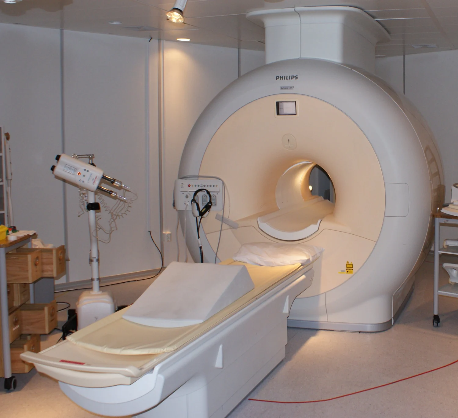
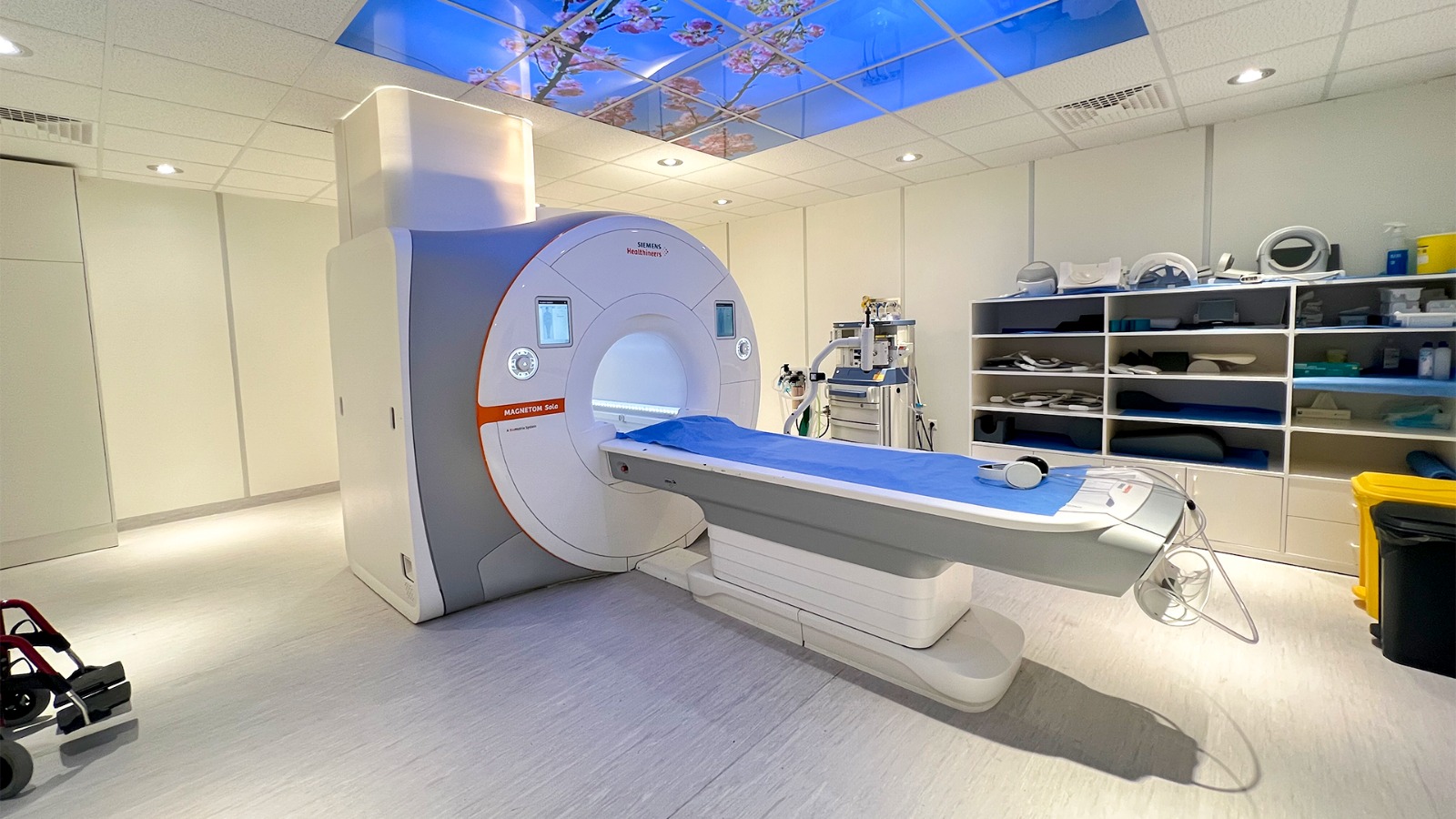
Follow the signal from a small imbalance in nuclear spins to spatial encoding and reconstruction, with worked examples showing why frequency, resolution, field of view, noise, and scan time are different quantities.
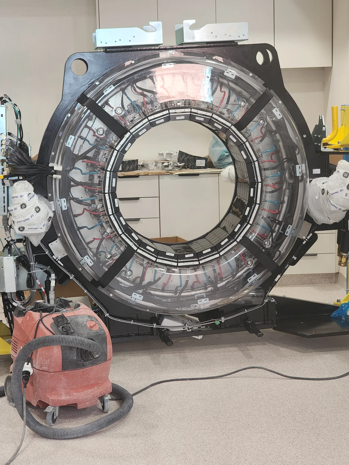
A June 2026 study used physics-constrained machine learning to design an MRI metasurface. Here is what the simulated field gains mean, how RF exposure constrains the design, and what remains untested.
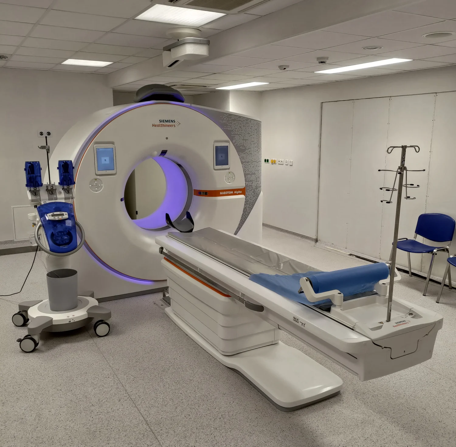
Photon-counting CT converts individual X-ray interactions into electrical pulses and sorts those pulses with energy thresholds. That preserves spectral information, while charge sharing, pulse overlap and calibration still shape the measurement.
An October 2026 study separates cross-channel interference from slower signal drift in ring-array photoacoustic tomography. Its finger and zebrafish tests suggest a useful signal-processing strategy, while leaving clinical performance untested.
A 27 October 2026 study reconstructed richly sampled arterial spin labeling MRI from data undersampled across several dimensions. Its eight-volunteer results preserve research maps, but do not yet establish a faster clinical protocol.
MRI receiver arrays do more than collect signal: their different spatial sensitivities can help reconstruct an image from fewer measurements. The speed gain depends on coil geometry, noise, and how aggressively data are undersampled.
A study published 16 October 2026 compared four photon-counting CT acquisition modes for coronary stenosis assessment. Ultra-high resolution improved image ratings but carried a higher reported dose; a 41-examination retrospective comparison cannot establish a universal best protocol.
A 2026 pilot study tested iodine-sensitive photon-counting CT measurements in tissue near a resected pancreas. Its signal is promising, but a small retrospective comparison cannot establish a diagnostic cutoff for routine care.
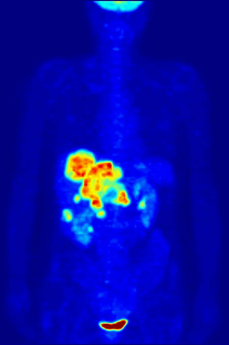
A standardized uptake value (SUV) is a normalized measure of PET tracer concentration, not a count of tumor cells. Its meaning depends on tracer, calculation method, scan protocol, and the question being asked.
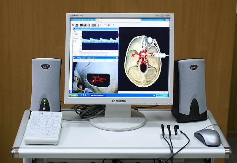
Pulsed Doppler estimates motion from returning echoes sampled at intervals. If the frequency shift outruns that sampling rate, the displayed signal folds back into the available range—a measurement limit, not a sudden reversal of flow.
An October 2026 phantom study tested whether spectral photon-counting CT could distinguish and quantify eight high-atomic-number elements in a lung-shaped model. The work supports a measurement concept, not a validated lung test or clinical benefit.
At the 18 October 2026 EANM congress, a small EQUIP study compared short reconstructions from a long-axial-field PET/CT scanner with conventional PET images. Stronger reader ratings are a promising image-quality result, not yet proof of equal diagnostic accuracy.
CTDIvol and DLP make scanner output comparable, but they describe reference conditions. Patient size, anatomy, scan coverage, and the quantity being estimated all matter.
A prospective study published 15 October 2026 compared low-dose photon-counting CT with standard-dose scans for measuring interstitial lung disease in adults with inflammatory myopathy. Its close agreement is promising feasibility evidence, not proof of long-term monitoring or improved care.
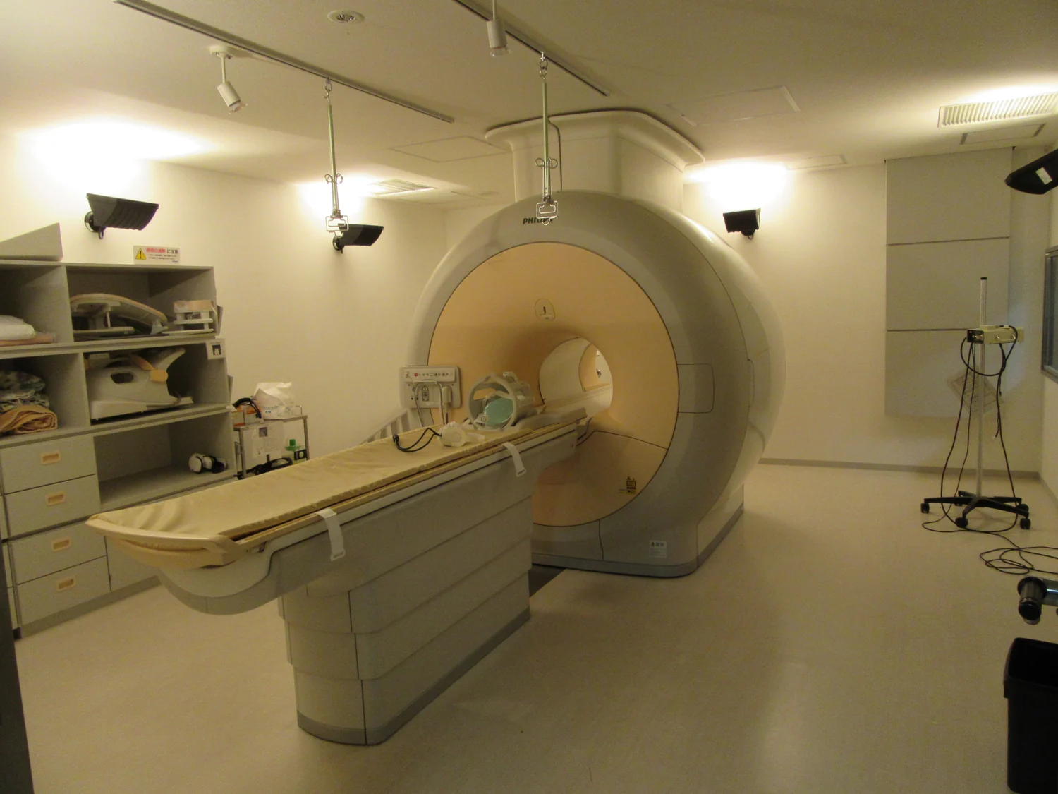
MRI converts tiny changes in hydrogen-proton behavior into spatial detail. Magnetic fields align the signal, radiofrequency pulses disturb it, and carefully timed measurements distinguish tissue environments without x-rays.
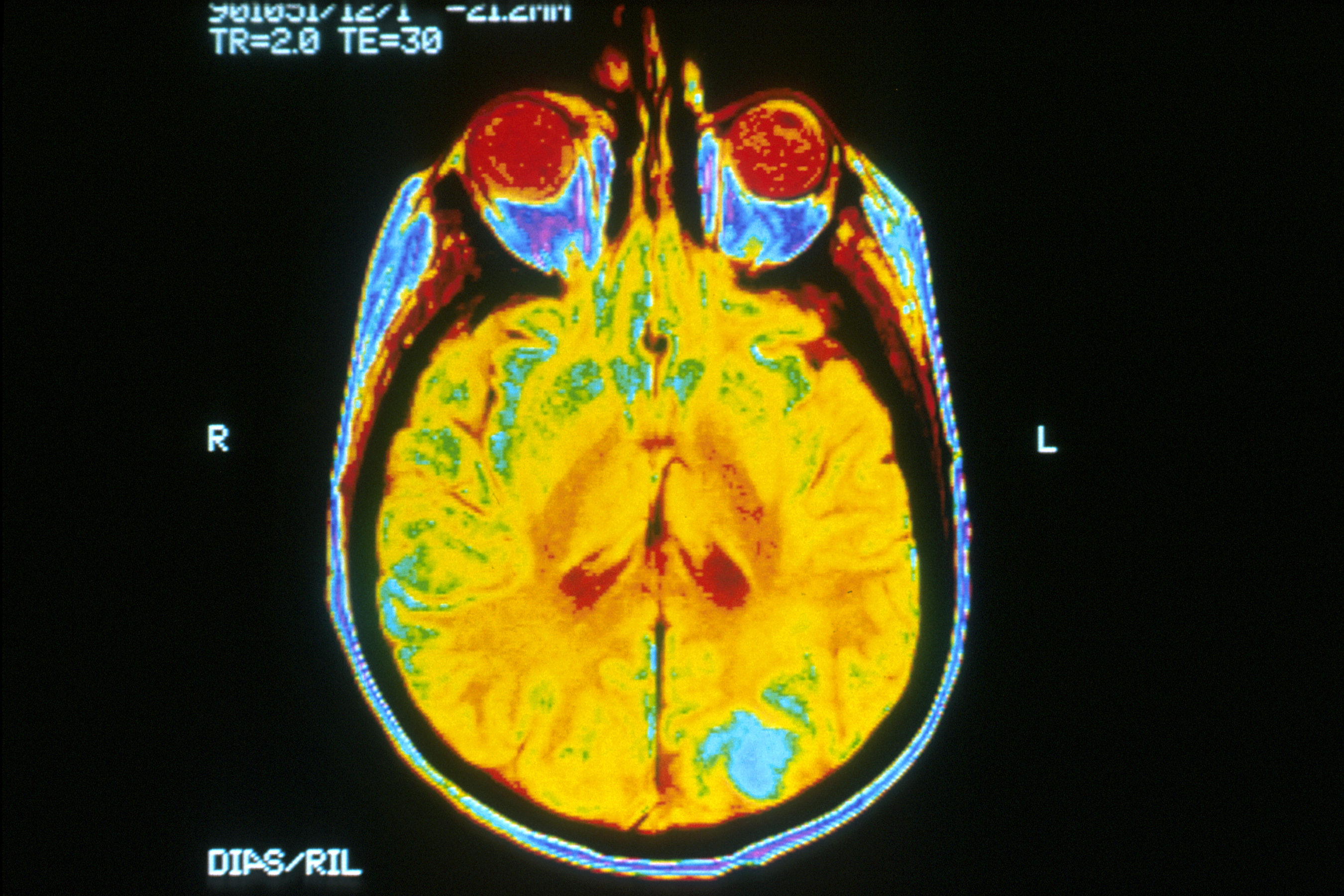
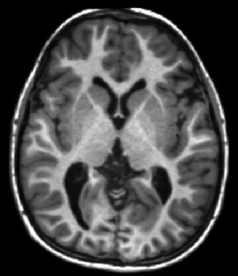
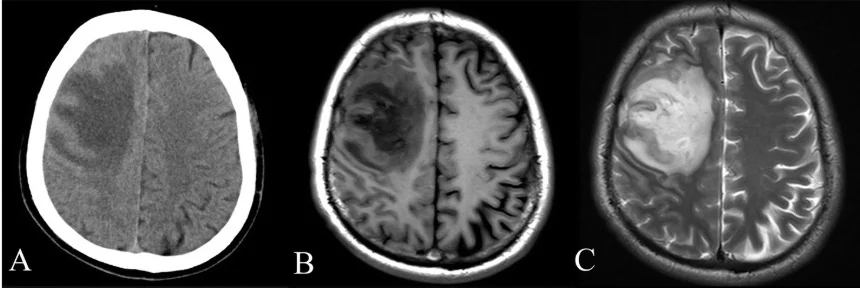
MRI contrast comes from how protons respond and recover in different tissue environments, as well as from scan settings and image processing. Optical imaging measures light interactions and can add complementary information at other scales.
Children’s imaging asks a practical physics question: how can a scanner gather enough information to answer a clinical question while accounting for a smaller body and a longer lifetime ahead?
A radiotracer combines a radioactive atom with a carrier that helps determine where it travels. The resulting signal can reveal biological activity, while the carrier and isotope shape what the scan can show.