A scan lasting one minute sounds like a simple efficiency upgrade. In positron emission tomography (PET), however, acquisition time affects how many radioactive-decay events the scanner can use to build an image. A recent study presentation offers a useful case study in the distinction between a sharper-looking image and proof that a scan answers a clinical question just as well.
Why a longer detector can change the timing
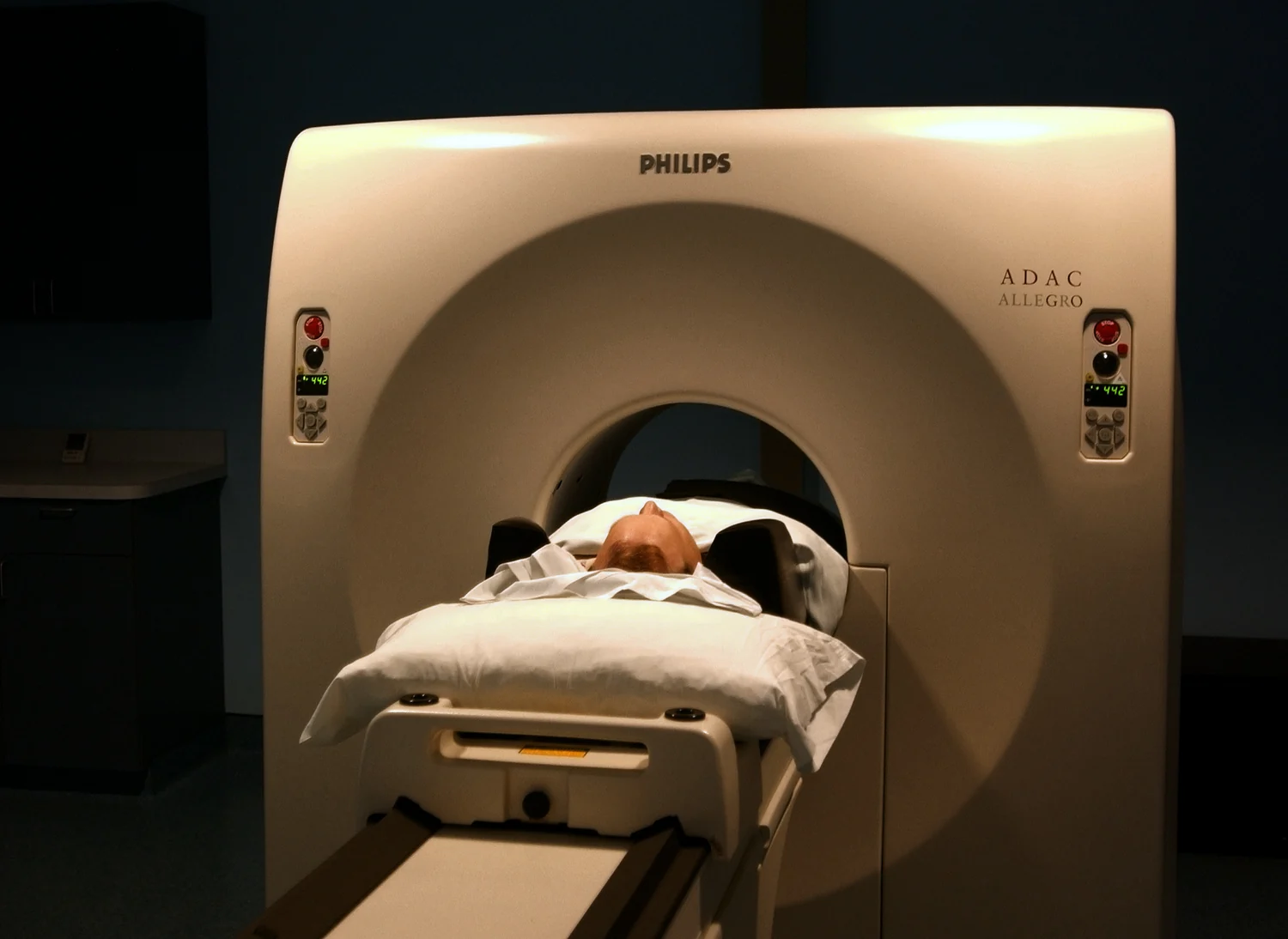
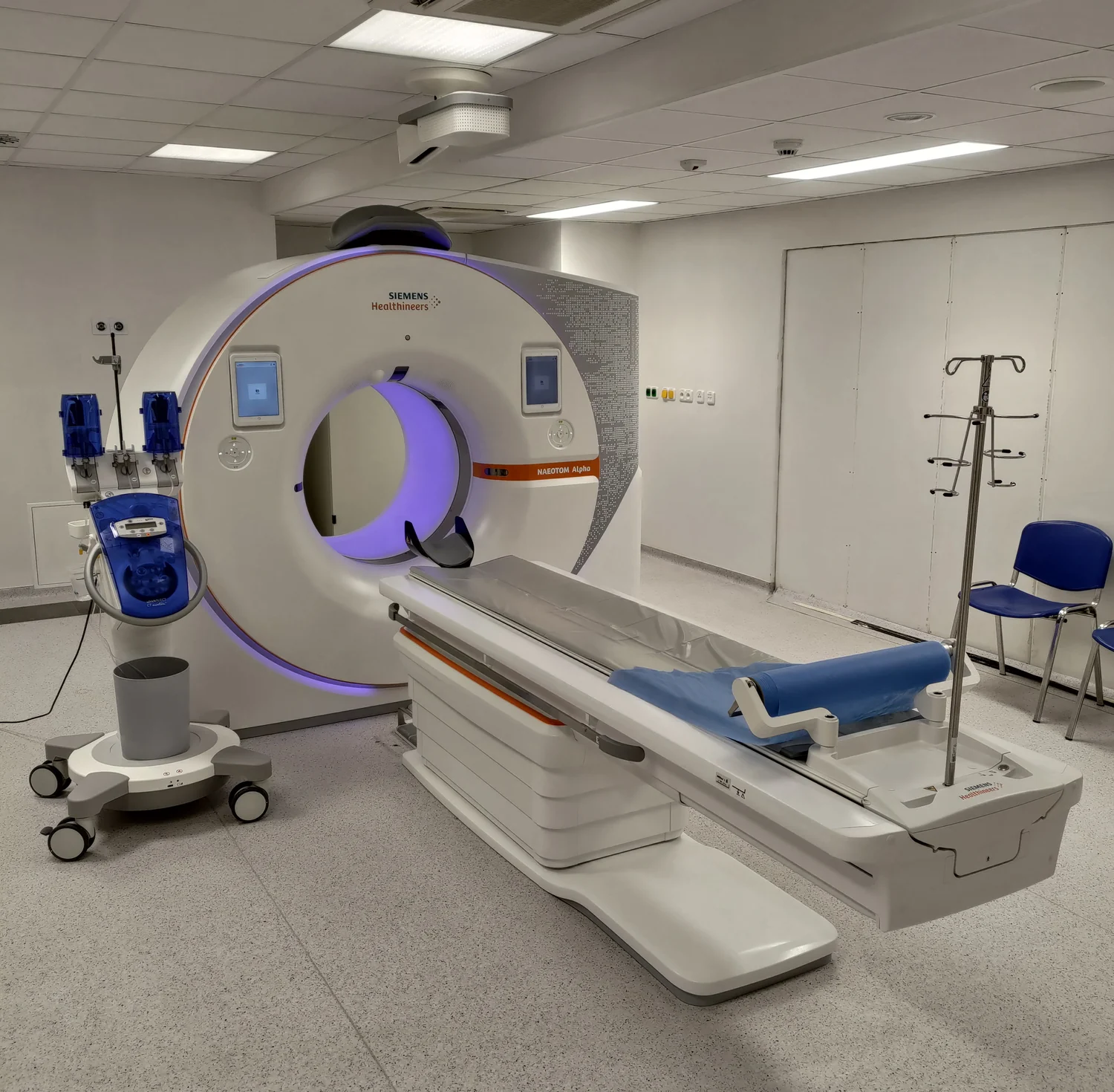
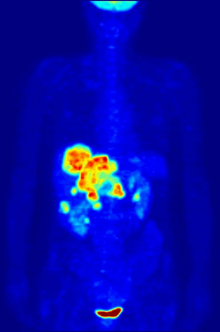
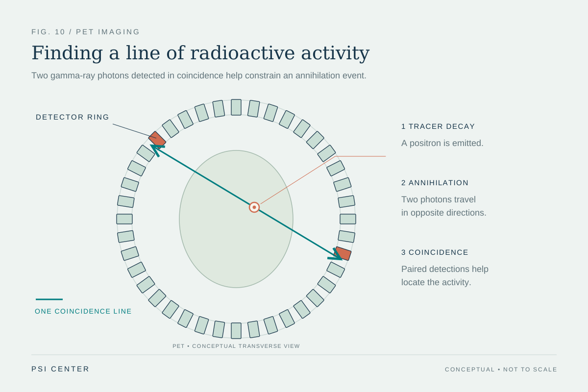
PET detects pairs of gamma photons produced when a positron from a radiotracer meets an electron. The scanner uses many detected pairs to estimate where tracer-related activity is distributed. A conventional short-axial scanner covers only a limited length of the body at once, so whole-body imaging generally requires moving the table through several positions. A long-axial-field (LA-FOV) system covers much more of the body in one position and can register more of the emitted signal. That higher sensitivity creates options: use the counts to shorten acquisition, reduce administered activity, or improve image statistics. It does not make those choices interchangeable or guarantee that every task tolerates the same trade-off [source].
The EQUIP study compared routine scans on a conventional short-axial-field system with scans on a 128-centimeter LA-FOV PET/CT system. Twenty-four participants, who had received one of five radiotracers, underwent a 10-minute LA-FOV acquisition. Investigators reconstructed subsets using 5, 3, 2, and 1 minute of data. Two nuclear medicine specialists who did not know the acquisition times rated image quality, sharpness, lesion conspicuity, and noise [1].
A strong rating is a specific kind of result
On a five-point scale, average overall image-quality ratings for LA-FOV images were 5.0 at 10 and 5 minutes, 4.9 at 3 and 2 minutes, and 4.5 at 1 minute. The conventional scanner's average was 3.9. The one-minute images were therefore rated more favorably on average in this comparison, although scores declined as acquisition shortened, particularly for noise. A reader score records expert judgment about image appearance and usability under the study's assessment method. It is not a direct count of correctly detected cancers, nor does it tell us how often a finding changed a diagnosis.
That distinction is familiar from measurement science: a tool may produce repeatable, legible measurements while still needing validation for a particular decision. An image-quality scale helps compare images under defined conditions. Diagnostic accuracy asks whether the images correctly identify disease against an appropriate reference. EQUIP's reported ratings do not answer all of those questions [1].
Counts, noise, and a useful thought experiment
A simplified count model helps explain why speed is not free. If detected events are independent and the expected count falls from N to N/2, the relative statistical fluctuation grows roughly as 1/√(N/2), or √2 times the fluctuation at N. Real PET reconstruction is more complicated: corrections, detector geometry, tracer distribution, patient motion, and reconstruction settings also matter. The relationship is a teaching approximation, not a prediction of this scanner's performance. It explains why using a tenth of the acquisition time can increase noise even when the scanner captures more signal than a shorter-field system.
The researchers also reported broadly consistent signal-to-noise ratios in healthy liver tissue between the 10-minute and 1-minute data for the tracers assessed. That is a quantitative image measure in one tissue region, not evidence that every small or faint lesion remains visible. A large high-contrast feature and a subtle low-contrast feature place different demands on the signal. Good-looking averages can coexist with a meaningful risk of losing detail in selected cases [1].
What the comparison did—and did not—test
This was a small, single-center comparison involving 24 people and five tracers on one LA-FOV system. The long-axial images were reconstructed from data collected during a 10-minute scan; the one-minute set represents a shorter portion of that acquisition. The design therefore tests image appearance under shortened data collection. It does not, by itself, establish the performance of a one-minute protocol across scanner models, patient groups, tracers, or clinical indications. Nor does a conference presentation carry the same evidentiary detail as a complete peer-reviewed report. The EANM presentation record and EQUIP registry identify the research context; the registry is not a substitute for published results [2] [3].
The trade-off is a choice, not a universal benefit
Higher sensitivity may let a department choose faster imaging, lower radiotracer activity, or greater counting statistics. Which option matters depends on the clinical question, patient, tracer, scanner, and local protocol. A one-minute acquisition could be valuable for someone who struggles to remain still, but an image appearance study alone cannot demonstrate improved comfort or fewer motion-related failures. Likewise, a shorter scan does not automatically mean a lower radiation burden if administered activity and the CT component are unchanged.
Before routine use is justified for a specific task, studies should test whether shortened acquisitions preserve lesion detection and diagnostic decisions, including small and low-contrast findings, and should report performance across more patients and settings. Image-quality scoring is a useful early screen: it can show that a technical approach deserves further evaluation. It is one step in a chain of evidence, not the final clinical outcome [1].
How to read the headline
The defensible takeaway is that a 128-centimeter long-axial PET/CT system produced one-minute reconstructions that two blinded readers rated higher on average than conventional scans in this small comparison. That is a notable feasibility signal. The stronger claim—that one-minute whole-body PET is diagnostically equivalent or ready for every routine examination—was not established by the reported reader ratings. The physics explains why the possibility is real; careful task-specific validation will determine where the speed is useful.
Explore the physics: an explanatory diagram
Sources & further reading
- Next-generation total-body PET scanner can produce diagnostic-quality images in as little as 1 minute European Association of Nuclear Medicine (via MedicalXpress)
- EANM’26 Abstracts Book European Association of Nuclear Medicine
- EQUIP: Evaluation of Quality of Imaging on a Next-generation Total Body PET Scan in Comparison to Conventional PET ClinicalTrials.gov; study sponsor: Peter MacCallum Cancer Centre
Sources checked: 2 November 2026. The calculations illustrate physical models; they are not clinical settings.
Prepared for the independent PSI Center reading library. About edition dates and source checking.
This explainer describes scientific principles. It does not assess individual treatment choices.