How can a camera show not only the shape of an organ, but what its cells are doing? Nuclear medicine answers by tracking a radioactive tracer: a molecule selected to travel through or interact with a particular process in the body. Its emitted signal maps activity, while methods such as CT can supply the anatomical landmarks that explain where that activity lies.
A molecule becomes a moving marker
A radiotracer combines a carrier molecule with a radioactive atom. The carrier is chosen for its biological behavior: it may bind to a target, participate in a process, or travel with a particular kind of cell. The radioactive atom makes the carrier detectable from outside the body. The resulting image therefore reflects the tracer’s distribution, which can provide evidence about a bodily function rather than simply outline an organ [1].
Consider red blood cells labeled with a radioactive atom and returned to the bloodstream. A gamma camera can follow their path; an unusual accumulation in the intestine may help locate bleeding. The camera does not see blood as a colored fluid. It detects emitted gamma rays and reconstructs where the labeled cells have traveled. What the image means depends on the tracer and the question being asked.
Two routes to a three-dimensional map
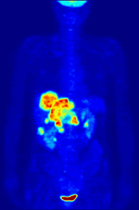
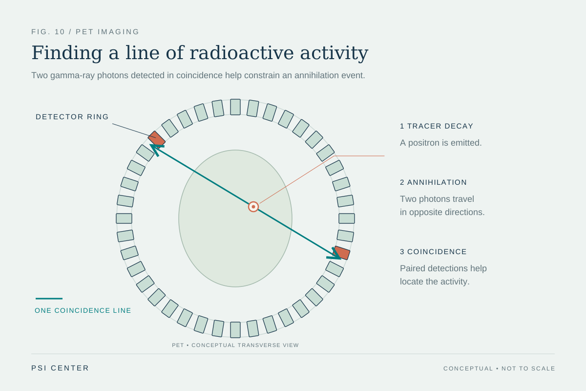
SPECT and PET both use rotating or surrounding detectors and computer reconstruction to create three-dimensional maps, but their signals arise differently. In SPECT, detectors register gamma rays emitted by the tracer. In PET, a positron emitted by the radionuclide meets an electron in the body; their annihilation produces two gamma photons traveling in opposite directions. Detecting those paired photons helps the scanner infer where the event occurred [1].
The distinction is not that one method shows anatomy and the other shows function: both map radiotracer distribution. Their tracers and detection physics differ. A selected tracer can make a process visible because its uptake or movement relates to that process. For example, FDG is a radiolabeled glucose-like molecule used in many cancer studies; tissues with high glucose use can take up more of it, though uptake alone does not establish a diagnosis.
Activity needs an address
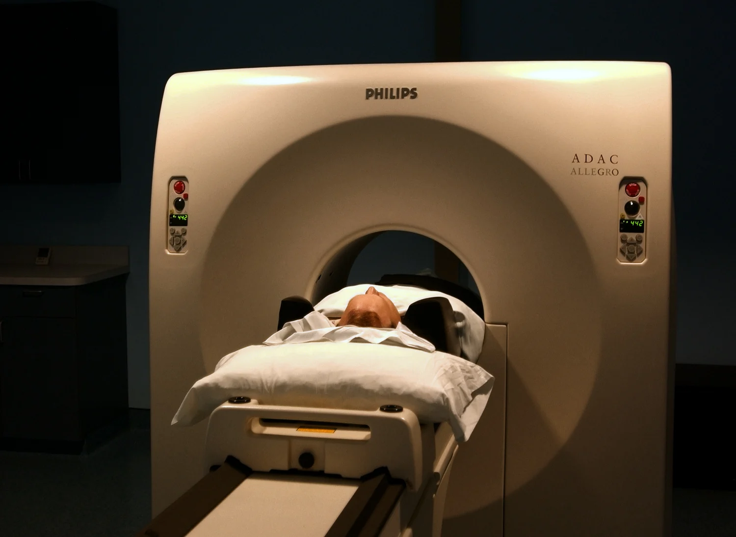
A functional signal becomes easier to interpret when it is aligned with a structural map. A PET/CT system acquires both types of information for corresponding body regions. The PET component indicates where the tracer is concentrated; CT provides anatomical detail that helps locate that signal. Combining them can clarify whether an active region corresponds to a particular structure or lesion [1].
This is like seeing a city’s traffic intensity overlaid on its street map. The traffic pattern answers where movement is concentrated, while the map identifies the roads and buildings. The analogy has limits: tracer uptake is not a direct measure of every cellular action, and image interpretation depends on the tracer, timing, and clinical context. A structural image alone and a tracer map alone answer different questions.
Imaging and treatment share a targeting idea
Some radiopharmaceuticals are used for therapy as well as diagnosis. In a radiotheranostic approach, a diagnostic tracer may first show whether a molecule reaches a relevant target. A related therapeutic compound can carry a different radioactive atom, selected for emissions that deposit energy in tissue. A medical physicist may use diagnostic images in dosimetry, estimating radiation absorbed by organs [1].
Radioactive emissions are ionizing radiation, which has enough energy to damage DNA; that property is relevant to both potential risk and therapeutic effect. NCI notes that radiation exposure from medical procedures can cause cell damage, while the risks from medically indicated imaging are generally small relative to expected benefit [2]. The practical lesson is that an image is not simply a photograph of the body: it is a measurement shaped by a chosen molecule, its physical signal, and the anatomical context used to interpret it.
Explore the physics: an explanatory diagram
Sources & further reading
- Nuclear Medicine NIBIB
- Radiation National Cancer Institute
Prepared for the independent PSI Center reading library. About edition dates and source checking.
This explainer describes scientific principles. It does not assess individual treatment choices.