How can a scan reveal both where a tumor is and what its cells are doing? PET and CT answer different parts of that question. One follows a radioactive tracer through biological activity; the other uses x-rays to map internal structure. Placing the two images together can make a signal easier to interpret because function gains an anatomical address.
PET follows a molecular signal
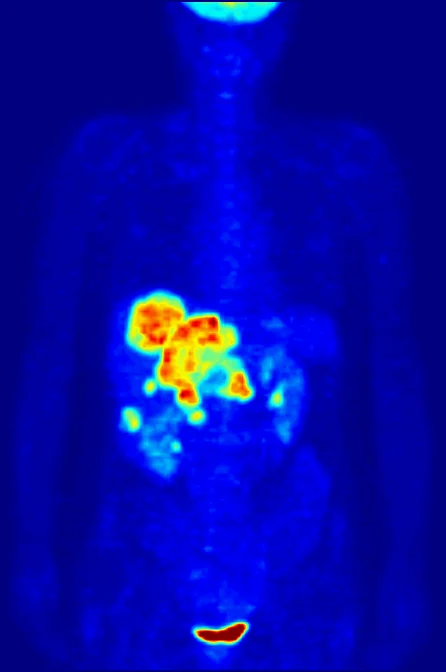
Positron emission tomography, or PET, is a form of nuclear medicine imaging. A radiotracer combines a carrier molecule with a radioactive atom. The carrier is chosen to participate in or bind to a process of interest, so the tracer’s distribution can provide information about function rather than simply outlining an organ. [1]
In a common cancer imaging example, a tracer based on a modified glucose molecule can accumulate more in tissue using glucose intensely. Many rapidly dividing cancer cells have high glucose use, so the resulting signal can help reveal a tumor or spread. But uptake is a biological clue, not a photograph of cancer cells themselves; interpretation depends on the tracer, the body region, and the clinical question.
CT supplies the map
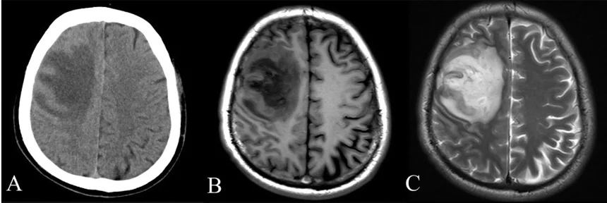
Computed tomography (CT) makes cross-sectional images with rotating x-ray beams. Detectors measure how much x-ray signal emerges after passing through the body, and a computer reconstructs slices that can be stacked into a three-dimensional view. Bone and other dense structures are especially visible, while contrast agents can help distinguish some soft tissues and blood vessels. [2]
A mental picture is a glowing street seen on a city map. PET is like marking the places where a particular activity is concentrated; CT provides streets, buildings, and landmarks. Without the map, a PET signal may be hard to place precisely. Without the activity marker, CT may show an abnormal shape without indicating the cellular process associated with it.
Registration makes signals local
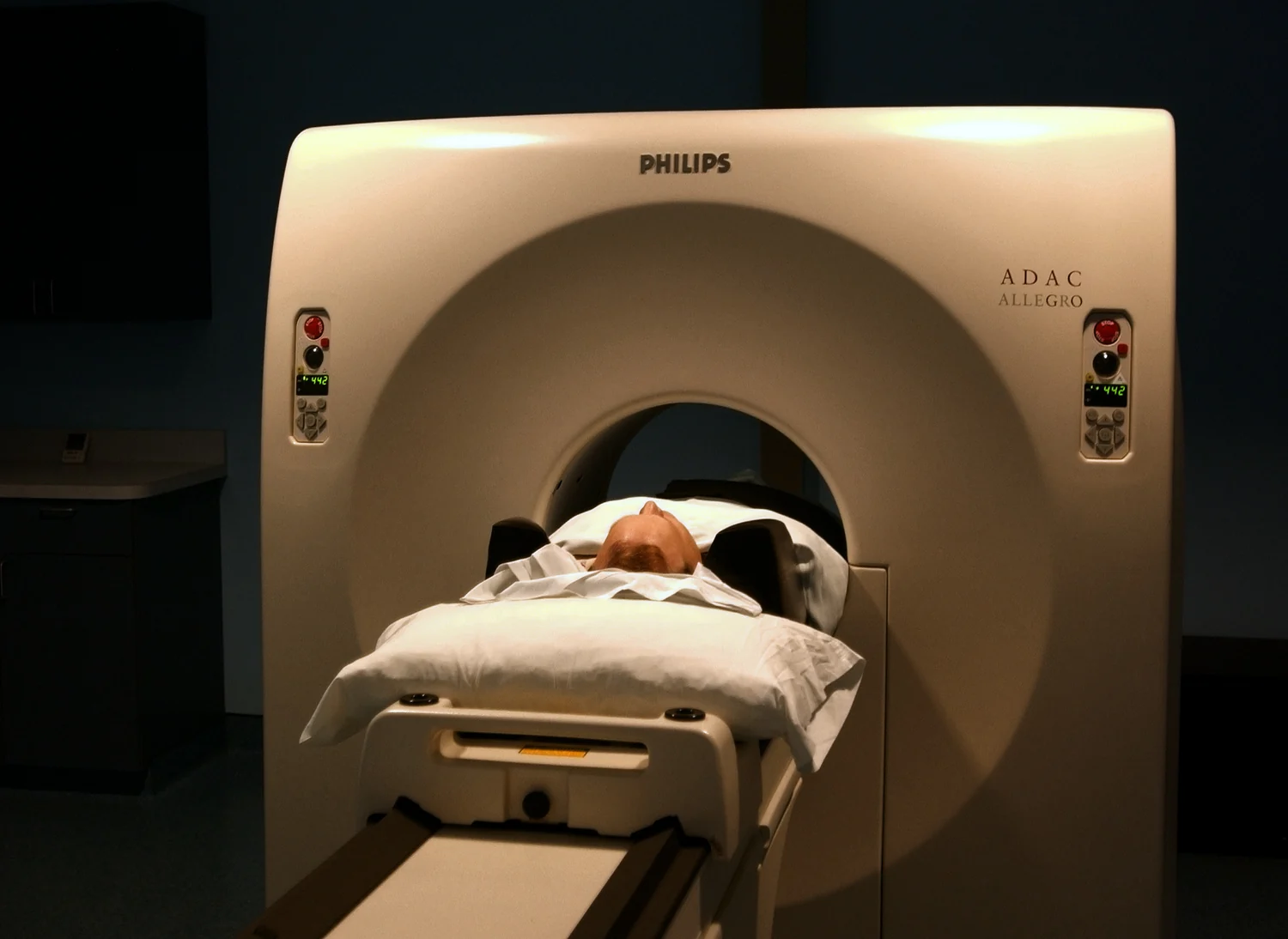
A combined PET/CT scanner acquires both kinds of information for the same body region in one examination. Software aligns or fuses the images so a reader can see tracer uptake against CT anatomy. The result can clarify whether a functional hotspot lies in a lymph node, bone, or another structure, and PET/CT is widely used in cancer staging and monitoring. [1]
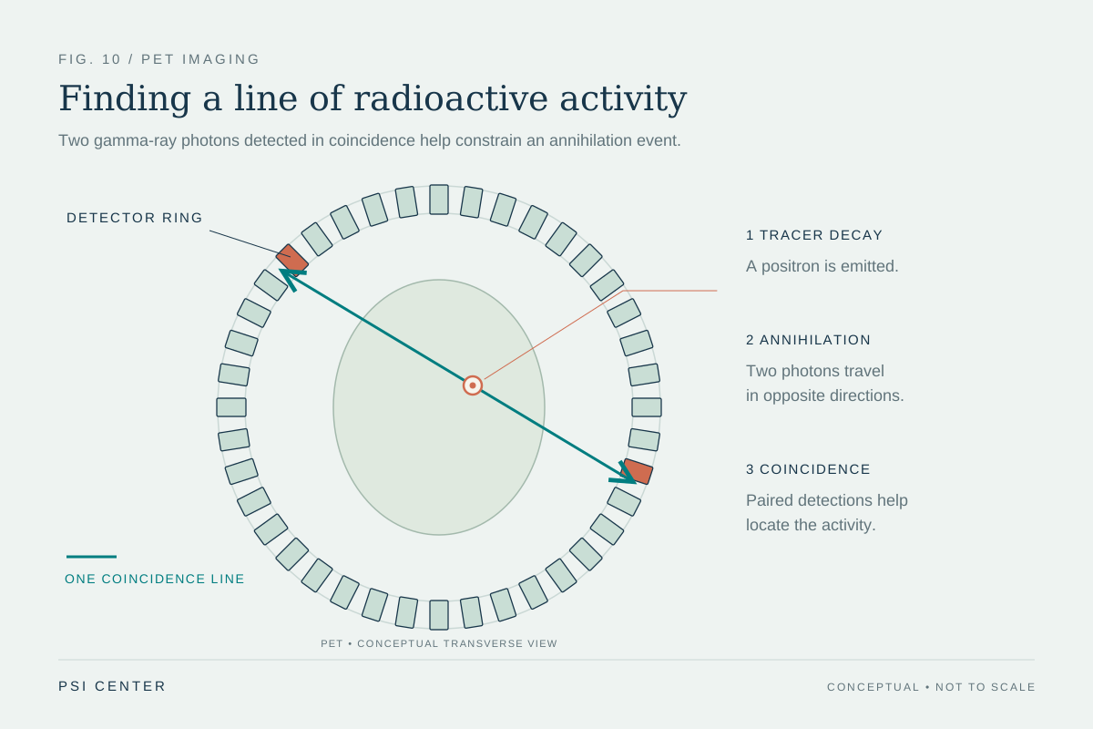
The two components remain distinct measurements. PET detects paired gamma photons produced after positrons from the tracer meet electrons in tissue. CT detects transmitted x-rays. Their physics, sources of contrast, and limitations differ, even when the display overlays them. The overlay is useful precisely because the modalities complement one another rather than duplicating the same measurement.
What the combined image cannot decide
A bright PET region does not by itself establish a diagnosis, and CT anatomy does not reveal every molecular behavior. Tracer uptake can reflect more than one biological cause, while a structural abnormality may not be active in the same way as nearby tissue. The scan is interpreted alongside other information to answer a defined question.
Both modalities also involve tradeoffs. CT uses ionizing x-rays, and PET involves a radioactive tracer; the amount and type of exposure depend on the examination. Combining images can improve localization and provide a more complete view, but it does not turn a scan into certainty. The central advantage is practical: PET contributes a map of selected biological activity, and CT tells clinicians where that activity sits in the body.
Explore the physics: an explanatory diagram
Sources & further reading
- Nuclear Medicine NIBIB
- Computed Tomography NIBIB
Prepared for the independent PSI Center reading library. About edition dates and source checking.
This explainer describes scientific principles. It does not assess individual treatment choices.