How can an injected substance make one medical image brighter without doing the same thing in another scanner? The answer depends on what the scanner measures. MRI listens to signals from hydrogen protons in tissue after radiofrequency pulses disturb their alignment in a magnetic field. CT measures how much X-ray radiation passes through the body. Contrast agents change those signals by different physical routes, so the word “contrast” describes a purpose, not a single material or mechanism.
MRI reads proton behavior
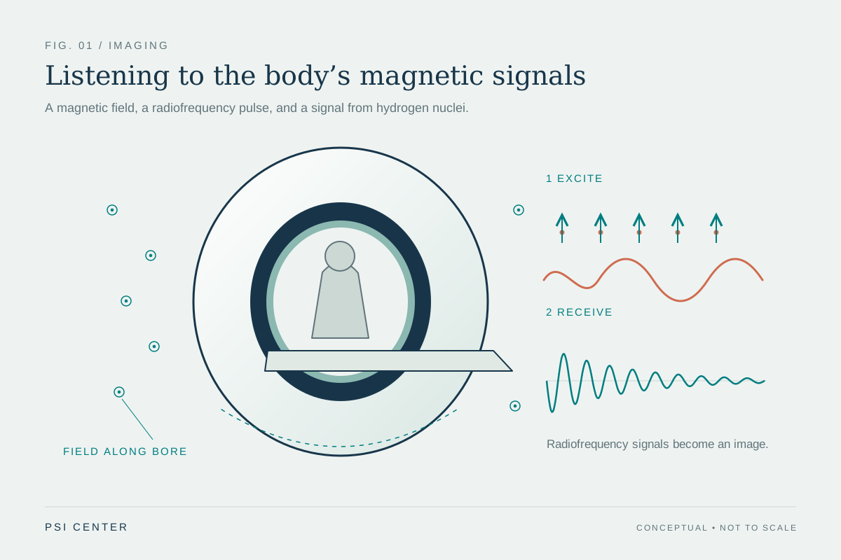
Most of the body's water contains hydrogen, whose nucleus is a proton. In an MRI scanner, a strong magnetic field encourages many of these protons to align. A radiofrequency pulse tips them away from alignment; when the pulse ends, they move back and release energy that receiver coils detect. The return time and signal depend on the protons’ molecular surroundings, allowing different tissues to appear distinct. MRI uses magnetic fields and radio waves, not plasma or ionizing X-rays. [1]
An MRI contrast agent, often containing gadolinium, changes the magnetic environment around nearby protons and can speed their realignment. That changes the signal and may make structures appear brighter on particular images. It is not a dye that simply paints a vessel in a visible color. Its effect comes through proton behavior, and the resulting appearance depends on how the scan is configured. [1]
CT tracks X-ray transmission
CT uses a rotating X-ray source and detectors on the opposite side of the patient. As X-rays pass through the body, different materials absorb or scatter different amounts. A computer combines measurements taken from many angles into cross-sectional slices. Dense bone stands out naturally, while some soft tissues differ only subtly in their ability to stop X-rays. [2]
For some CT examinations, an iodine-based agent in the bloodstream makes vessels more visible because iodine attenuates X-rays strongly. Barium-based agents taken by mouth can help outline parts of the digestive tract. These agents change the X-ray measurements directly: more radiation is stopped along the path through the contrast-containing region, so the reconstructed image shows it differently. That is fundamentally unlike changing the magnetic relaxation of hydrogen protons in MRI. [2]
Same goal, different signal
Imagine tracing a branching blood vessel. In a CT image, iodine in the blood changes the amount of X-ray signal reaching the detectors, helping the vessel stand out against surrounding tissue. In an MRI image, a gadolinium-containing agent can alter the timing of nearby proton signals, changing image brightness. Both approaches can help reveal anatomy, but the scanner’s underlying measurement explains why the agents are not interchangeable.
The distinction also clarifies why “contrast” does not mean a single universal enhancement. MRI contrast works within a method that does not use ionizing radiation; CT contrast works within an X-ray method that does. CT can provide detailed slices and is useful for many questions, including complex bone injury, while MRI is particularly well suited to soft tissues such as ligaments, muscles and brain structures. Which image is useful depends on the clinical question, not just on whether contrast is given. [1] [2]
Contrast changes the safety picture too
A contrast agent adds considerations beyond the scan’s basic imaging physics. The CT reference notes that some patients can have allergic reactions to contrast agents and that intravenous agents may pose kidney risks, especially with abnormal kidney function. Those concerns are tied to the particular agent and patient circumstances; a visible image enhancement is not a reason to assume the substance is risk-free. [2]
MRI avoids the ionizing X-rays used in CT, but it still uses a powerful magnetic field and has separate safety issues. Implants and magnetizable objects need careful screening. The MRI reference also describes a rare, serious illness that may be linked to certain gadolinium agents in people with severe renal failure who require dialysis. Thus, the two contrast strategies differ both in how they alter an image and in the questions clinicians consider when deciding whether to use them. [1]
Explore the physics: an explanatory diagram
Sources & further reading
- Magnetic Resonance Imaging NIBIB
- Computed Tomography NIBIB
Prepared for the independent PSI Center reading library. About edition dates and source checking.
This explainer describes scientific principles. It does not assess individual treatment choices.


