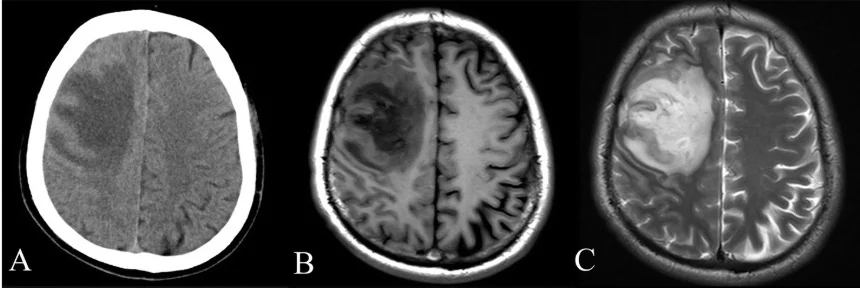
If a clinician needs to see inside the body, why choose one scan over another? The answer starts with the question being asked. Imaging methods do not reveal the same features in the same way: computed tomography measures how x-rays pass through tissues, while magnetic resonance imaging detects signals from protons responding to magnetic fields and radio waves. Their physics shapes their strengths and limits.
CT turns x-ray paths into slices
A CT scanner sends a narrow x-ray beam through the body while the x-ray source rotates around the patient. Detectors on the opposite side record how much of the beam emerges. A computer uses measurements from many angles to reconstruct cross-sectional slices; those slices can then be stacked into a three-dimensional view [1]. The core idea is like inferring the inside of an object from many projections rather than relying on one shadow.
Because dense structures such as bone stop x-rays effectively, CT can show complex fractures and other bony changes in detail. It can also help locate conditions such as bleeding, clots, tumors, or lung abnormalities, depending on the clinical question [1]. Contrast agents can make selected structures more visible: iodine-based contrast in the bloodstream can highlight vessels, while barium-based agents can help display parts of the digestive tract [1].
MRI listens to changing proton signals
MRI begins with the hydrogen protons abundant in body water. A strong magnetic field encourages them to align; a radiofrequency pulse disturbs that alignment. When the pulse ends, protons return toward their earlier state and release energy that sensors detect. The timing and amount of signal depend on the local molecular environment, allowing different tissues to produce distinguishable signals [2]. MRI therefore builds images from magnetic behavior rather than x-ray attenuation.
This makes MRI particularly useful for many soft tissues, including the brain, spinal cord, nerves, muscles, ligaments, and tendons. A knee injury, for instance, may require information about structures that a conventional x-ray depicts less clearly. MRI does not use ionizing x-rays, a distinction from CT, and it is often used when repeated imaging is needed. It generally takes longer and costs more than x-ray imaging or CT, so its advantages must fit the task [2].
The clinical question sets the tradeoff
Imagine a person with a suspected complex bone fracture. CT’s detailed cross-sections may make the fracture’s geometry easier to assess. Now imagine a question about a ligament or a brain structure: MRI’s contrast among soft tissues may provide more useful information. These examples are not a universal ranking. The best method depends on which structures matter, how quickly an answer is needed, and whether the expected image will change the next clinical decision.
CT uses ionizing radiation, and the risk from x-ray exposure generally increases with cumulative exposure, although the risk from an individual scan is generally small [1]. Children are more sensitive to ionizing radiation than adults, which is one reason scan settings and the need for the examination matter [1]. MRI avoids that radiation but brings different considerations, including a powerful magnetic field that can interact dangerously with some magnetizable objects and implants [2].
Images are measurements, not the whole decision
No scan simply displays “the disease.” Each method measures physical signals, processes them into images, and requires interpretation in context. A CT image can be enhanced with contrast to emphasize vessels or digestive structures; an MRI may use a contrast agent to alter proton relaxation and image brightness [1] [2]. These choices affect what is visible and should be linked to the clinical question, rather than treated as routine decorations.
A practical way to compare options is to ask three things: What structure or feature needs to be seen? Which physical method gives useful information about it? What tradeoffs—such as radiation exposure, magnetic-field constraints, time, or contrast—are relevant? CT and MRI are complementary tools built on different physics. Their value comes from matching a measurement to a question, then interpreting the resulting image alongside the rest of the clinical picture.
Sources & further reading
- Computed Tomography NIBIB
- Magnetic Resonance Imaging NIBIB
Prepared for the independent PSI Center reading library. About edition dates and source checking.
This explainer describes scientific principles. It does not assess individual treatment choices.


