What does one bright square in a medical image represent: a patch of tissue, a measurement, or both? A pixel is a two-dimensional picture element; a voxel is its three-dimensional counterpart, a small volume assigned an image value. Neither is a tiny photograph of a cell. Each summarizes information collected by a scanner and reconstructed into a grid, so image detail depends on the acquisition as well as the display.
A grid gives measurements a place
An image is made from values arranged in a grid. Each pixel marks a position in a flat image, while a voxel extends that position through a thickness, giving it volume. The value assigned to a location represents a measurement processed by the imaging system. A stack of two-dimensional slices can be viewed in sequence or combined to represent three-dimensional anatomy.
The word “resolution” often describes how finely an image can distinguish spatial features, but a smaller displayed square does not automatically mean the scanner measured more detail. Image quality also depends on the data collected, the reconstruction, motion, and the contrast between tissues. A useful mental picture is a map made from measurements: increasing the number of squares helps only if the underlying survey contains reliable information at that scale.
CT builds slices from X-ray measurements
Computed tomography, or CT, uses a rotating X-ray source and detectors on the opposite side of the body. As the scanner collects measurements from many angles, a computer reconstructs cross-sectional slices. These can be digitally stacked into a three-dimensional image [1]. The CT image is therefore computed from how X-rays pass through the body, not a direct optical photograph.
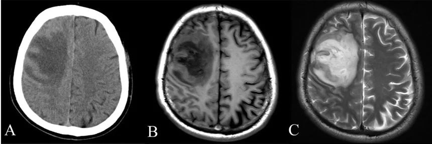
Dense structures such as bone stop X-rays readily and are usually clear, while soft tissues can be harder to distinguish. Contrast agents can make selected structures more visible; for example, iodine-based contrast in the bloodstream can help show vessels [1]. CT uses ionizing X-rays, so the imaging method involves a different physical interaction from MRI.
MRI listens to proton responses
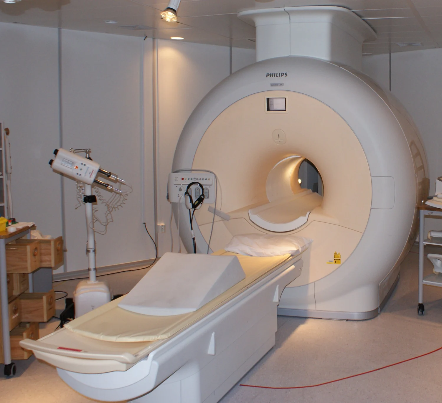
Magnetic resonance imaging, or MRI, uses strong magnetic fields and radiofrequency pulses. The magnetic field aligns protons found in water in living tissues; a radiofrequency pulse disturbs that alignment, and sensors detect energy released as the protons return toward alignment. The response varies with the molecules’ local environment, allowing tissue types to appear differently [2]. MRI does not use ionizing X-rays [2].
The contrast in an MRI image is thus connected to proton behavior and the tissue environment, rather than to the way X-rays are absorbed. An MRI scan must also account for movement: staying still helps avoid blur [2]. The scanner’s measurements are reconstructed into image elements, but the underlying signal and the physical cause of contrast differ from CT.
A voxel is not a promise of microscopic detail
The three-dimensional grid can make it easier to locate structures, compare slices, or examine anatomy from different viewpoints. CT computers can stack successive slices to create a rotatable 3D representation [1]. MRI also produces detailed three-dimensional anatomical images, often useful for soft tissues such as muscles, ligaments, and tendons [2]. In either case, the image remains a model derived from signals, not a literal view of every microscopic feature.
It helps to distinguish sampling from certainty. A small voxel represents a smaller region in the reconstructed grid, but the scanner’s physics and signal quality determine what distinctions can be made. CT and MRI answer different imaging questions through different signals, and each has strengths and limits. Pixels and voxels describe where image values sit; they do not, by themselves, tell us what tissue is present or how confidently it can be identified.
Sources & further reading
- Computed Tomography NIBIB
- Magnetic Resonance Imaging NIBIB
Prepared for the independent PSI Center reading library. About edition dates and source checking.
This explainer describes scientific principles. It does not assess individual treatment choices.