 PHOTOGRAPH
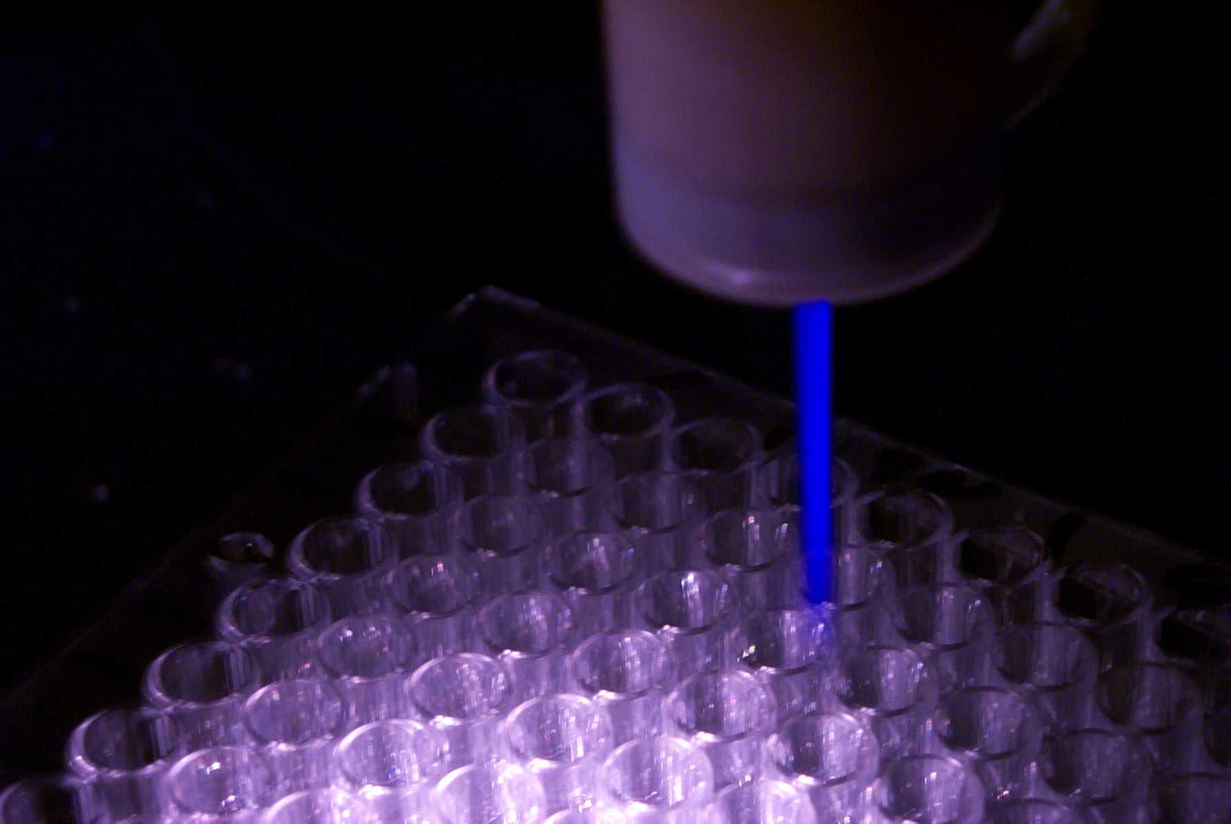
Plasma Pencil
A laboratory plasma pencil produces a low-temperature plasma plume. The photograph illustrates a research source; it does not document a clinical treatment.
Entropi5 / Wikimedia Commons. Original source ↗
CC BY-SA 3.0. None. Original file retained. Display may use CSS cropping. Display layout may crop the image.
 PHOTOGRAPH
Brain MRI
A color-coded brain MRI released by the National Cancer Institute. The source describes a blue-highlighted metastasis in the occipital lobe. Color in this image is a visualization choice.
National Cancer Institute / Dr. Leon Kaufman, University of California, San Francisco. Original source ↗
Public domain (NIH work, per NCI reuse statement reproduced on Commons). None. Original file retained. Display layout may crop the image.
 MEDICAL IMAGE
T1-weighted-MRI
A T1-weighted axial brain MRI illustrates the contrast between tissue types. The image is an example scan and does not diagnose a condition.
Mim.cis / Johns Hopkins University, via Wikimedia Commons. Original source ↗
Public domain (US medical imaging without added copyrightable graphics). None. Original file retained. Display layout may crop the image.
 MEDICAL IMAGE
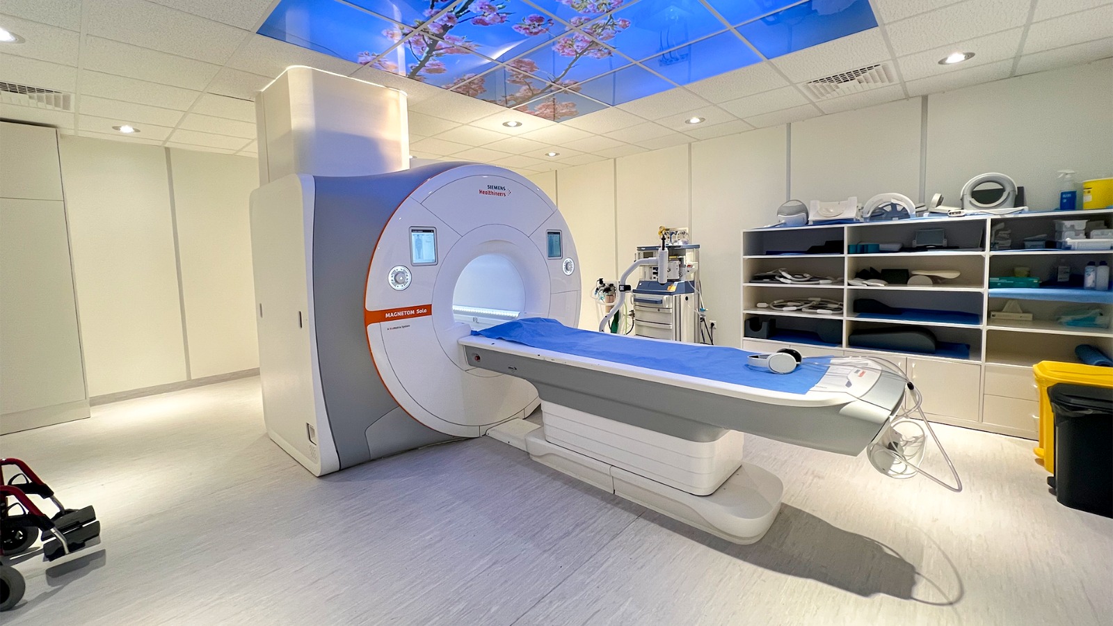
MRI Saudi Hospital
An MRI system in a hospital imaging room. Its magnetic field and radiofrequency equipment produce images without ionizing X-rays.
Yazan.shawahneh / Wikimedia Commons. Original source ↗
CC0 1.0. None. Original file retained. Display layout may crop the image.
 MEDICAL IMAGE
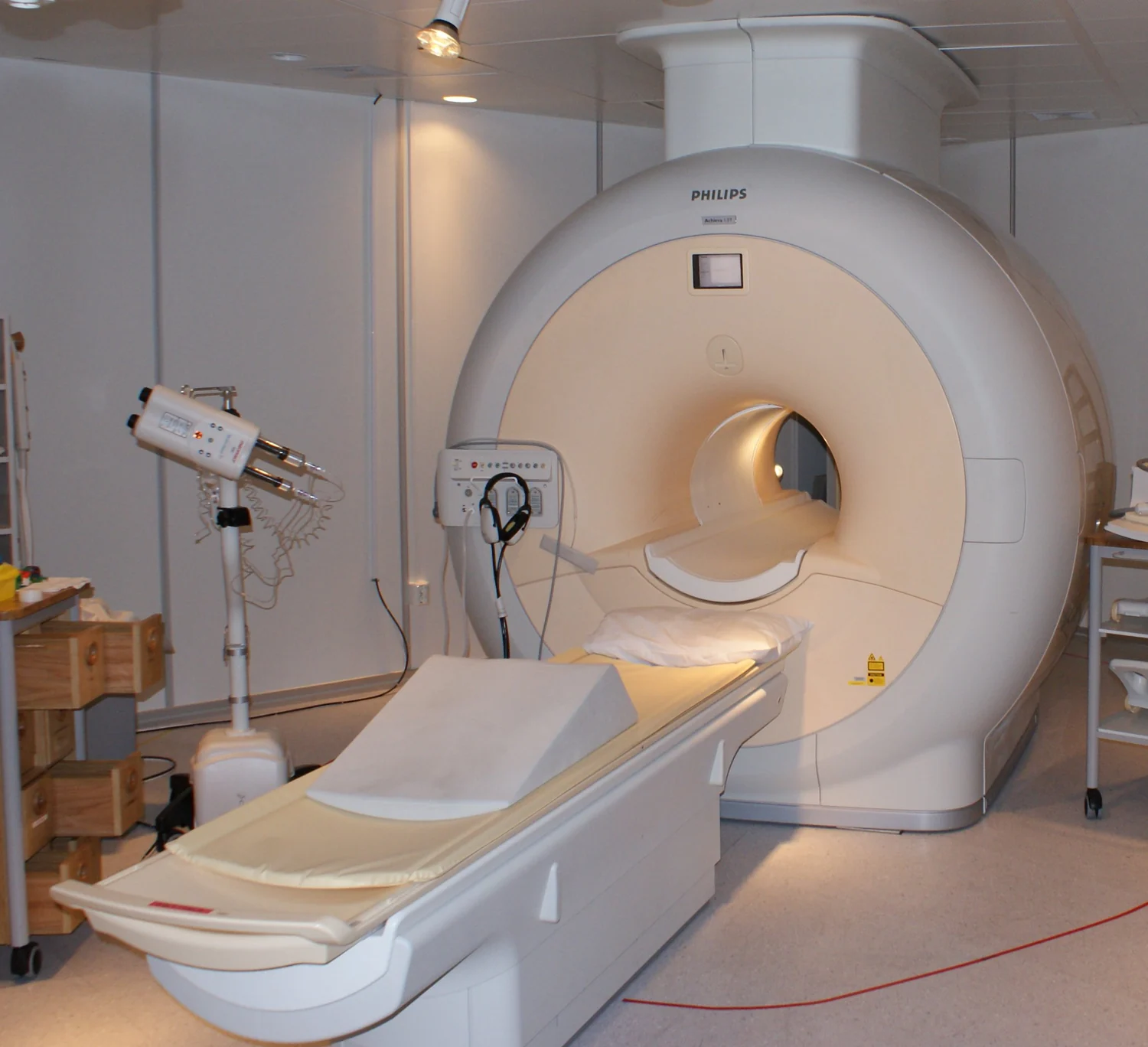
MRI-Philips
An MRI system at Sahlgrenska University Hospital in Gothenburg, Sweden.
Jan Ainali / Wikimedia Commons. Original source ↗
CC BY 3.0. Resized to a maximum dimension of 1500 pixels and encoded as WebP; no content changes. Display layout may crop the image.
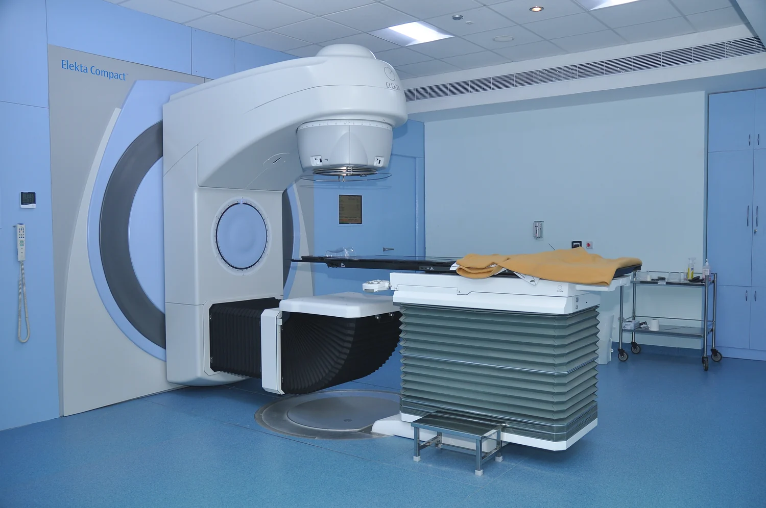 PHOTOGRAPH
Linear Accelerator - Elekta Compact model
An Elekta Compact linear accelerator used for radiotherapy at a hospital in Mysore, India.
Narenfox / Wikimedia Commons. Original source ↗
CC BY-SA 4.0. Resized to a maximum dimension of 1500 pixels and encoded as WebP; no content changes. Display layout may crop the image.
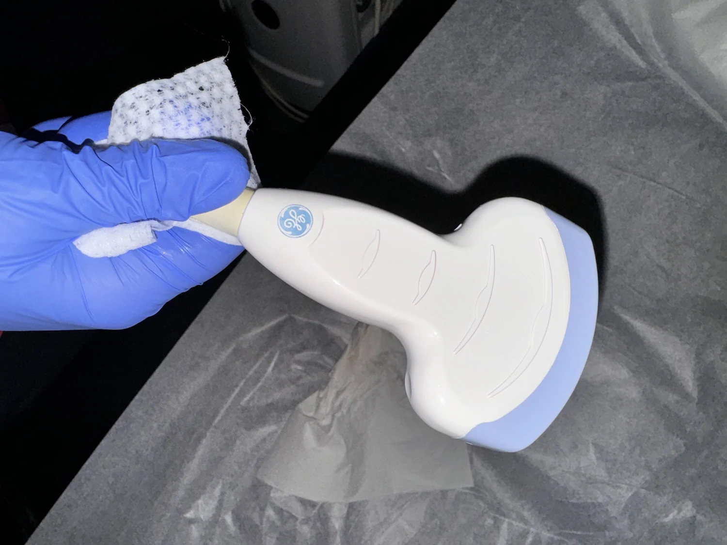 PHOTOGRAPH
A medical ultrasound linear array probe, scan head, transducer
A medical ultrasound probe; its contact surface couples sound between the transducer and the body.
Harrison Keely / Wikimedia Commons. Original source ↗
CC BY 4.0. Resized to a maximum dimension of 1500 pixels and encoded as WebP; no content changes. Display layout may crop the image.
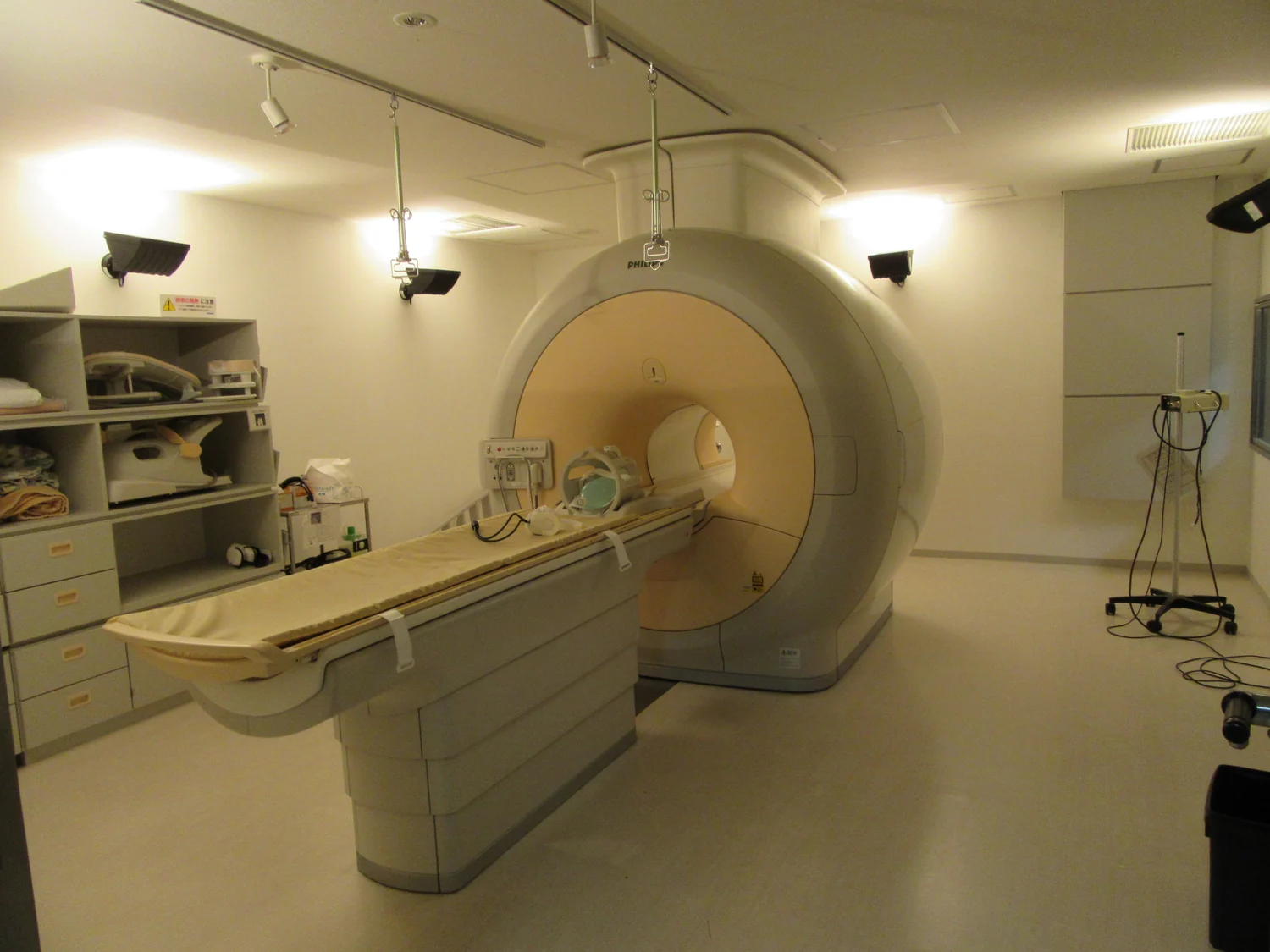 MEDICAL IMAGE
An MRI scanner
A hospital MRI scanner photographed as a piece of imaging equipment.
Walter Davies / Wikimedia Commons. Original source ↗
CC BY-SA 4.0. Resized to a maximum dimension of 1500 pixels and encoded as WebP; no content changes. Display layout may crop the image.
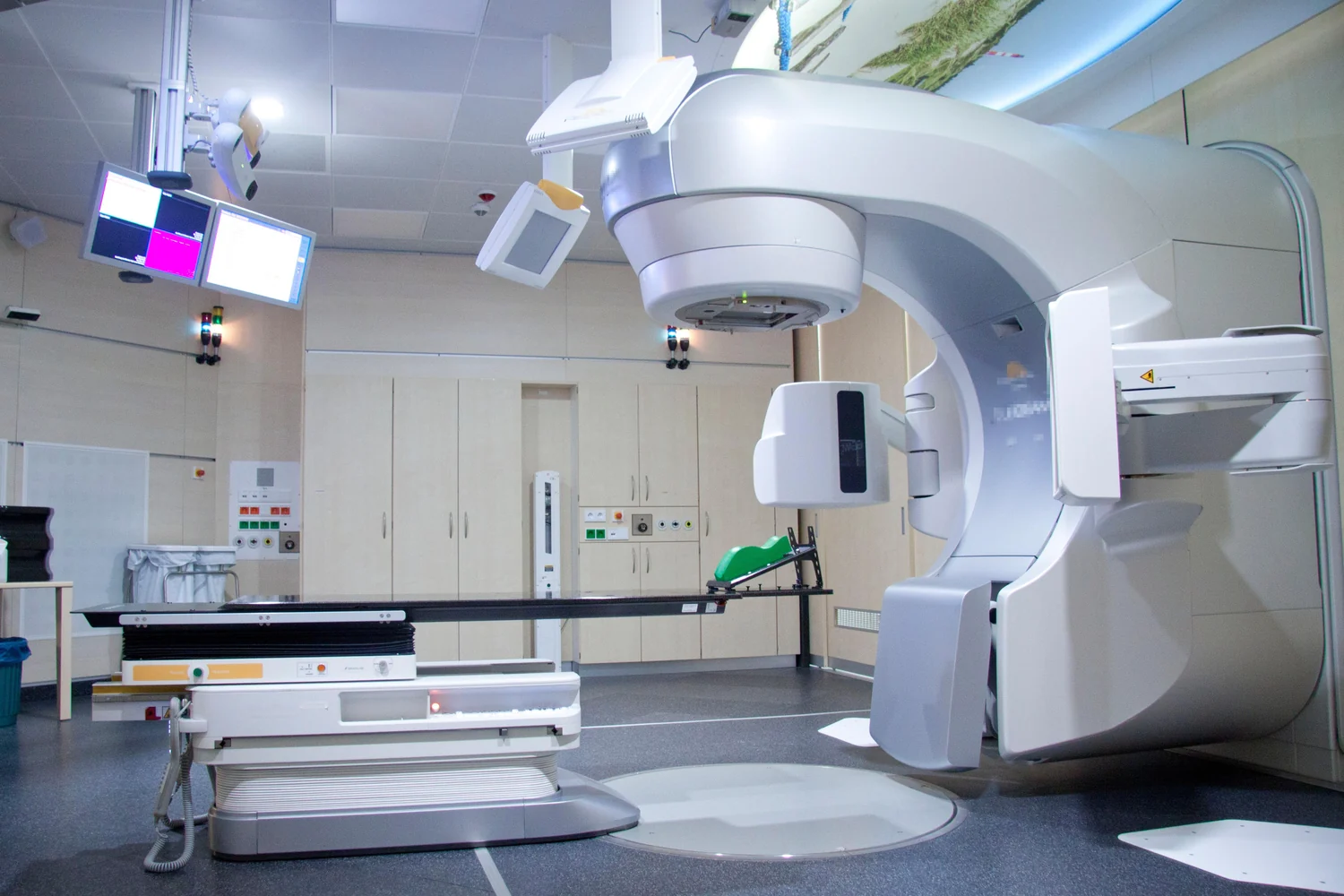 PHOTOGRAPH
Linearbeschleuniger Kiel
A linear accelerator for cancer radiotherapy at University Medical Center Schleswig-Holstein in Kiel, Germany.
Universitätsklinikum Schleswig-Holstein, Stabsstelle Integrierte Kommunikation / Wikimedia Commons. Original source ↗
CC BY-SA 3.0 de. Resized to a maximum dimension of 1500 pixels and encoded as WebP; no content changes. Display layout may crop the image.
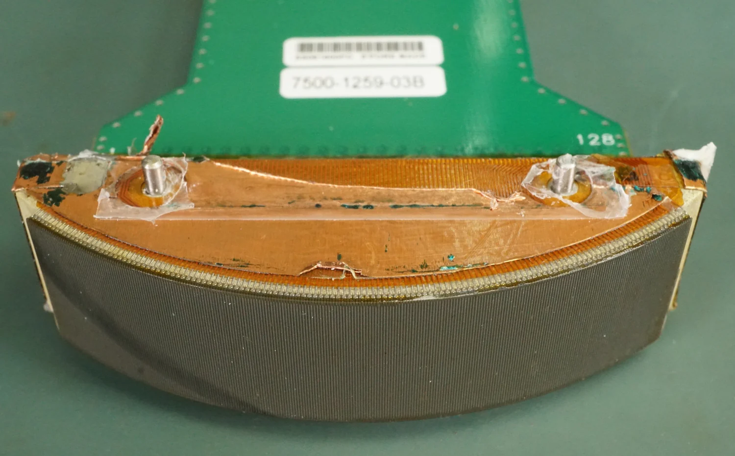 PHOTOGRAPH
Curved Array Ultrasound Sensor Construction
The opened construction of a Philips C5-2 curved-array ultrasound sensor reveals the probe’s internal components.
Binarysequence / Wikimedia Commons. Original source ↗
CC BY-SA 4.0. Resized to a maximum dimension of 1500 pixels and encoded as WebP; no content changes. Display layout may crop the image.
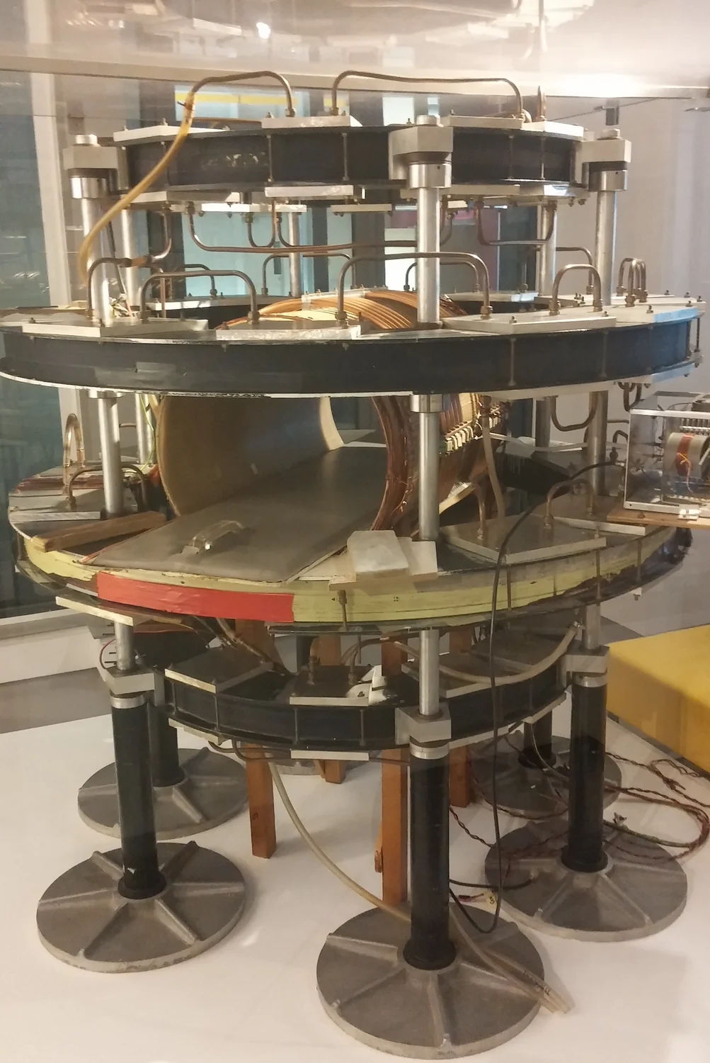 MEDICAL IMAGE
MRI Scanner Mark One
The Mark One MRI scanner on display in Scotland, documenting an early stage in MRI engineering.
AndyGaskell / Wikimedia Commons. Original source ↗
CC BY-SA 4.0. Resized to a maximum dimension of 1500 pixels and encoded as WebP; no content changes. Display layout may crop the image.
 MEDICAL IMAGE
Moderní výpočetní tomografie s přímo digitální detekcí rentgenového záření
A CT scanner at University Hospital Lochotín in Pilsen, Czech Republic.
Tomáš Vendiš / Wikimedia Commons. Original source ↗
CC BY-SA 4.0. Resized to a maximum dimension of 1500 pixels and encoded as WebP; no content changes. Display layout may crop the image.
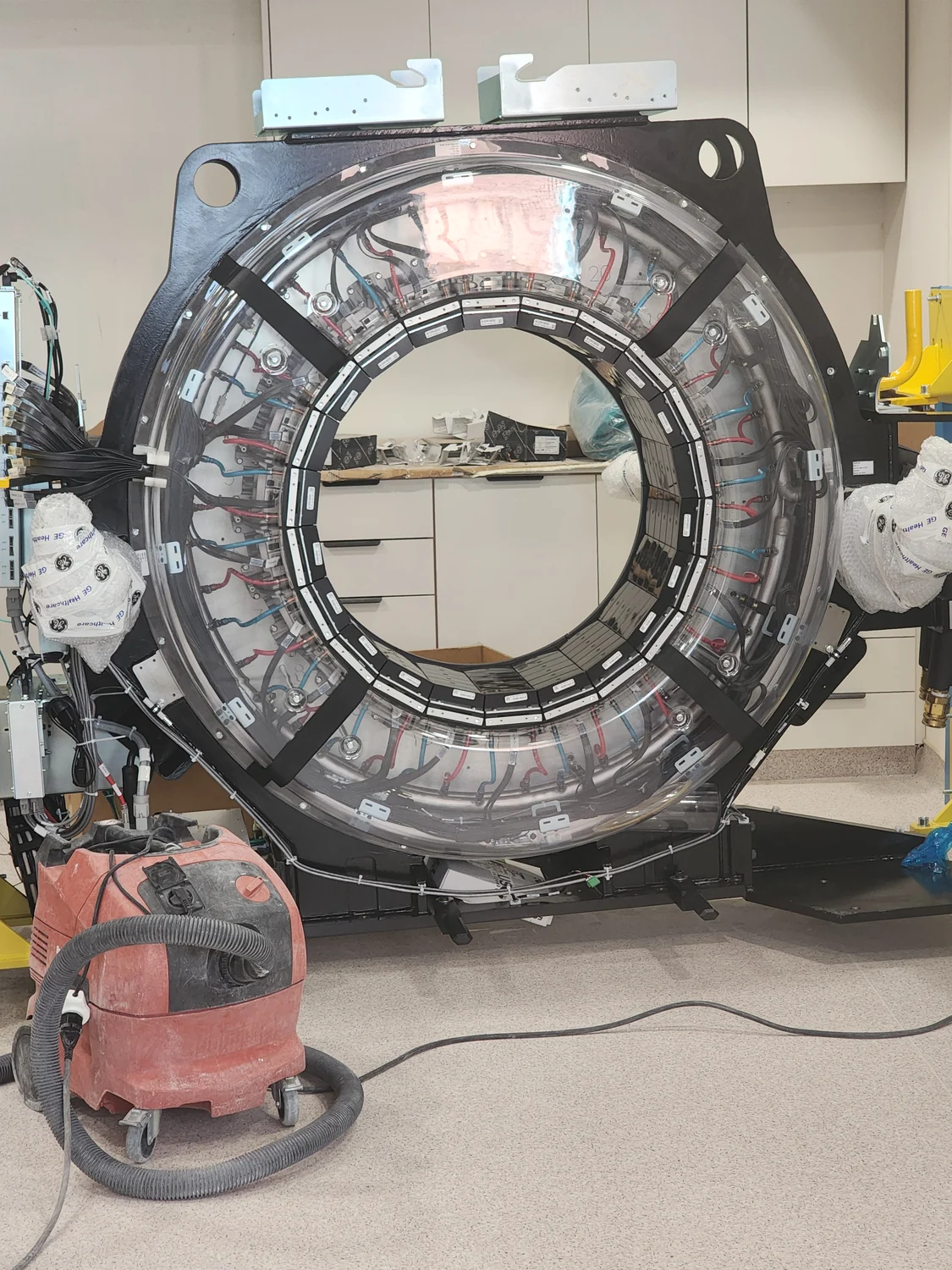 PHOTOGRAPH
The inner parts of a CT scanner
A view inside a CT scanner shows the hardware surrounding its circular patient opening.
Nettadi / Wikimedia Commons. Original source ↗
CC BY 4.0. Resized to a maximum dimension of 1500 pixels and encoded as WebP; no content changes. Display layout may crop the image.
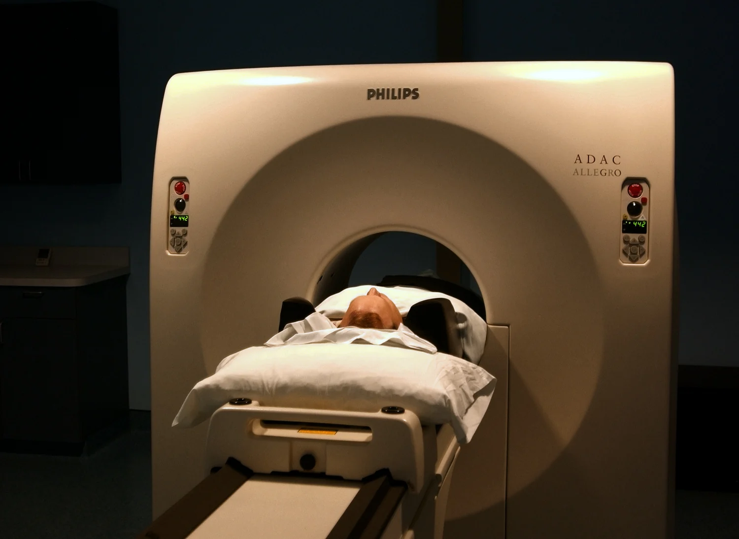 MEDICAL IMAGE
US Navy 030819-N-9593R-151 A patient goes through Positiron Emission Tomography (PET) at the National Naval Medical Center in Bethesda, Maryland
A PET examination photographed at the National Naval Medical Center in Bethesda, Maryland, in 2003.
U.S. Navy photo by Chief Warrant Officer 4 Seth Rossman. / Wikimedia Commons. Original source ↗
Public domain. Resized to a maximum dimension of 1500 pixels and encoded as WebP; no content changes. Display layout may crop the image.
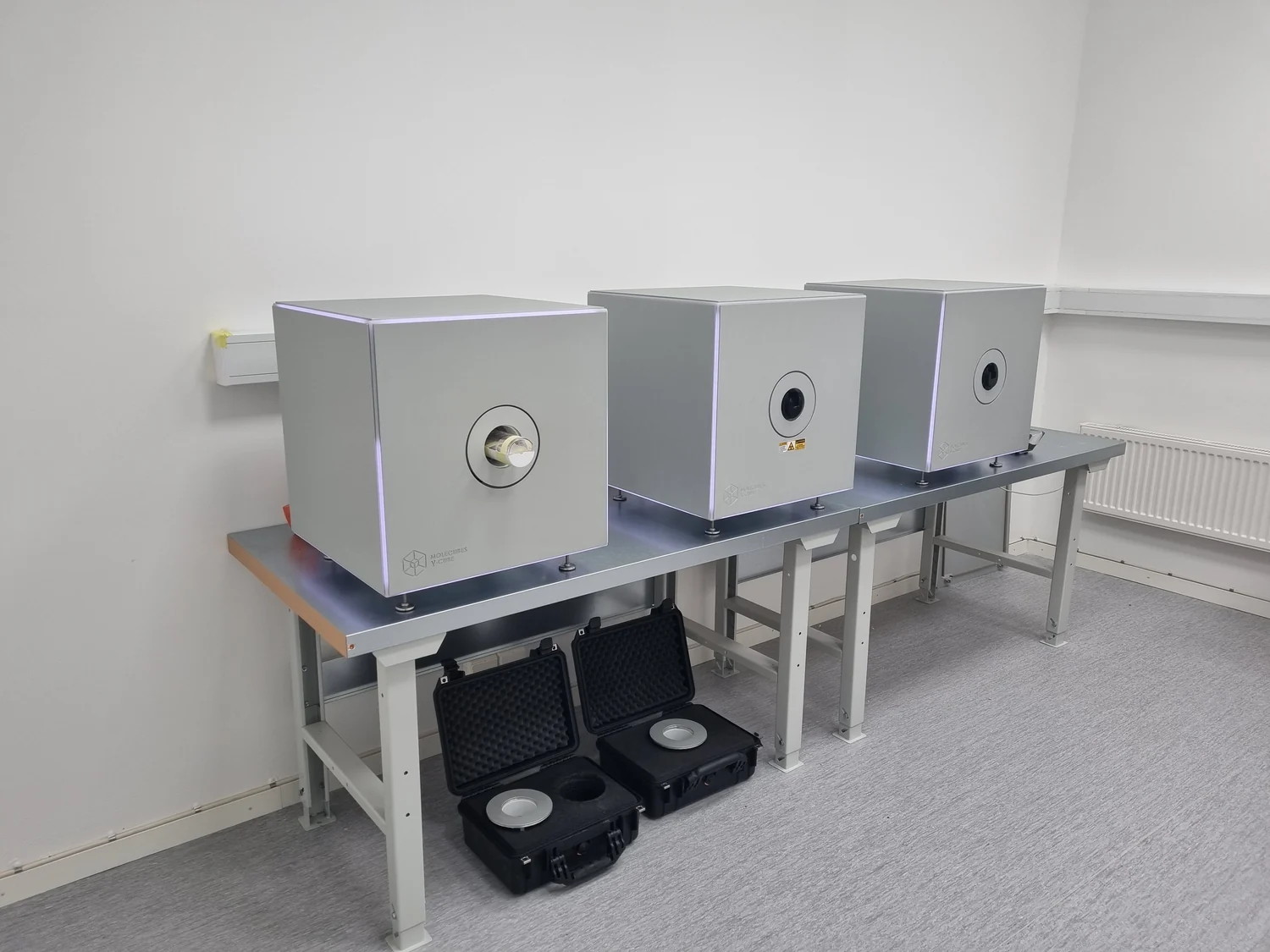 MEDICAL IMAGE
Molecubes SPECT uCT and PET
Benchtop imaging systems for preclinical SPECT, micro-CT, and PET research.
Adville / Wikimedia Commons. Original source ↗
CC BY-SA 4.0. Resized to a maximum dimension of 1500 pixels and encoded as WebP; no content changes. Display layout may crop the image.
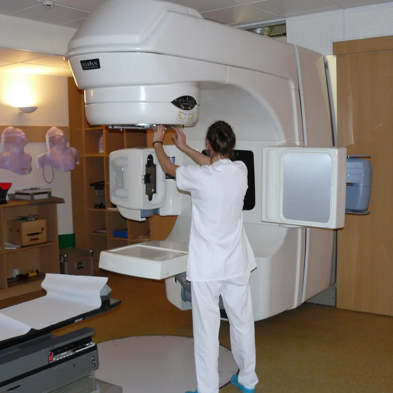 PHOTOGRAPH
Radiotherapie
A Varian Clinac linear accelerator used for external-beam radiotherapy.
Guy Lebègue / Wikimedia Commons. Original source ↗
CC BY-SA 3.0. Resized to a maximum dimension of 1500 pixels and encoded as WebP; no content changes. Display layout may crop the image.
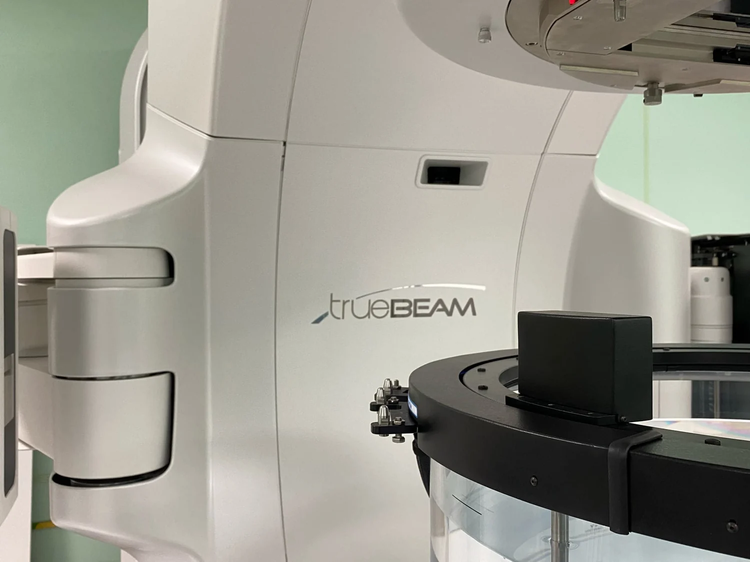 PHOTOGRAPH
Комплекс оборудования (линейный ускоритель) TrueBeam STx для проведения лучевой терапии
A TrueBeam STx treatment system photographed at the Tomsk Oncology Centre.
YuliaNiki / Wikimedia Commons. Original source ↗
CC BY-SA 4.0. Resized to a maximum dimension of 1500 pixels and encoded as WebP; no content changes. Display layout may crop the image.
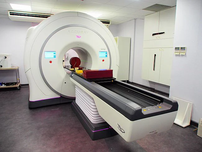 PHOTOGRAPH
Accuray TomoTherapy HDA System
A TomoTherapy HDA radiotherapy system at Centre Oscar Lambret in Lille, France.
Tetrazene / Wikimedia Commons. Original source ↗
CC BY-SA 3.0. Resized to a maximum dimension of 1500 pixels and encoded as WebP; no content changes. Display layout may crop the image.
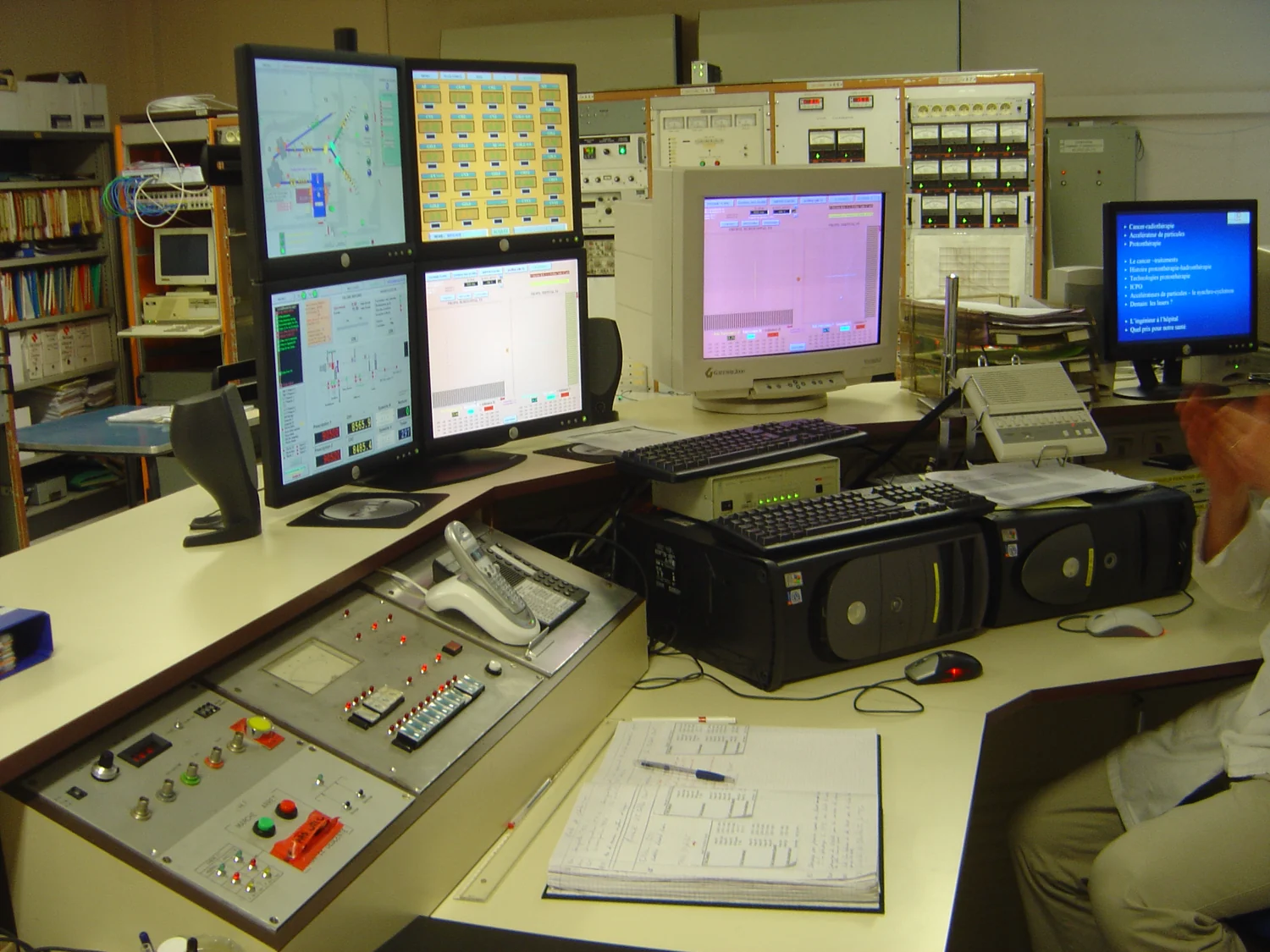 PHOTOGRAPH
Orsay proton therapy dsc04460
The synchrocyclotron control panel at the Orsay proton-therapy center, photographed in 2005.
David Monniaux / Wikimedia Commons. Original source ↗
CC BY-SA 3.0. Resized to a maximum dimension of 1500 pixels and encoded as WebP; no content changes. Display layout may crop the image.
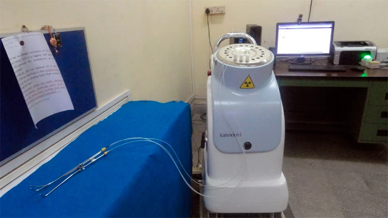 PHOTOGRAPH
Karknidon - Indigenous Brachytherapy Equipment developed by BRIT, DAE, India
Brachytherapy equipment developed by India’s Board of Radiation and Isotope Technology.
Sudip Bhunia / Wikimedia Commons. Original source ↗
CC BY-SA 4.0. Resized to a maximum dimension of 1500 pixels and encoded as WebP; no content changes. Display layout may crop the image.
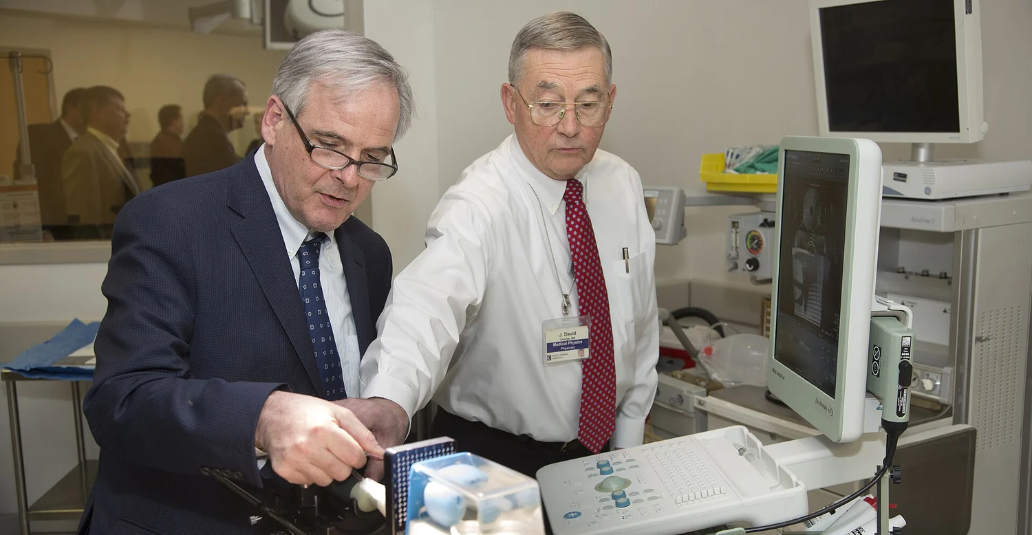 PHOTOGRAPH
NRC Chairman visits INOVA Fairfax Hospital (25940327411)
Medical physicist J. David George demonstrates brachytherapy equipment at INOVA Fairfax Hospital during an NRC visit.
Nuclear Regulatory Commission from US / Wikimedia Commons. Original source ↗
CC BY 2.0. Resized to a maximum dimension of 1500 pixels and encoded as WebP; no content changes. Display layout may crop the image.
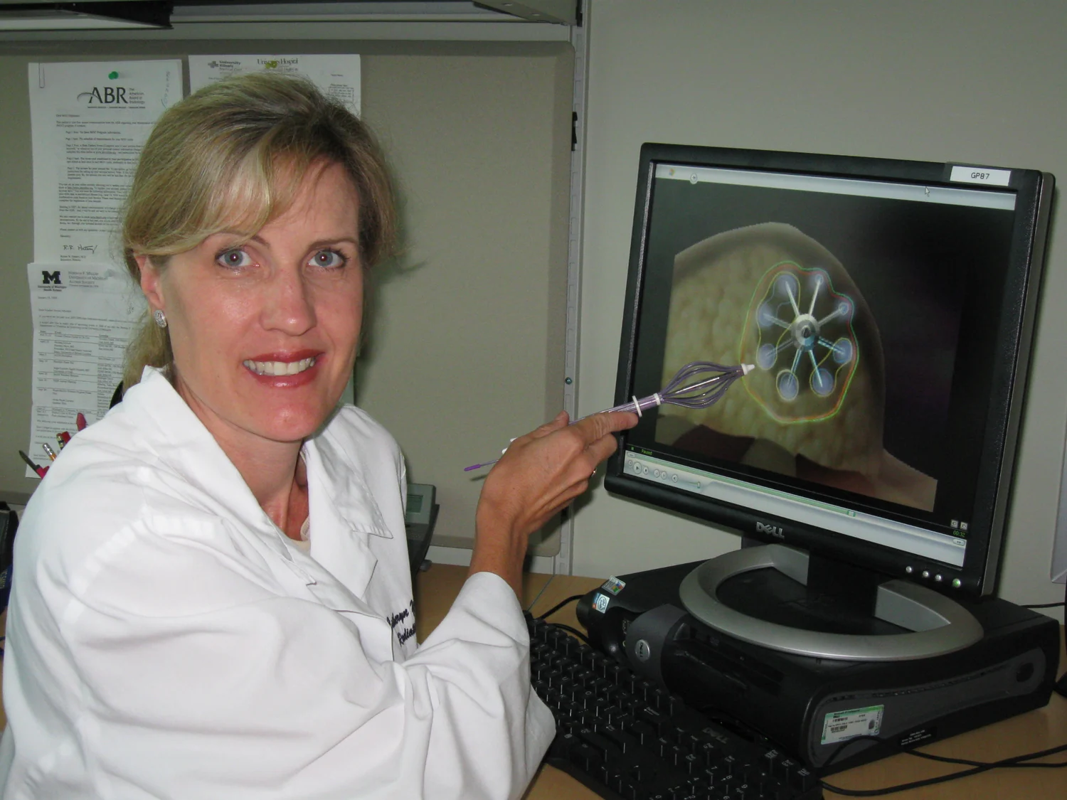 PHOTOGRAPH
1 applicator
A professional demonstrates a SAVI breast brachytherapy applicator beside an image of its arrangement in tissue.
Brian Driscoll, Vice President/Marketing, Cianna Medical, Inc. / Wikimedia Commons. Original source ↗
CC BY-SA 3.0. Resized to a maximum dimension of 1500 pixels and encoded as WebP; no content changes. Display layout may crop the image.
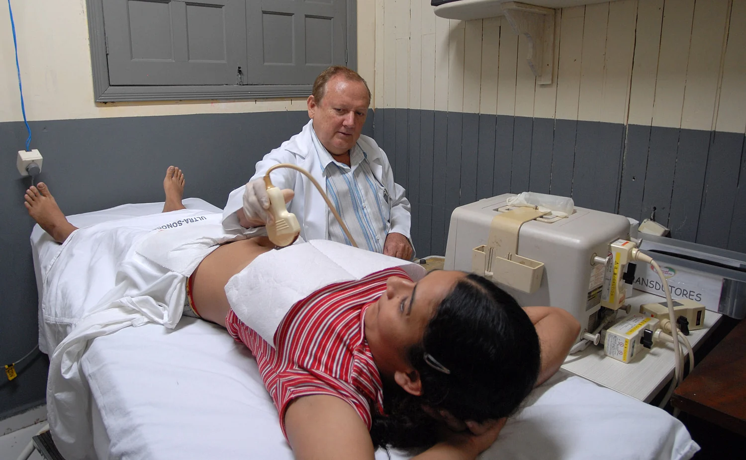 PHOTOGRAPH
Medical ultrasound examination, Brazil
An ultrasound examination photographed during a healthcare program in Acre, Brazil.
Agência de Notícias do Acre / Wikimedia Commons. Original source ↗
CC BY 2.0. Resized to a maximum dimension of 1500 pixels and encoded as WebP; no content changes. Display layout may crop the image.
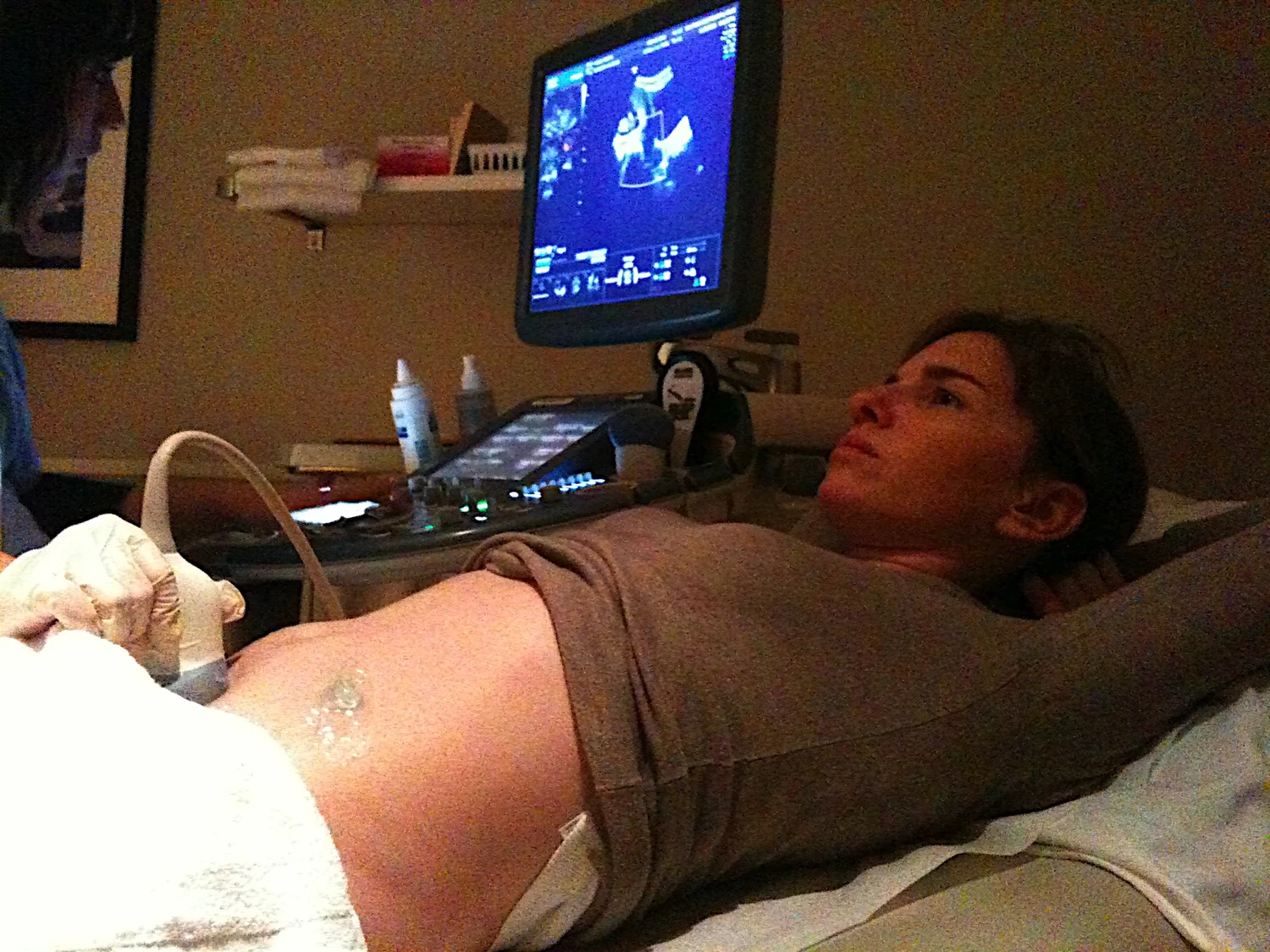 PHOTOGRAPH
Ultrasound examination of woman
An obstetric ultrasound examination photographed in 2009.
Scott / Wikimedia Commons. Original source ↗
CC BY-SA 3.0. Resized to a maximum dimension of 1500 pixels and encoded as WebP; no content changes. Display layout may crop the image.
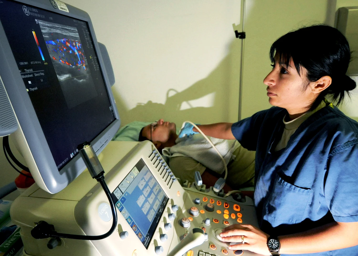 MEDICAL IMAGE
Medical ultrasound examination
A medical ultrasound examination photographed by Christopher Hubenthal of the U.S. Air Force.
Christopher Hubenthal, U.S. Air Force / Wikimedia Commons. Original source ↗
Public domain. Resized to a maximum dimension of 1500 pixels and encoded as WebP; no content changes. Display layout may crop the image.
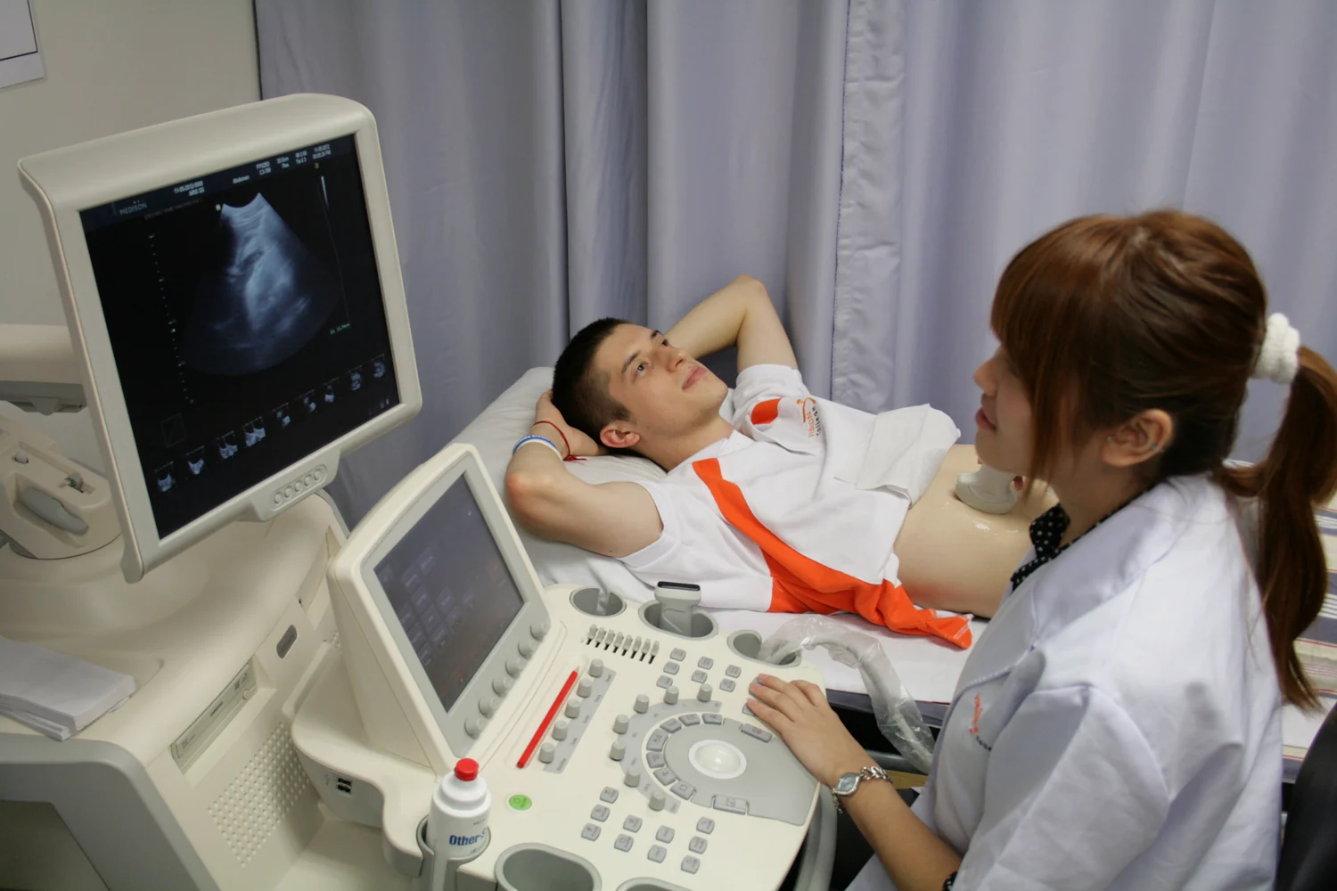 MEDICAL IMAGE
Ultrasound vision
Ultrasound equipment and an examination setup in a diagnostic clinic.
Aseev artem / Wikimedia Commons. Original source ↗
CC BY-SA 3.0. Resized to a maximum dimension of 1500 pixels and encoded as WebP; no content changes. Display layout may crop the image.
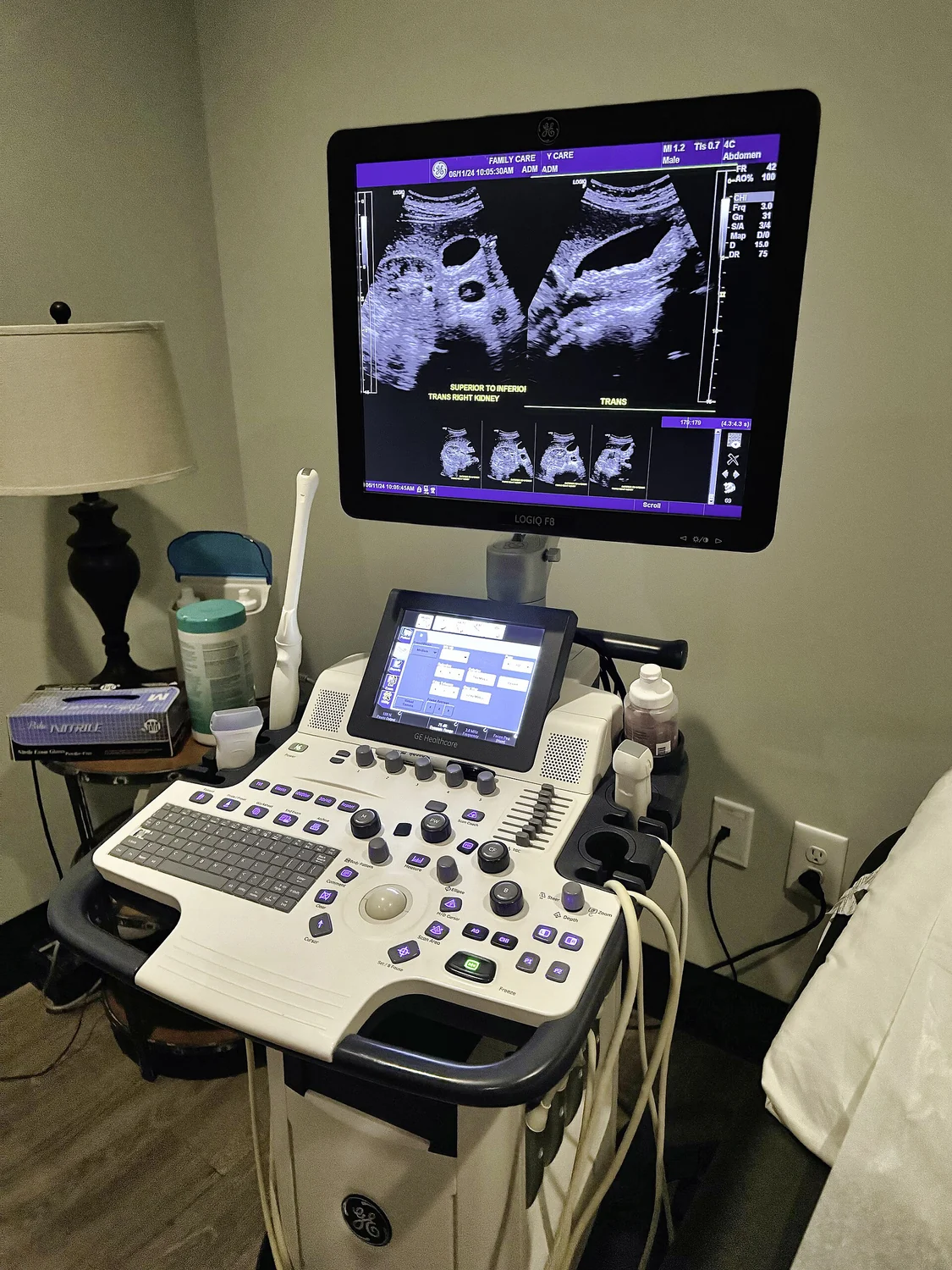 MEDICAL IMAGE
A modern medical ultrasound scanner
An ultrasound scanner photographed with its display and control console.
Harrison Keely / Wikimedia Commons. Original source ↗
CC BY 4.0. Resized to a maximum dimension of 1500 pixels and encoded as WebP; no content changes. Display layout may crop the image.
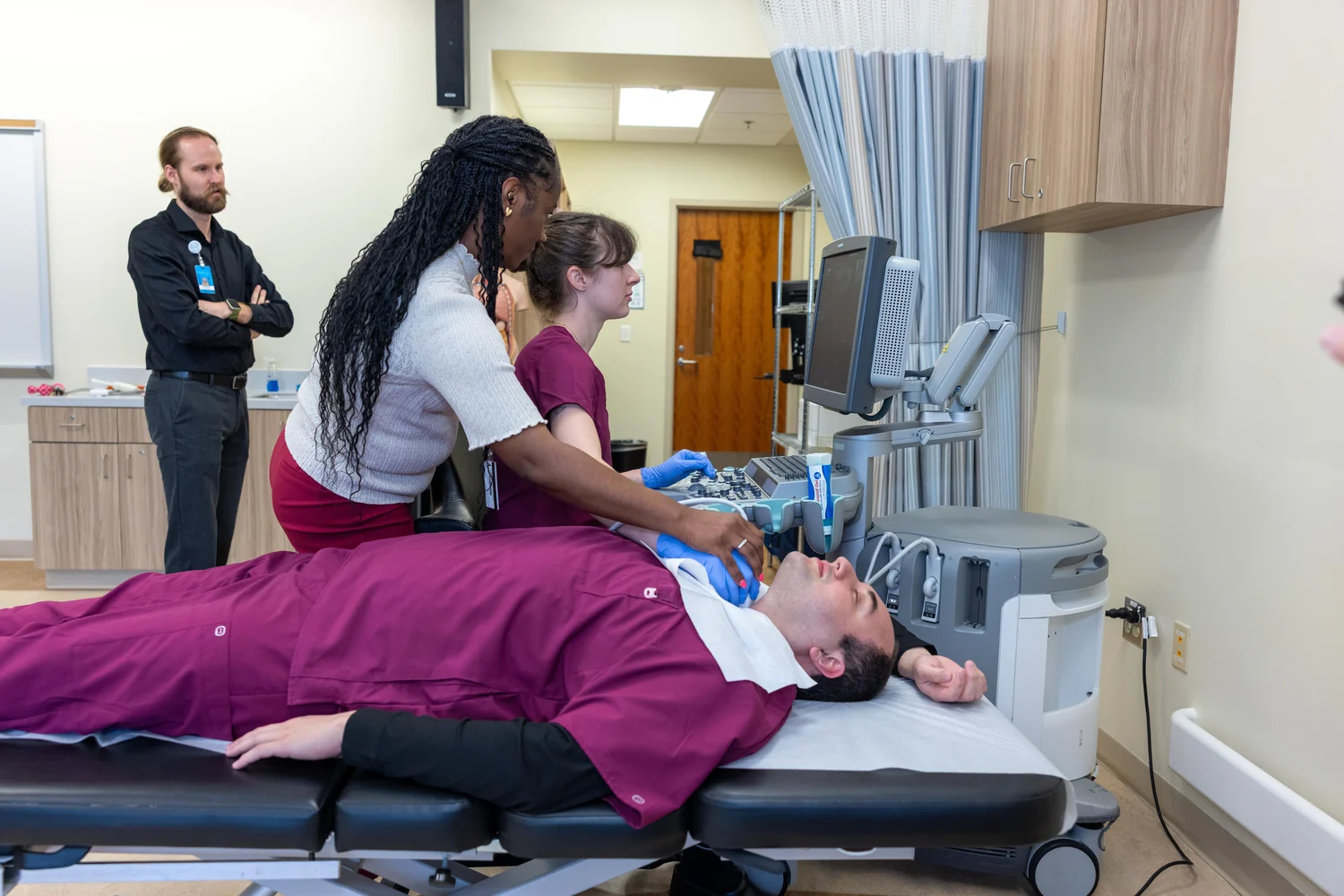 PHOTOGRAPH
Sonography Machine Donation from Raleigh Radiology-06
Sonography equipment photographed at Wake Technical Community College.
waketechcc / Wikimedia Commons. Original source ↗
CC0. Resized to a maximum dimension of 1500 pixels and encoded as WebP; no content changes. Display layout may crop the image.
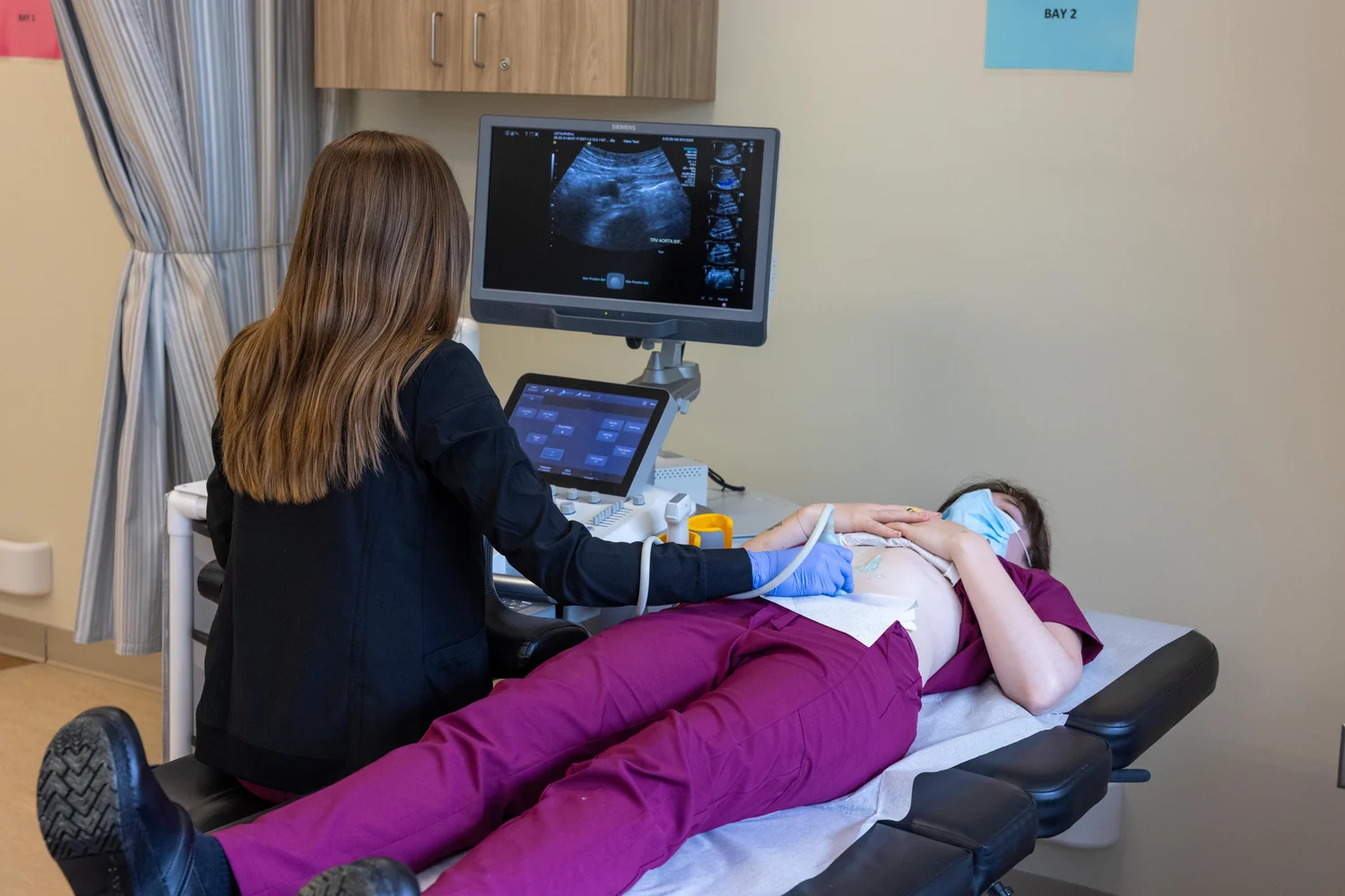 PHOTOGRAPH
Sonography Machine Donation from Raleigh Radiology-35
A sonography workstation at Wake Technical Community College, photographed during a donation from Raleigh Radiology.
waketechcc / Wikimedia Commons. Original source ↗
CC0. Resized to a maximum dimension of 1500 pixels and encoded as WebP; no content changes. Display layout may crop the image.
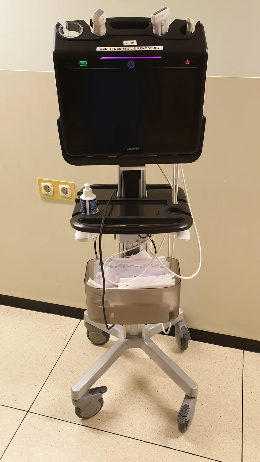 PHOTOGRAPH
Pocus (ultrasound) semi-portable
A semi-portable point-of-care ultrasound system provides a compact examination setup.
Dr Bowser / Wikimedia Commons. Original source ↗
CC BY-SA 4.0. Resized to a maximum dimension of 1500 pixels and encoded as WebP; no content changes. Display layout may crop the image.
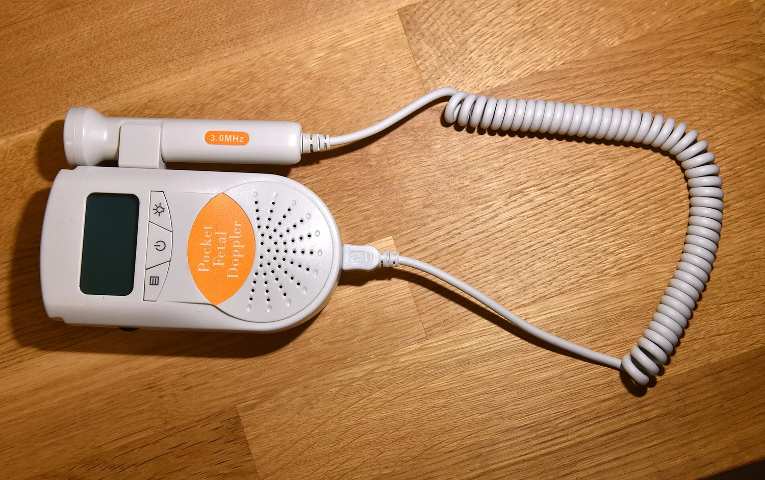 PHOTOGRAPH
Fetal doppler - Ultrasound - 5412
A handheld fetal Doppler ultrasound instrument photographed with its connected probe.
Amada44 / Wikimedia Commons. Original source ↗
CC BY-SA 3.0. Resized to a maximum dimension of 1500 pixels and encoded as WebP; no content changes. Display layout may crop the image.
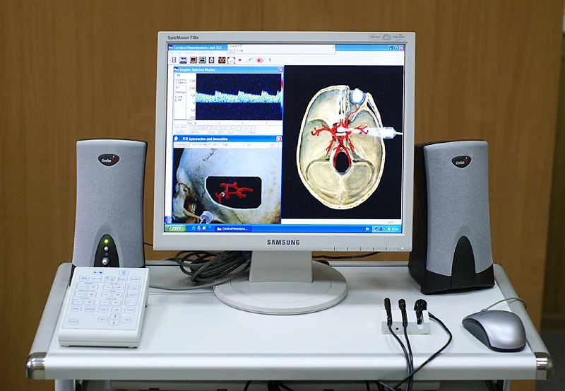 PHOTOGRAPH
Doppler ultrasound analyzer of blood velocity
A Doppler ultrasound analyzer of blood velocity manufactured by Bioss in Zelenograd, Russia.
Юрий Петрович Маслобоев / Yury Petrovich Masloboev / Wikimedia Commons. Original source ↗
CC BY-SA 3.0. Resized to a maximum dimension of 1500 pixels and encoded as WebP; no content changes. Display layout may crop the image.
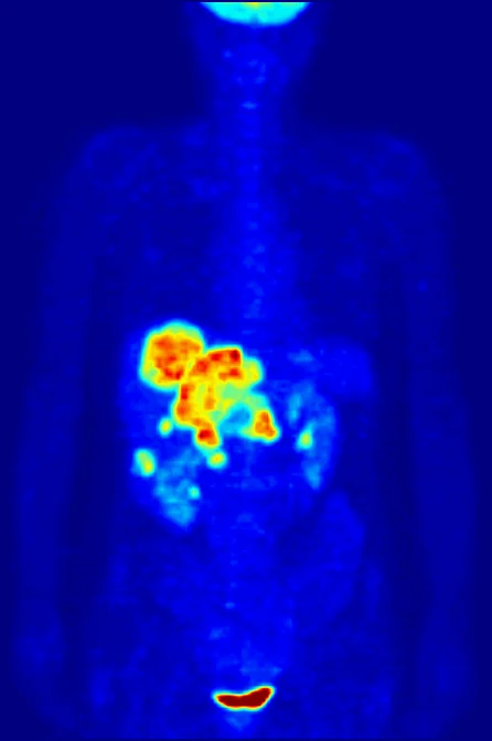 MEDICAL IMAGE
PET-MIPS-anim
A whole-body PET maximum-intensity projection shared as a clinical imaging example; tracer uptake is visualized rather than a photograph of surface anatomy.
Jens Maus ( https://jens-maus.de/ ) / Wikimedia Commons. Original source ↗
Public domain. First frame extracted from the original animated PET projection and encoded as WebP; no image content added. Display layout may crop the image.
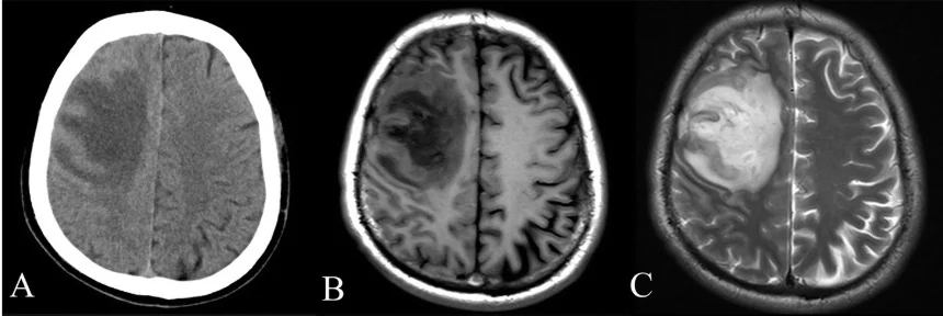 MEDICAL IMAGE
CT and MRI scan of the brain with melioidosis
CT and MRI brain images reproduced from an openly licensed research case report. The panel illustrates different imaging formats.
Wei-yuan Huang, Gang Wu, Feng Chen, Meng-meng Li and Jian-jun Li / Wikimedia Commons. Original source ↗
CC BY 4.0. Resized to a maximum dimension of 1500 pixels and encoded as WebP; no content changes. Display layout may crop the image.
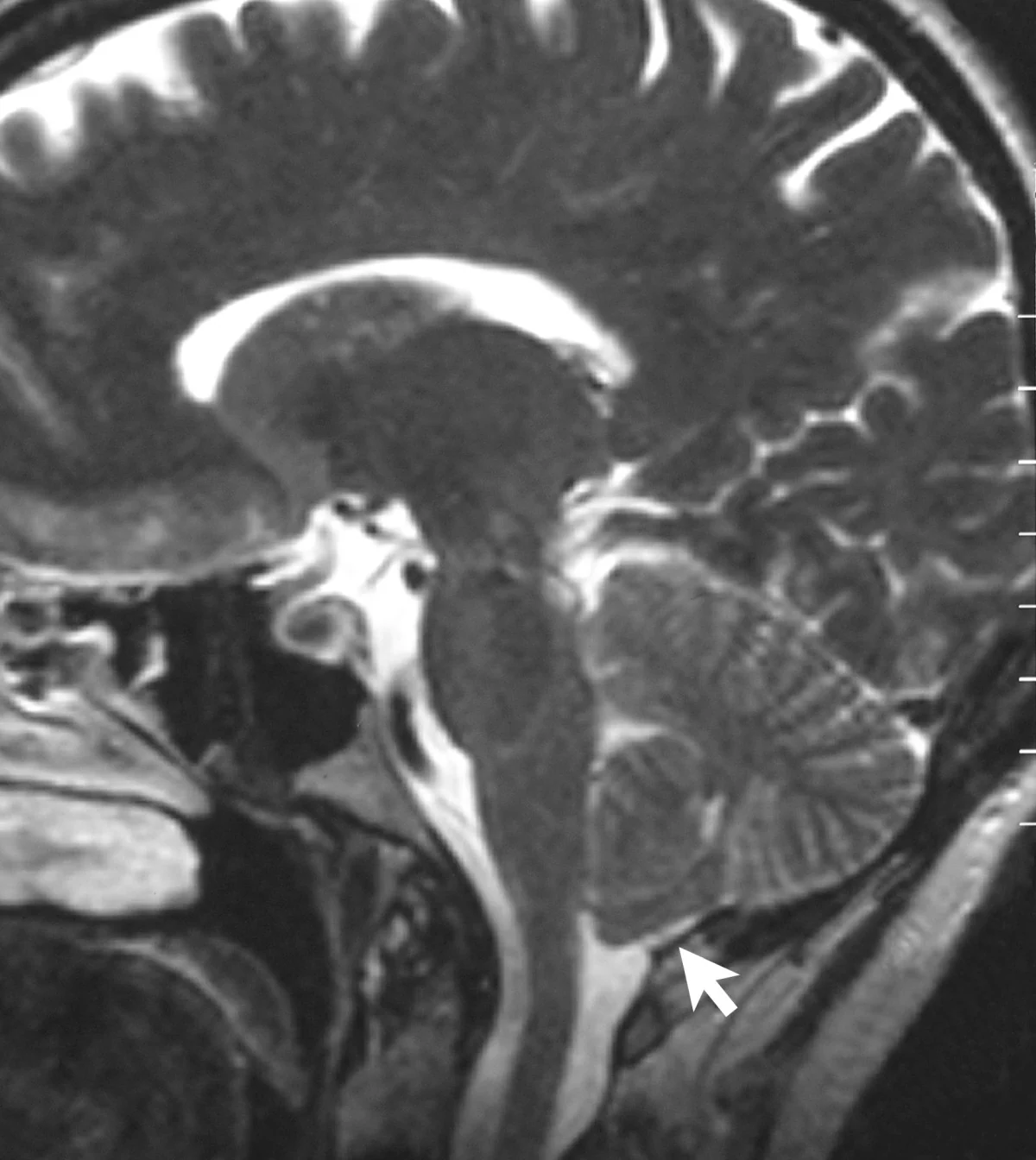 MEDICAL IMAGE
Sagittal MRI scan of brain of patient with Chiari malformation
A T2-weighted sagittal brain MRI shared as a medical-imaging example.
Raymond F Sekula Jr, Peter J Jannetta, Kenneth F Casey, Edward M Marchan, L Kathleen Sekula and Christine S McCrady / Wikimedia Commons. Original source ↗
CC BY 2.0. Resized to a maximum dimension of 1500 pixels and encoded as WebP; no content changes. Display layout may crop the image.
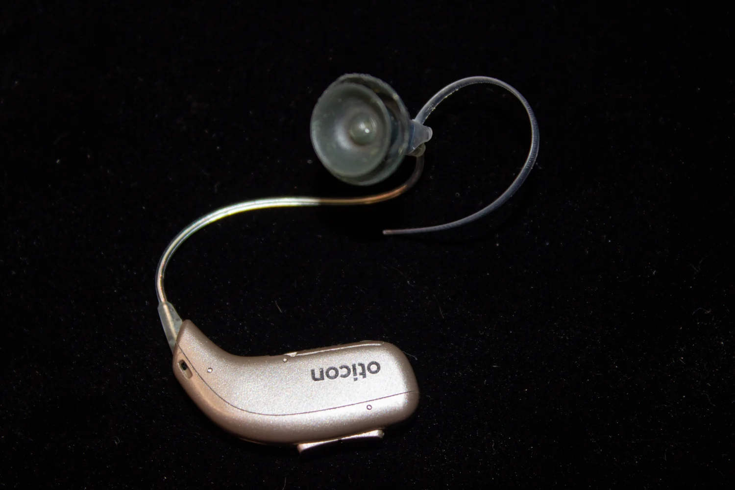 PHOTOGRAPH
Oticon hearing aid top view
A close view of an Oticon behind-the-ear hearing aid.
Bastique / Wikimedia Commons. Original source ↗
CC BY 4.0. Resized to a maximum dimension of 1500 pixels and encoded as WebP; no content changes. Display layout may crop the image.
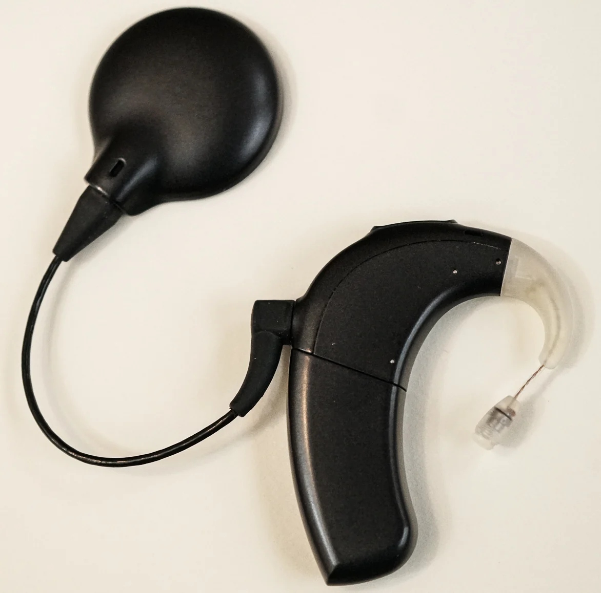 PHOTOGRAPH
Advanced Bionics cochlear implant processor, brand-named "Marvel" (fourth-generation behind-the-ear processor)
An Advanced Bionics Marvel external sound processor, photographed separately from an implanted component.
MikeCore9918 / Wikimedia Commons. Original source ↗
CC BY-SA 4.0. Resized to a maximum dimension of 1500 pixels and encoded as WebP; no content changes. Display layout may crop the image.
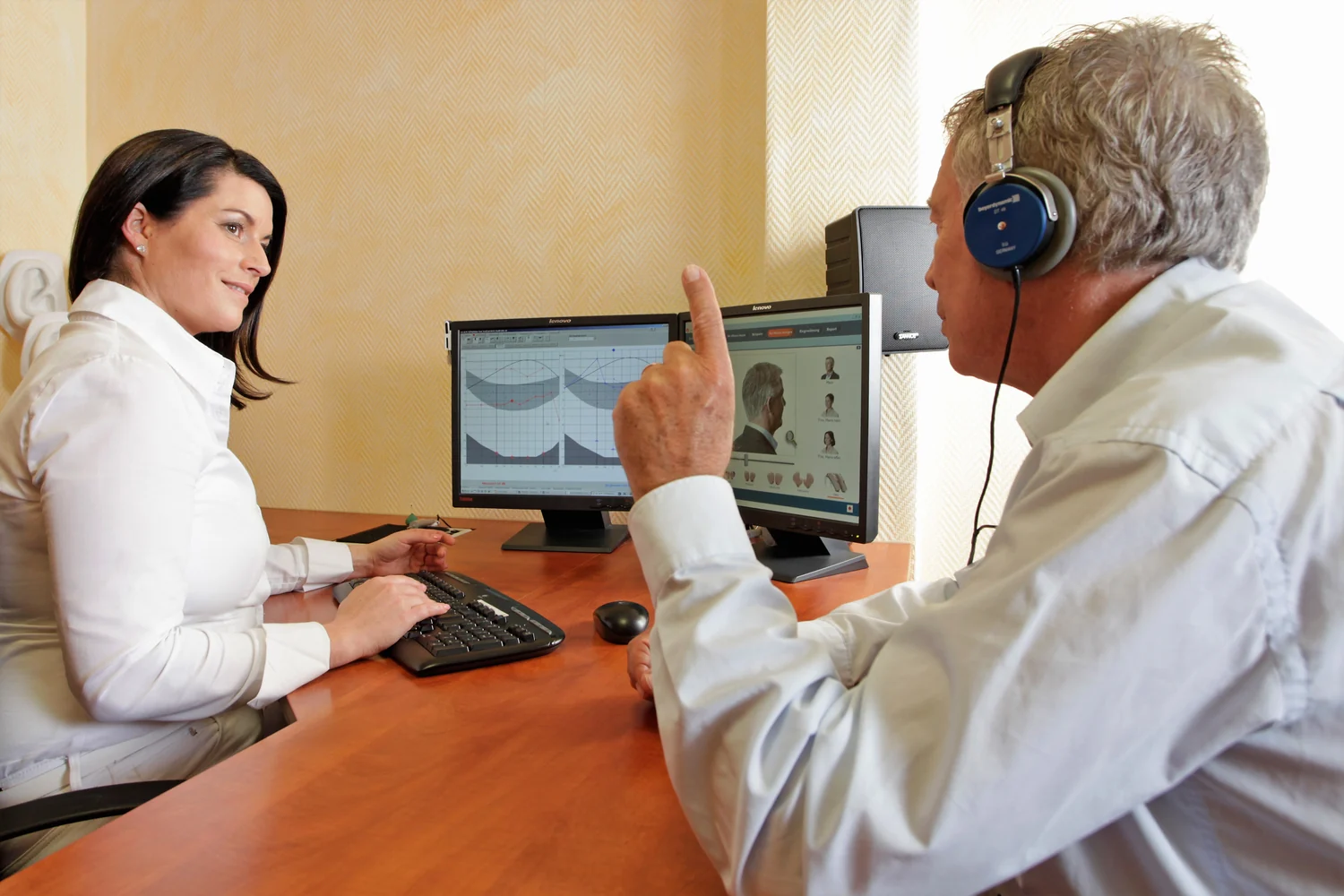 PHOTOGRAPH
Audiologist, 2015
A hearing-test session photographed in an audiology setting.
Bundesinnung Hörakustiker (bundesinnung_ha) / Wikimedia Commons. Original source ↗
CC BY-SA 2.0. Resized to a maximum dimension of 1500 pixels and encoded as WebP; no content changes. Display layout may crop the image.
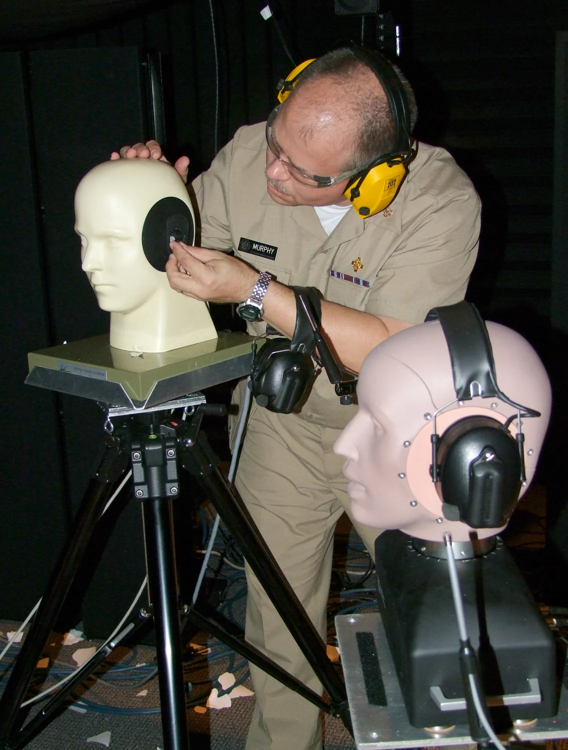 PHOTOGRAPH
Testing hearing protection devices using artificial head fixtures
Hearing-protection testing with artificial head fixtures in a NIOSH laboratory.
Chuck Kardous / Wikimedia Commons. Original source ↗
Public domain. Resized to a maximum dimension of 1500 pixels and encoded as WebP; no content changes. Display layout may crop the image.
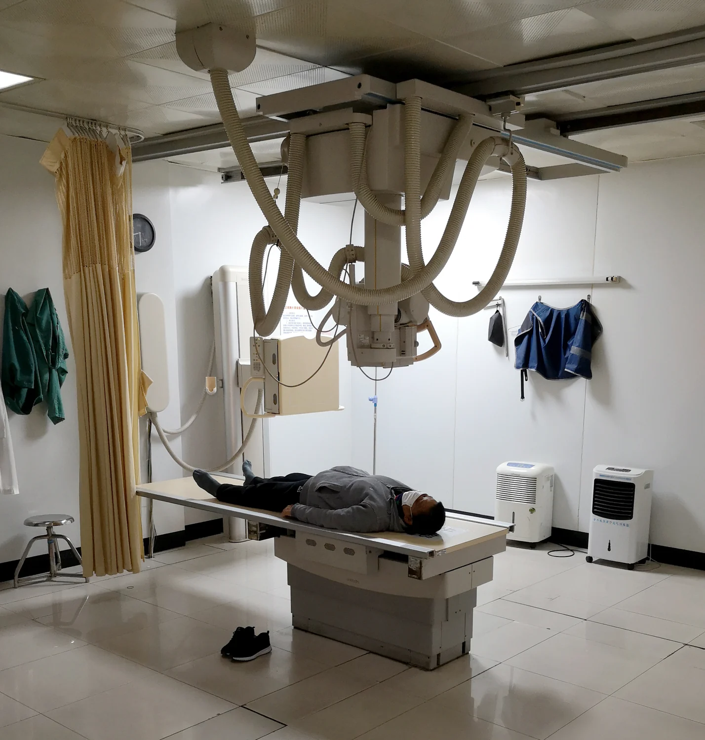 MEDICAL IMAGE
Hospital Radiology Room Philips DigitalDiagnost Digital Radiography System
A Philips DigitalDiagnost digital radiography system in a hospital X-ray room.
Ptrump16 / Wikimedia Commons. Original source ↗
CC BY-SA 4.0. Resized to a maximum dimension of 1500 pixels and encoded as WebP; no content changes. Display layout may crop the image.
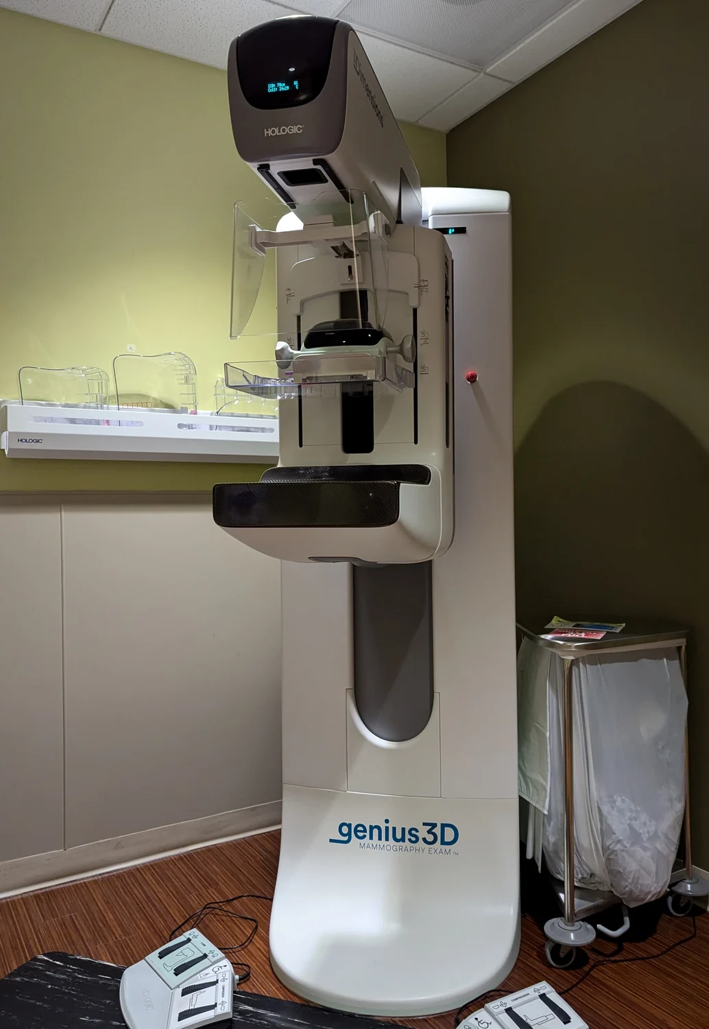 PHOTOGRAPH
Genius 3D mammogram machine
A Genius 3D mammography unit photographed at Kaiser Westside Medical Center in Oregon.
Aboutmovies / Wikimedia Commons. Original source ↗
CC BY-SA 4.0. Resized to a maximum dimension of 1500 pixels and encoded as WebP; no content changes. Display layout may crop the image.
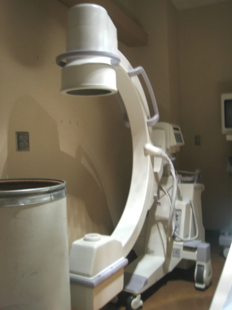 PHOTOGRAPH
Mobile X-ray machine
A mobile C-arm unit photographed as an example of fluoroscopy equipment.
Ben Stephenson from Beltsville, Maryland / Wikimedia Commons. Original source ↗
CC BY 2.0. Resized to a maximum dimension of 1500 pixels and encoded as WebP; no content changes. Display layout may crop the image.
 PHOTOGRAPH
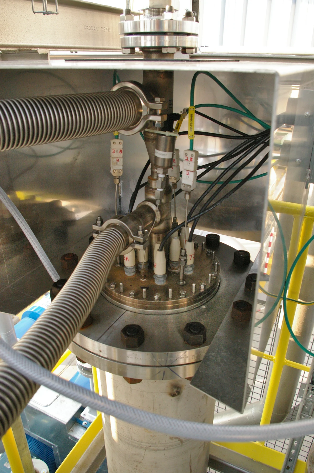
Plasma-Oxidiser-GlidArc-Poelkapelle-Belgium-2007
The head of a Glidarc cold-plasma oxidizer at an installation in Poelkapelle, Belgium.
Albin Czernichowski / Wikimedia Commons. Original source ↗
CC BY-SA 4.0. Resized from 2000 × 3008 to 1197 × 1800 pixels, EXIF orientation applied, and encoded as WebP. Original framing retained. Display layout may crop the image.
 PHOTOGRAPH
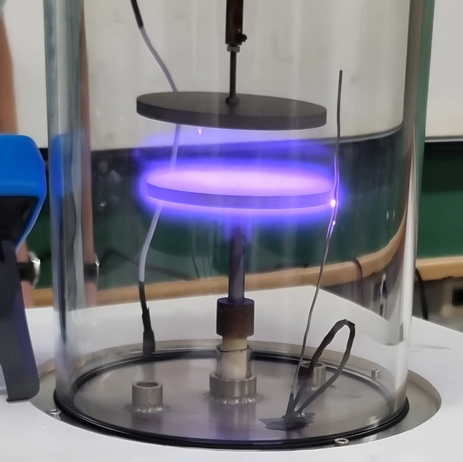
Low pressure plasma reactor
A low-pressure plasma reactor built for teaching. Chamber pressure, electrode geometry, and electrical power affect the discharge.
Lucasbortoletto1337 / Wikimedia Commons. Original source ↗
CC0 1.0. Resized from 1600 × 1596 to 1600 × 1596 pixels, EXIF orientation applied, and encoded as WebP. Original framing retained. Display layout may crop the image.
 PHOTOGRAPH
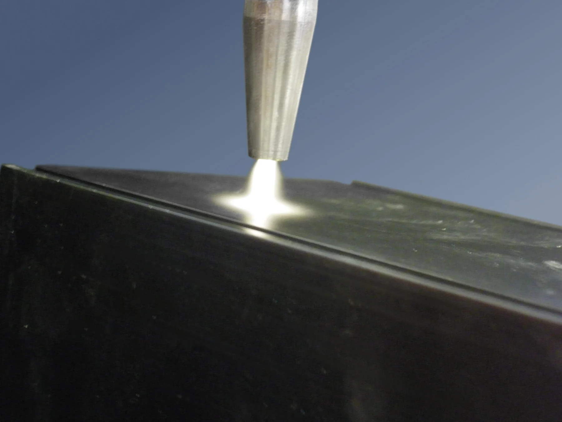
Buse plasma à pression atmosphérique
An atmospheric-pressure plasma jet at a material surface. Such sources are used to study how reactive gases interact with surfaces.
Acxys / Wikimedia Commons. Original source ↗
CC BY-SA 3.0. Resized from 3264 × 2448 to 1800 × 1350 pixels, EXIF orientation applied, and encoded as WebP. Original framing retained. Display layout may crop the image.
 PHOTOGRAPH
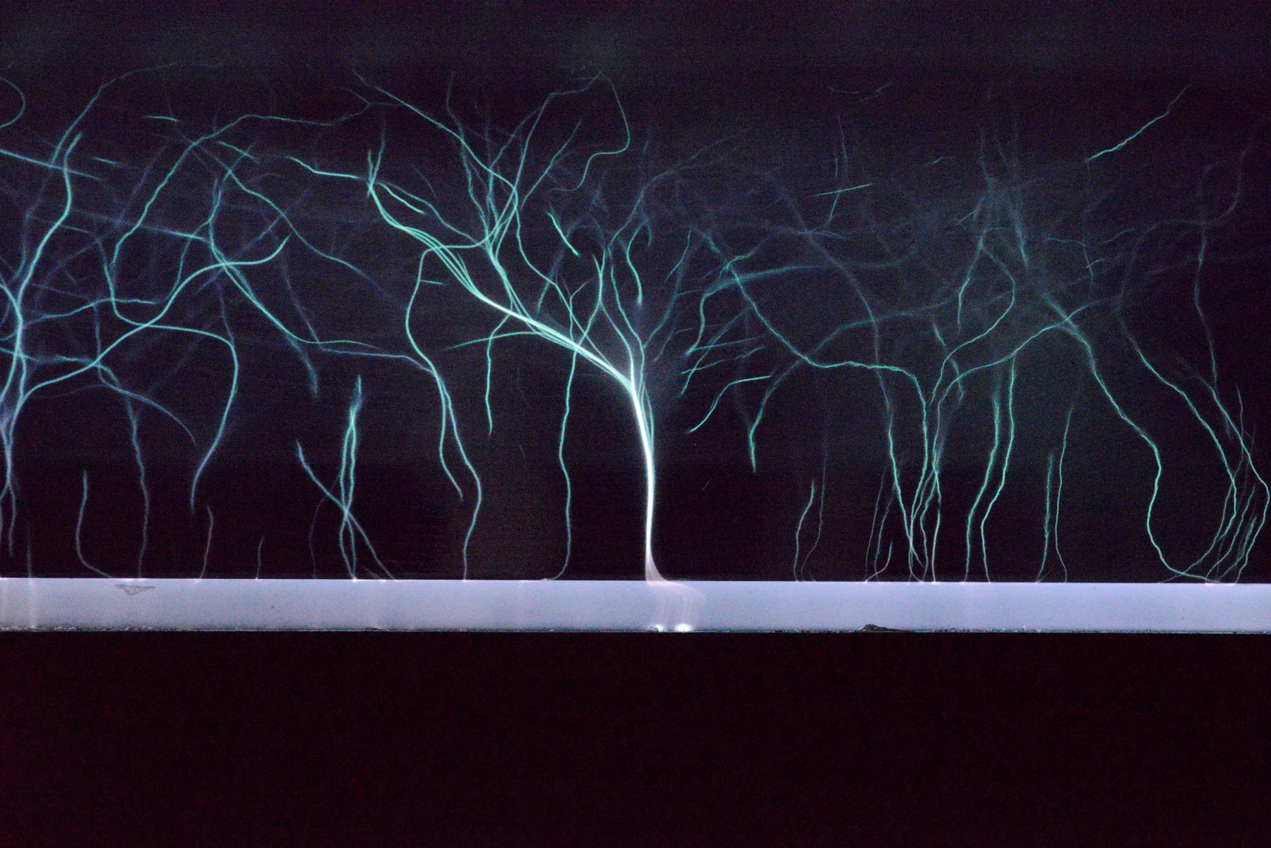
Plasma Trees
Atmospheric-pressure cold plasma photographed in the University of Bologna's industrial-plasma laboratory. Gas escaping around an electrode produces the branching pattern.
Themarnicle / Wikimedia Commons. Original source ↗
CC BY 4.0. Resized from 6670 × 4451 to 1800 × 1201 pixels, EXIF orientation applied, and encoded as WebP. Original framing retained. Display layout may crop the image.
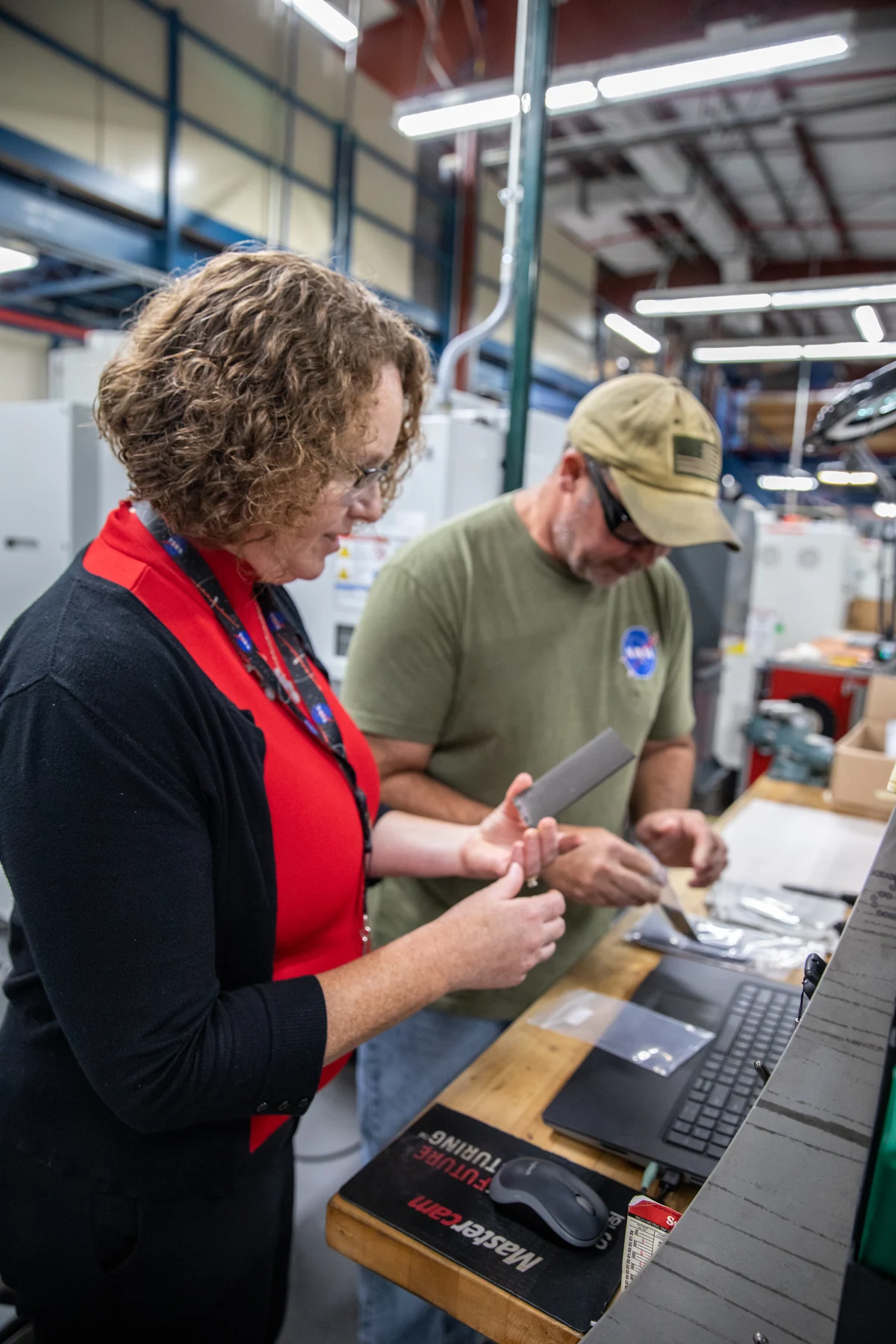 PHOTOGRAPH
Plasma Rapid Oxidation Technique for Extending Component Tenabil (KSC-20221102-PH-KLS01 0035)
NASA researchers inspect specimens for a plasma-electrolytic-oxidation experiment at Kennedy Space Center in 2022. The work concerns protective coatings on metals.
Kim Shiflett / NASA / Wikimedia Commons. Original source ↗
Public domain. Resized from 4480 × 6720 to 1200 × 1800 pixels, EXIF orientation applied, and encoded as WebP. Original framing retained. Display layout may crop the image.
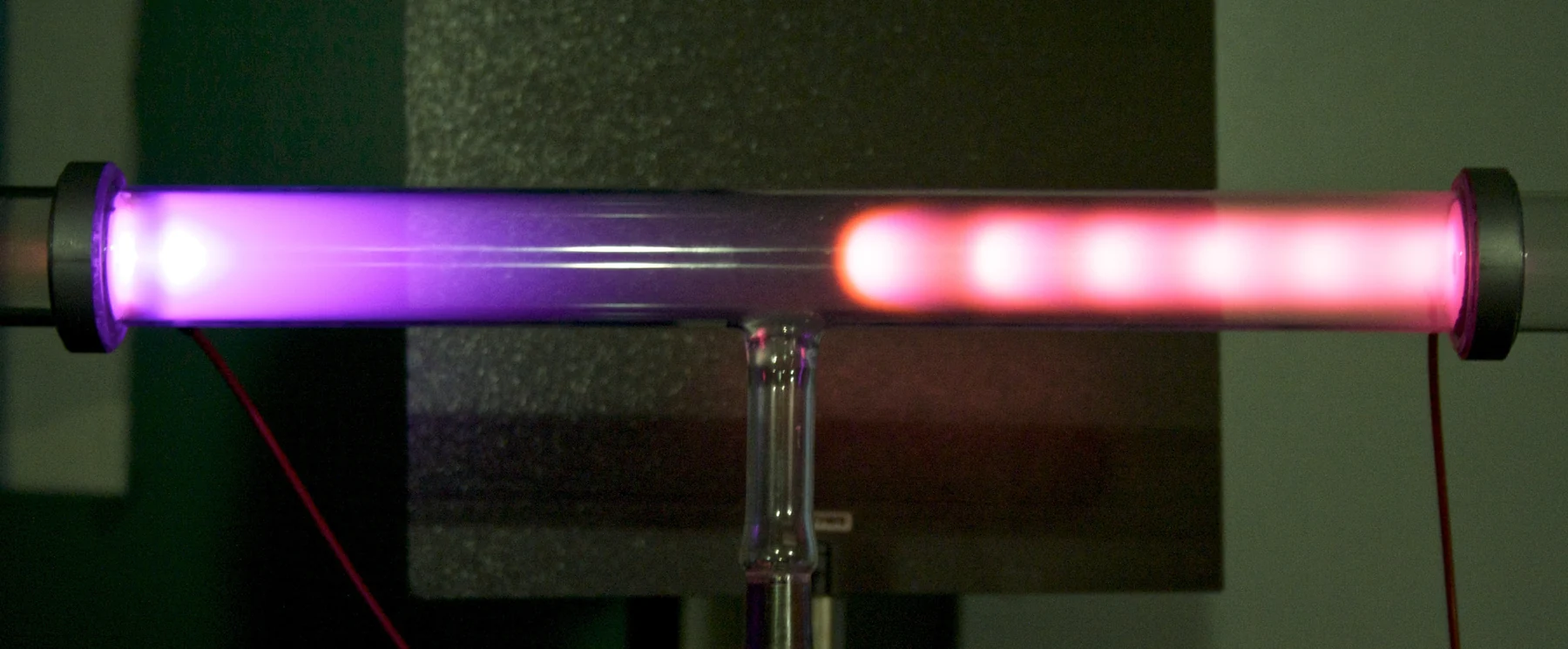 PHOTOGRAPH
Glow discharge regions
A glow discharge in an evacuated glass tube. The visible regions reflect how electrical energy is transferred to the gas.
Derived file: Chetvorno Original file: Jannis Andrija Schnitzer / Wikimedia Commons. Original source ↗
CC BY-SA 2.0. Resized from 3334 × 1380 to 1800 × 745 pixels, EXIF orientation applied, and encoded as WebP. Original framing retained. Display layout may crop the image.
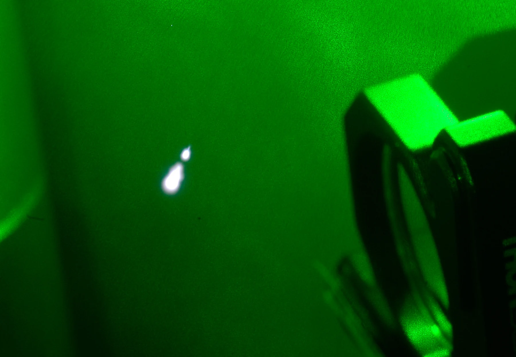 PHOTOGRAPH
Plasma generation in air
A focused pulsed laser produces a small plasma in air. This laboratory image concerns laser-driven ionization rather than a patient treatment.
Rcaravit / Wikimedia Commons. Original source ↗
CC BY 4.0. Resized from 2114 × 1464 to 1800 × 1247 pixels, EXIF orientation applied, and encoded as WebP. Original framing retained. Display layout may crop the image.
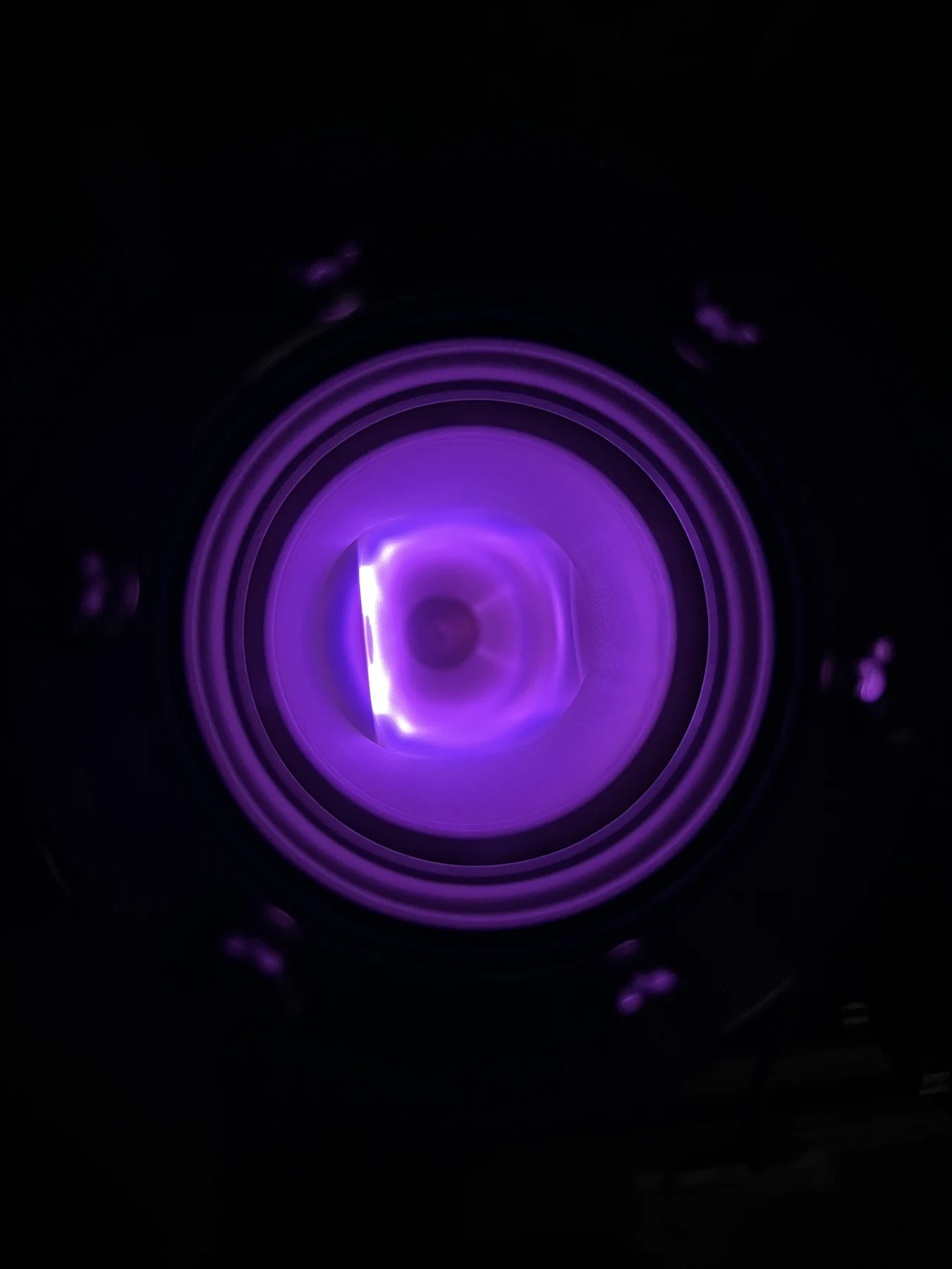 PHOTOGRAPH
Plasma source
A plasma source photographed in an MIT nuclear-physics laboratory. Ions from the source can be extracted into a beamline.
Driveraorona / Wikimedia Commons. Original source ↗
CC BY-SA 4.0. Resized from 3024 × 4032 to 1350 × 1800 pixels, EXIF orientation applied, and encoded as WebP. Original framing retained. Display layout may crop the image.
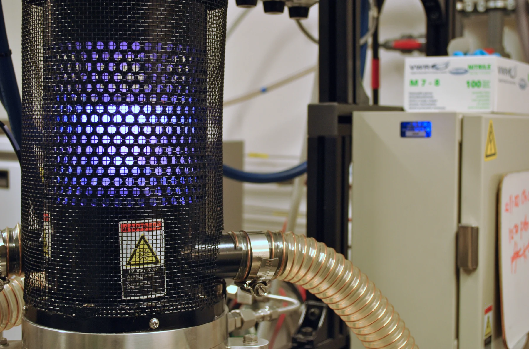 PHOTOGRAPH
Chemical vapour deposition machine in the LCN
A chemical-vapour-deposition system at the London Centre for Nanotechnology. The apparatus illustrates controlled material processing in a laboratory chamber.
O. Usher (UCL MAPS) / University College London Faculty of Mathematical & Physical Sciences / Wikimedia Commons. Original source ↗
CC BY 3.0. Resized from 3817 × 2524 to 1800 × 1190 pixels, EXIF orientation applied, and encoded as WebP. Original framing retained. Display layout may crop the image.
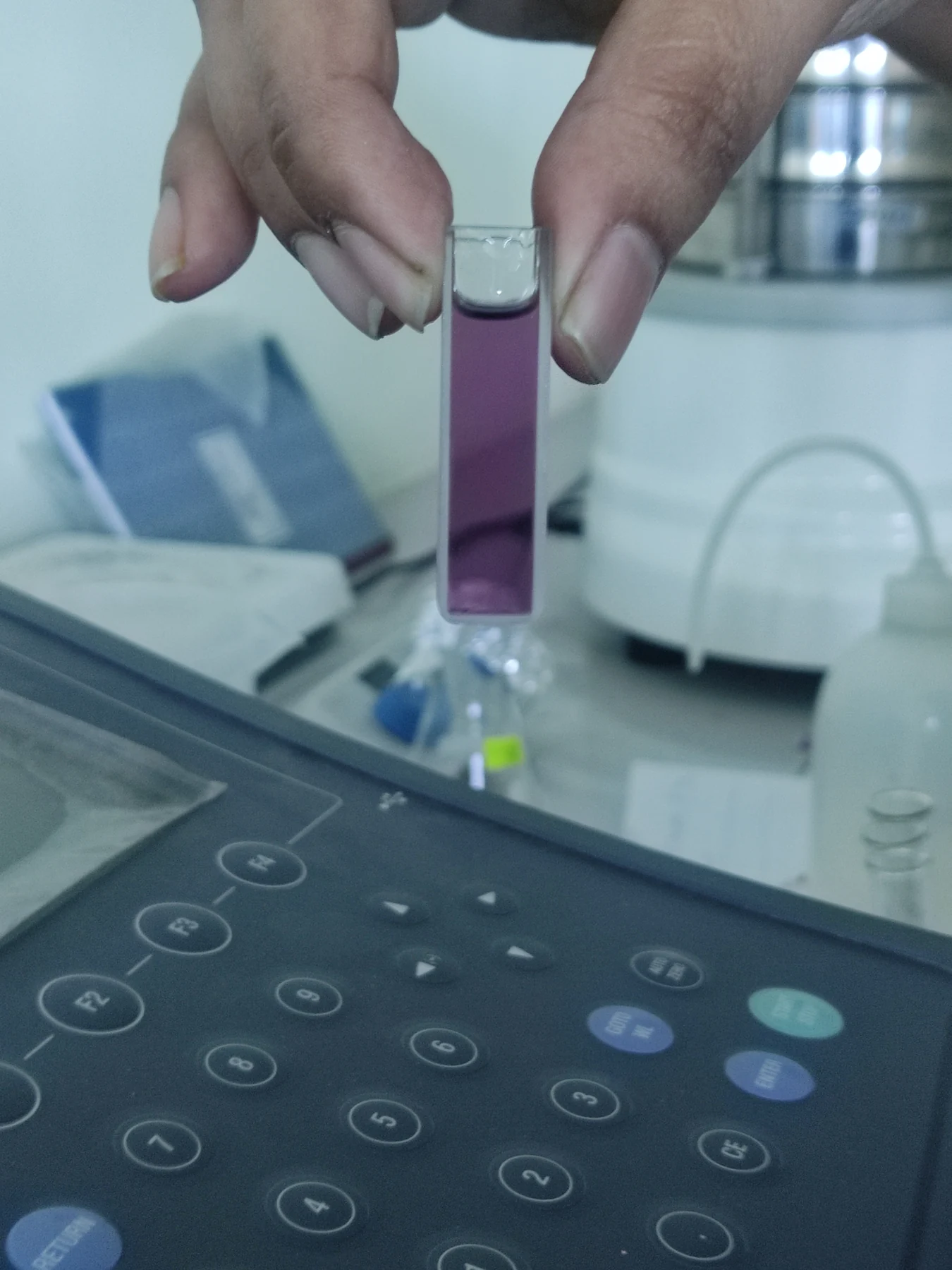 PHOTOGRAPH
Absorption measurement by UV Spectrometer in laboratory
A cuvette is prepared for an ultraviolet absorption measurement. Spectrometers compare how samples interact with different wavelengths of light.
Aurora Wild / Wikimedia Commons. Original source ↗
CC0 1.0. Resized from 3000 × 4000 to 1350 × 1800 pixels, EXIF orientation applied, and encoded as WebP. Original framing retained. Display layout may crop the image.
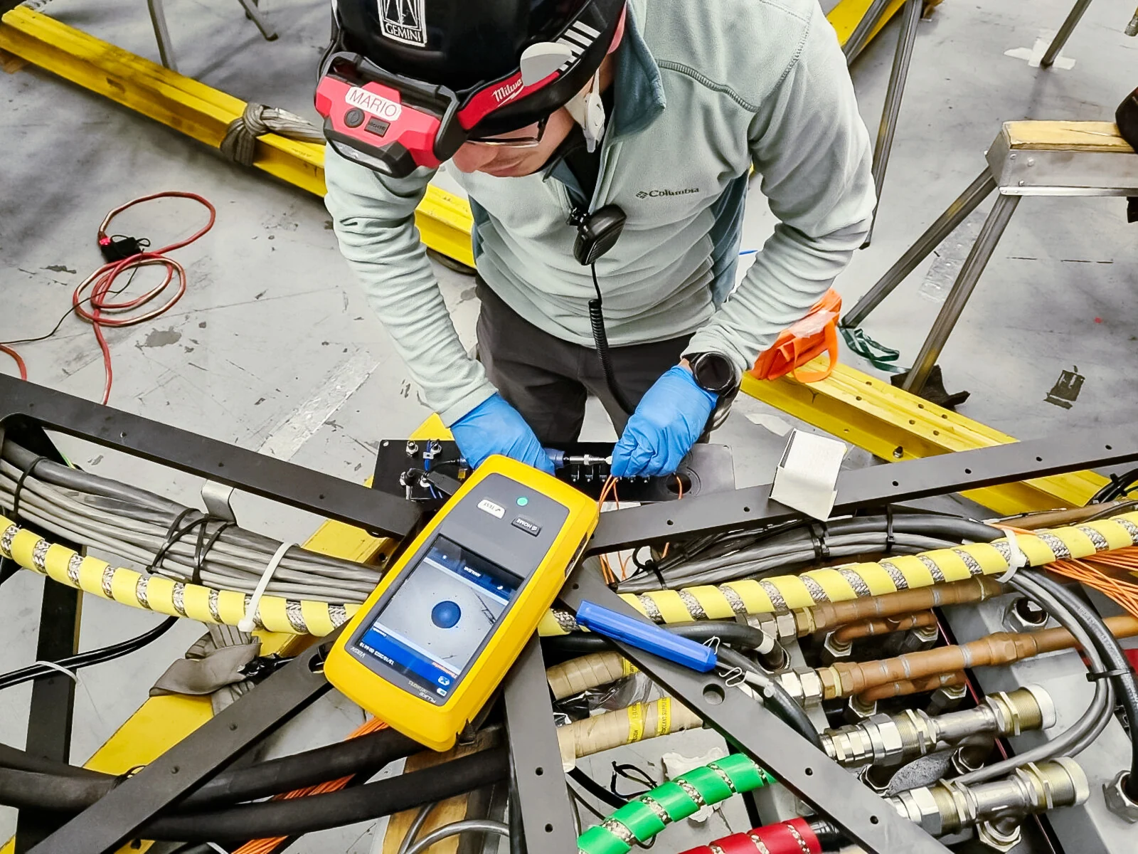 PHOTOGRAPH
Fiber optics verification (20231013 151223-CC)
Fibre-optic transmission is checked during work at the Gemini Observatory. Similar optical measurement principles are used in many scientific instruments.
International Gemini Observatory/NOIRLab/NSF/AURA / Wikimedia Commons. Original source ↗
CC BY 4.0. Resized from 1600 × 1200 to 1600 × 1200 pixels, EXIF orientation applied, and encoded as WebP. Original framing retained. Display layout may crop the image.
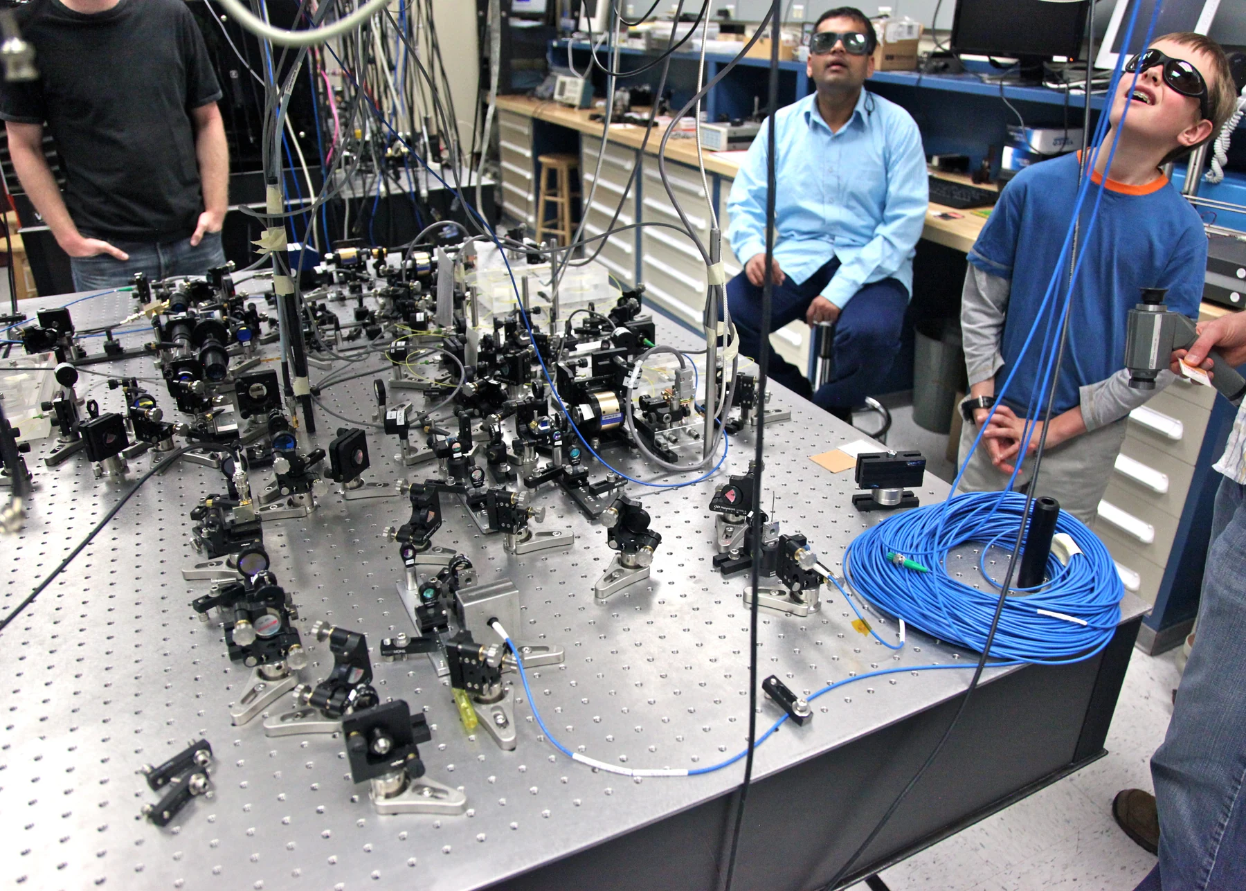 PHOTOGRAPH
Setup to generate an extremely precise Wavelength of Infrared light
An optical-table setup for generating a precise infrared wavelength. The beam path is controlled by many separate optical components.
Steve Jurvetson from Menlo Park, USA / Wikimedia Commons. Original source ↗
CC BY 2.0. Resized from 5236 × 3744 to 1800 × 1287 pixels, EXIF orientation applied, and encoded as WebP. Original framing retained. Display layout may crop the image.
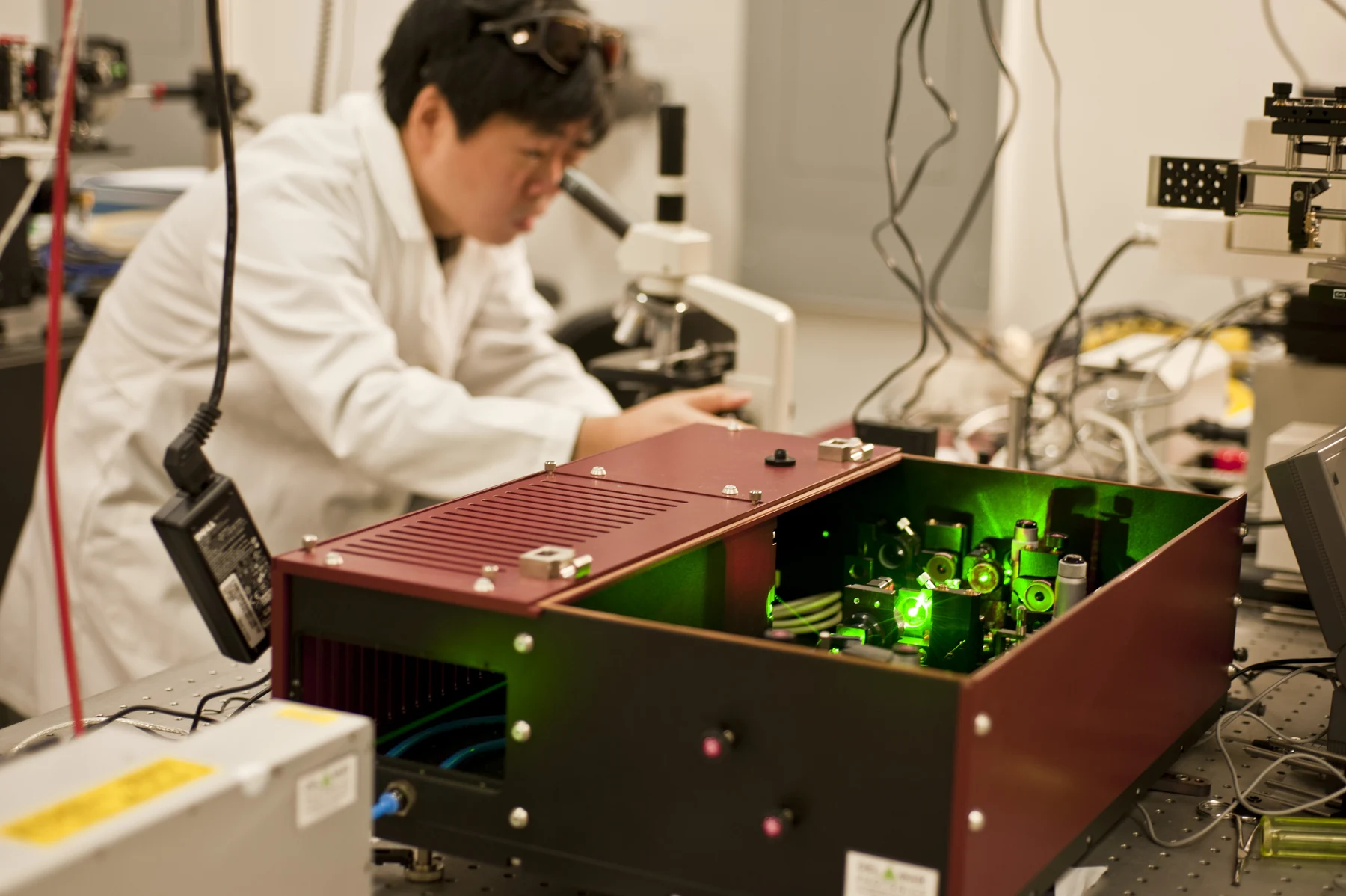 PHOTOGRAPH
Optical Therapeutics and Medical Nanophotonics Laboratory (5425575177)
Optical-therapy and nanophotonics research at the U.S. Food and Drug Administration, photographed by Joshua Budich.
Joshua Budich / U.S. Food and Drug Administration / Wikimedia Commons. Original source ↗
Public domain. Resized from 4256 × 2832 to 1800 × 1198 pixels, EXIF orientation applied, and encoded as WebP. Original framing retained. Display layout may crop the image.
 PHOTOGRAPH
Heelium Neoon laser
A helium-neon laser at the XLAB experimental laboratory in Göttingen. Gas lasers provide a visible example of stimulated light emission.
Thctamm / Wikimedia Commons. Original source ↗
CC BY-SA 3.0. Resized from 5472 × 3648 to 1800 × 1200 pixels, EXIF orientation applied, and encoded as WebP. Original framing retained. Display layout may crop the image.
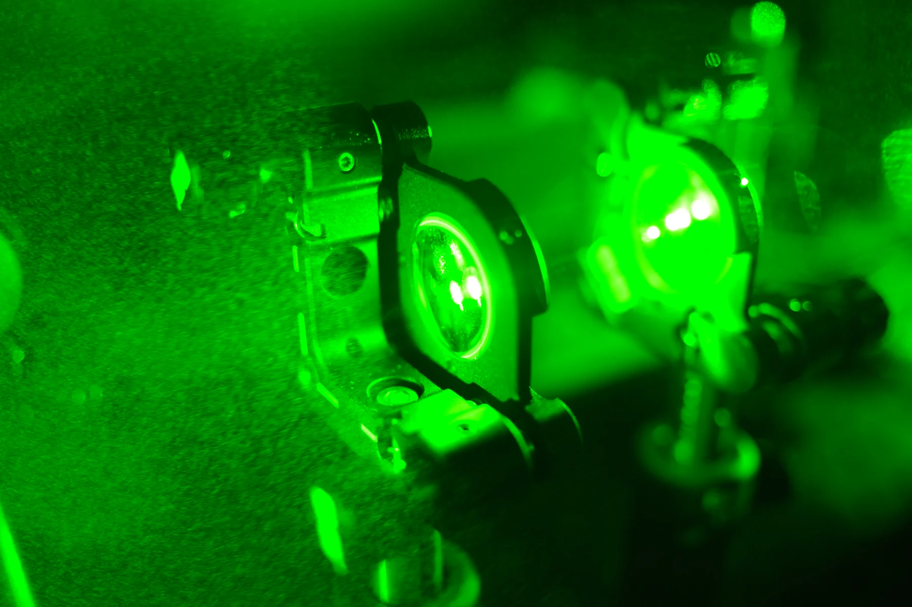 PHOTOGRAPH
Pfloutch 003
A laser beam passes through a lens doublet. Lenses shape the beam and help control where optical energy is delivered.
Pfloutch / Wikimedia Commons. Original source ↗
CC BY-SA 4.0. Resized from 4256 × 2832 to 1800 × 1198 pixels, EXIF orientation applied, and encoded as WebP. Original framing retained. Display layout may crop the image.
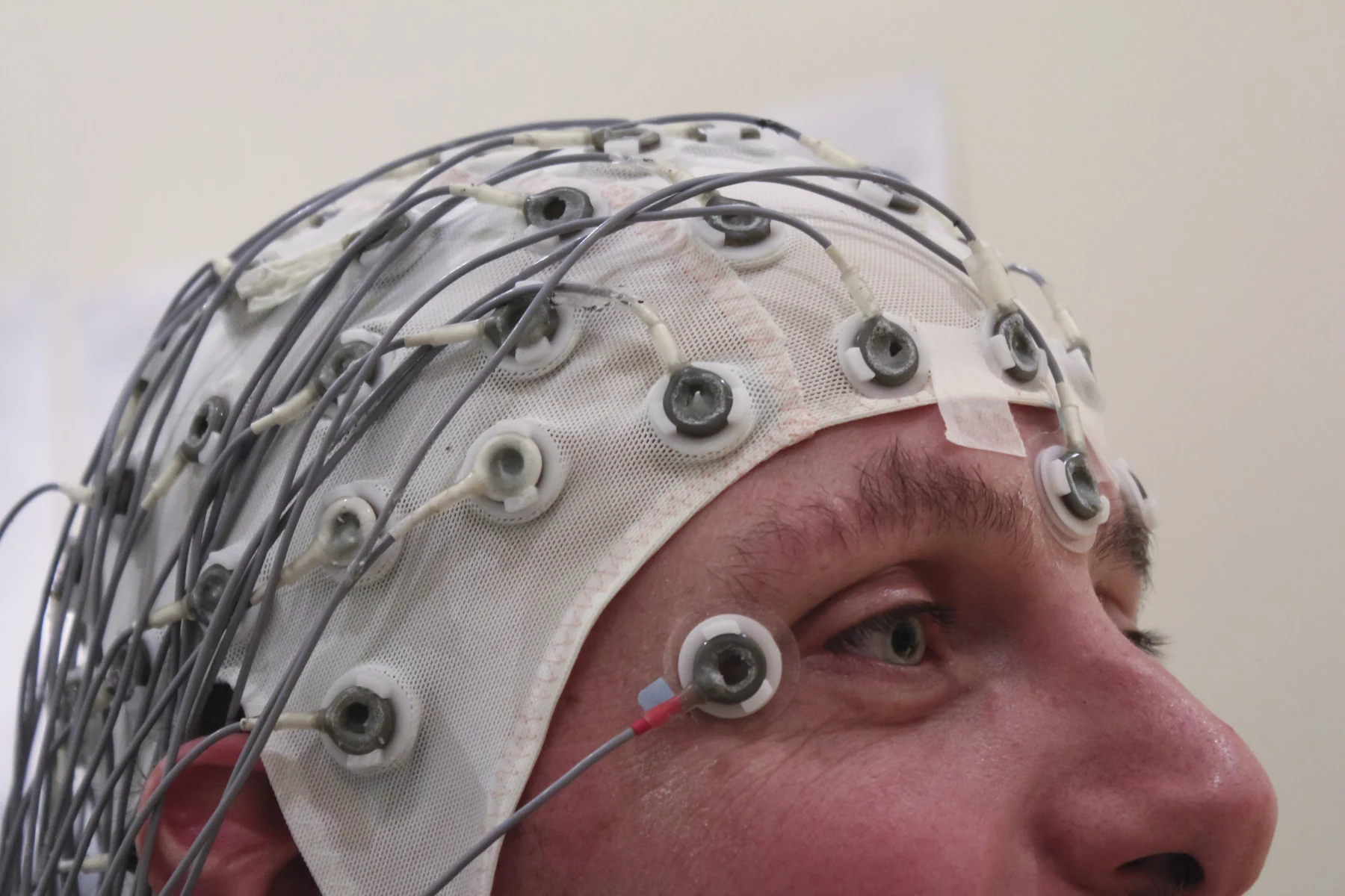 PHOTOGRAPH
EEG Recording Cap
An EEG recording cap positions multiple electrodes on the scalp to measure small voltage differences associated with brain activity.
Chris Hope / Wikimedia Commons. Original source ↗
CC BY 2.0. Resized from 5184 × 3456 to 1800 × 1200 pixels, EXIF orientation applied, and encoded as WebP. Original framing retained. Display layout may crop the image.
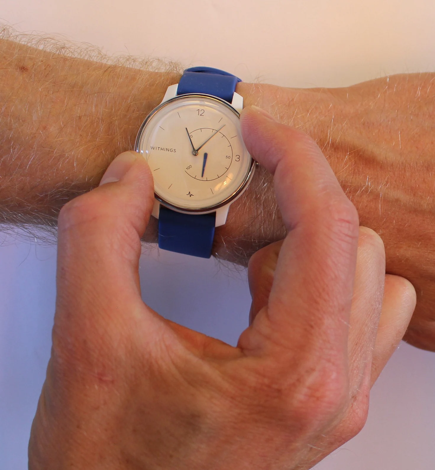 PHOTOGRAPH
Recording single-lead ECG using a Withings Move ECG watch
A single-lead ECG recording is made using a Withings Move ECG watch. Contact with both arms completes the measurement arrangement.
Peterhcharlton / Wikimedia Commons. Original source ↗
CC BY 4.0. Resized from 1471 × 1591 to 1471 × 1591 pixels, EXIF orientation applied, and encoded as WebP. Original framing retained. Display layout may crop the image.
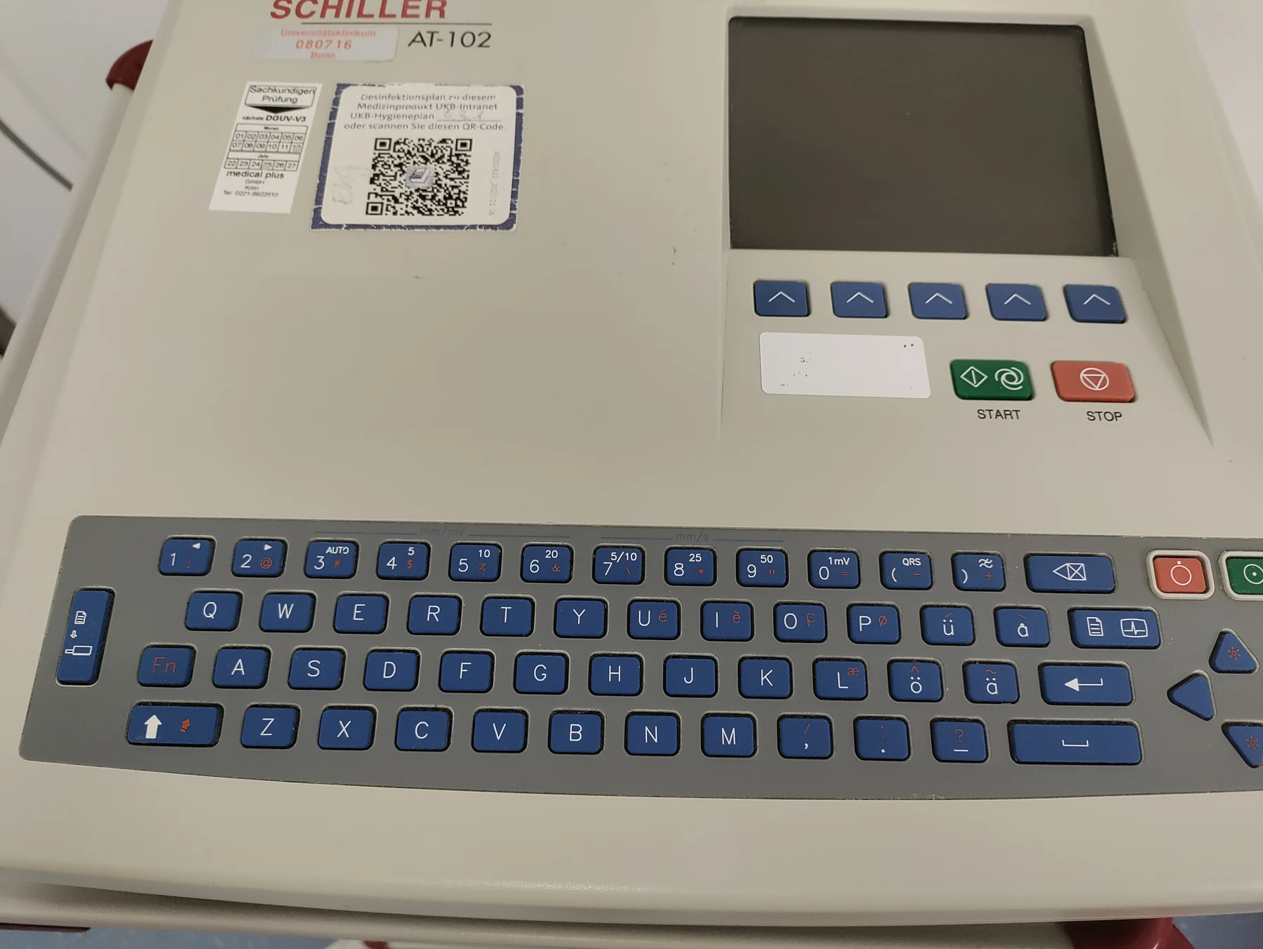 PHOTOGRAPH
Schiller EKG Gerät AT102 (35072)
A Schiller AT-102 electrocardiograph. ECG equipment records differences in electrical potential detected by surface electrodes.
Ibrahim Husain Meraj / Wikimedia Commons. Original source ↗
CC BY-SA 4.0. Resized from 4624 × 3472 to 1800 × 1352 pixels, EXIF orientation applied, and encoded as WebP. Original framing retained. Display layout may crop the image.
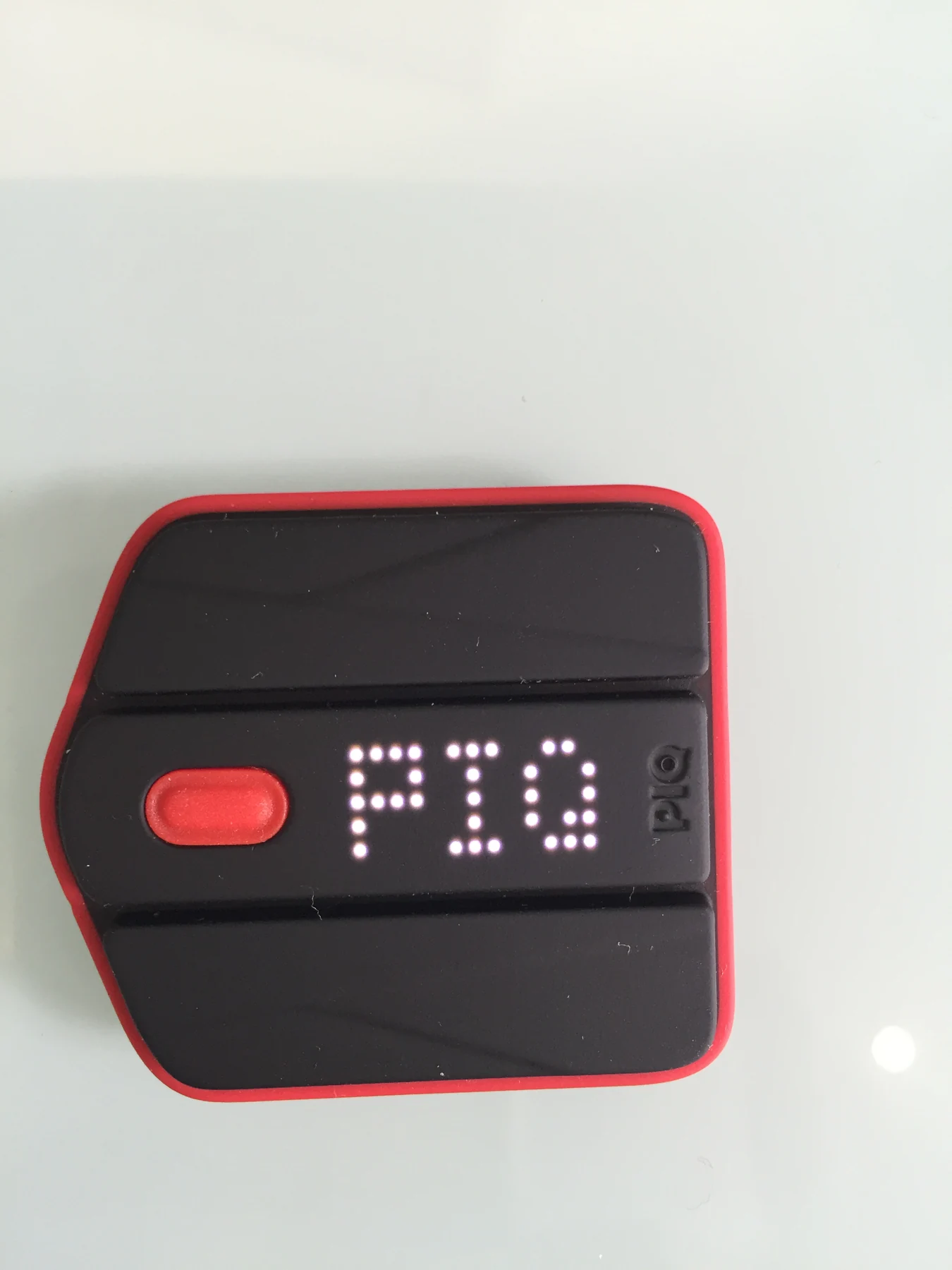 PHOTOGRAPH
Multi sport sensor PIQ
A PIQ multi-sport sensor. Small wearable instruments can capture movement for activity and performance analysis.
Johnspencer / Wikimedia Commons. Original source ↗
CC BY-SA 4.0. Resized from 2448 × 3264 to 1350 × 1800 pixels, EXIF orientation applied, and encoded as WebP. Original framing retained. Display layout may crop the image.
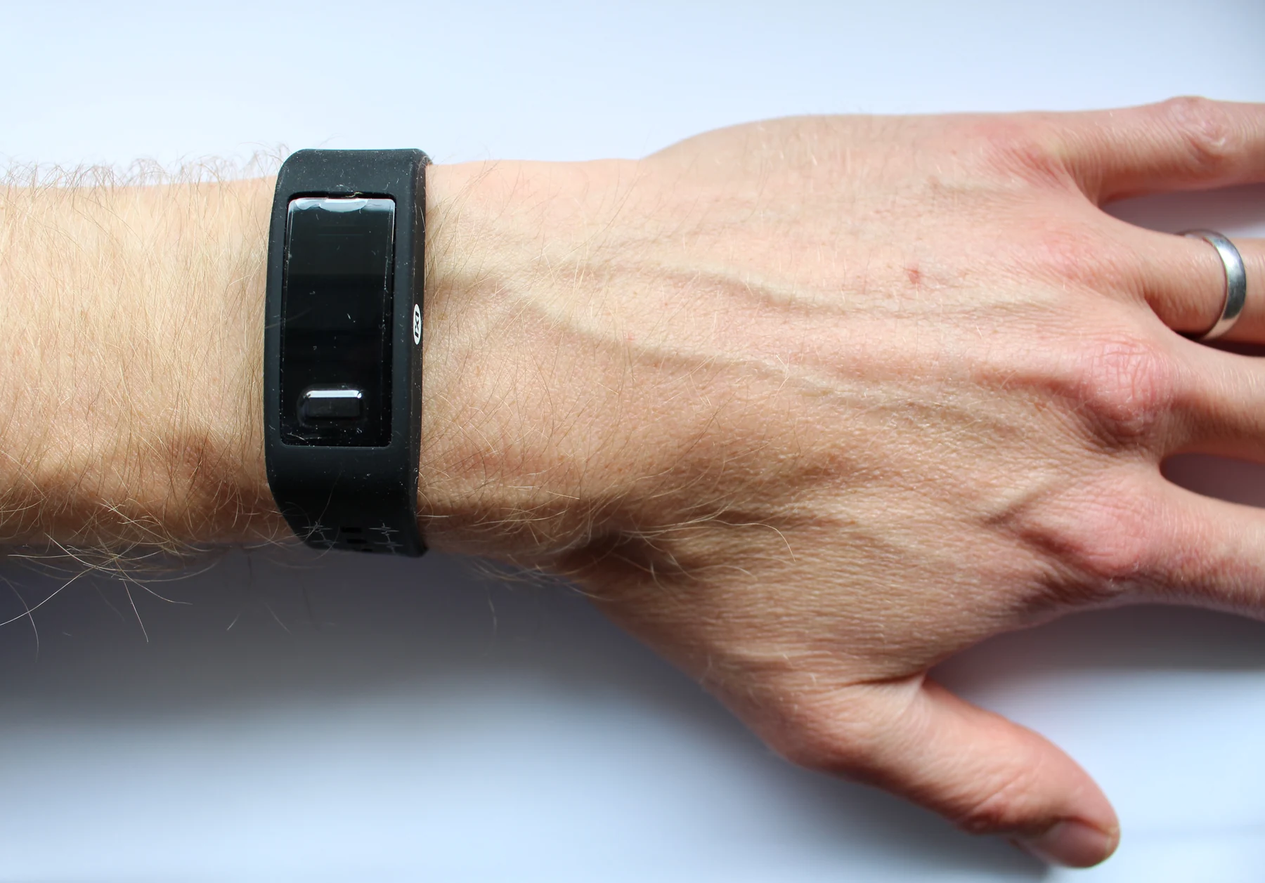 PHOTOGRAPH
Max Health Band
A wrist-worn health band illustrates the compact packaging of wearable sensors and their everyday contact with skin.
Peterhcharlton / Wikimedia Commons. Original source ↗
CC BY 4.0. Resized from 3298 × 2304 to 1800 × 1257 pixels, EXIF orientation applied, and encoded as WebP. Original framing retained. Display layout may crop the image.
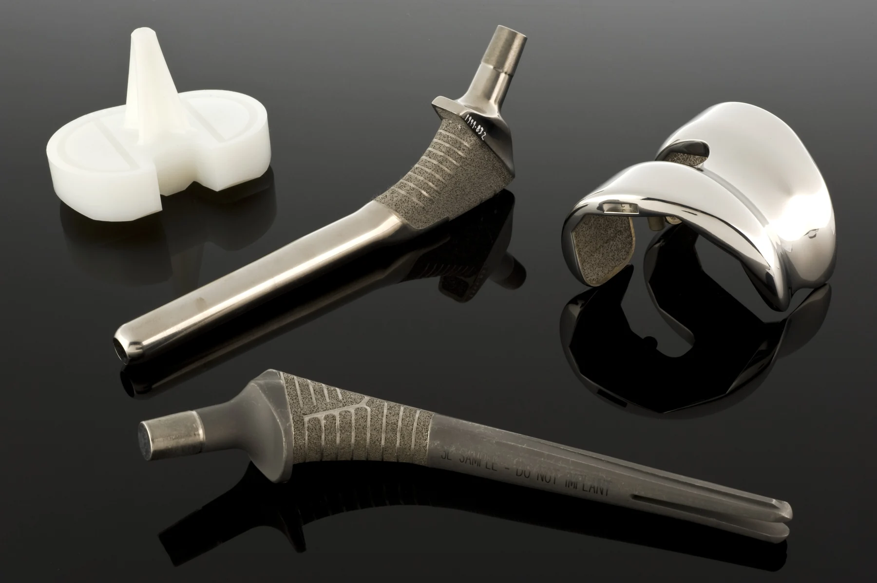 PHOTOGRAPH
Hip joint replacement, United States, 1998
Hip-joint replacement components from the Science Museum collection, photographed through Wellcome Images. Implant design must address both mechanical loading and interaction with the body.
Science Museum, London / Wellcome Collection / Wikimedia Commons. Original source ↗
CC BY 4.0. Resized from 4256 × 2832 to 1800 × 1198 pixels, EXIF orientation applied, and encoded as WebP. Original framing retained. Display layout may crop the image.
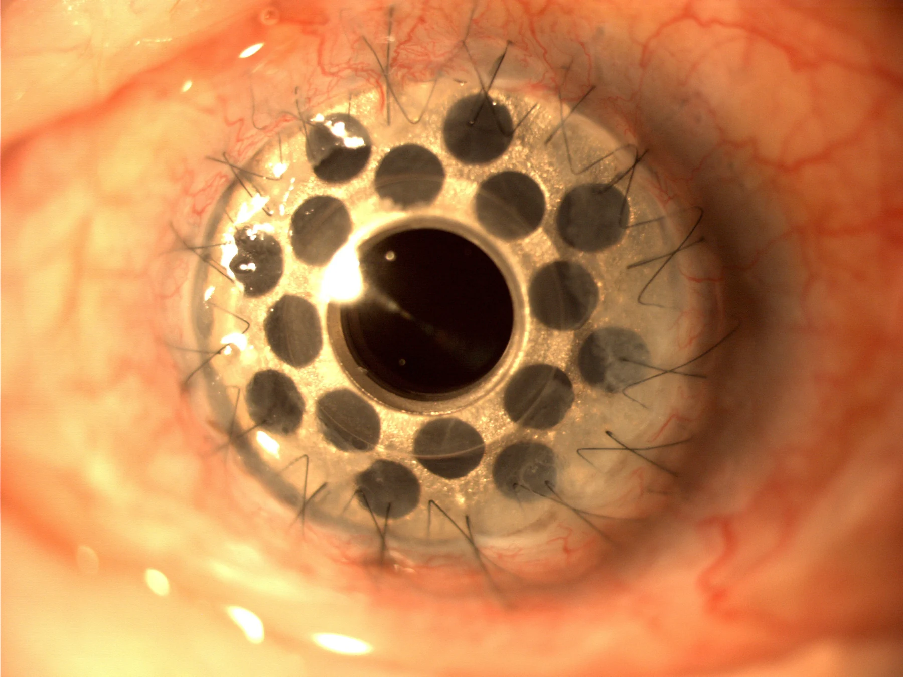 PHOTOGRAPH
Boston Kpro type 1 Titanium posterior plate
A Boston type-1 keratoprosthesis with a titanium posterior plate. The image shows a specific implanted artificial-cornea design.
Mariagessa / Wikimedia Commons. Original source ↗
CC BY-SA 4.0. Resized from 2048 × 1536 to 1800 × 1350 pixels, EXIF orientation applied, and encoded as WebP. Original framing retained. Display layout may crop the image.
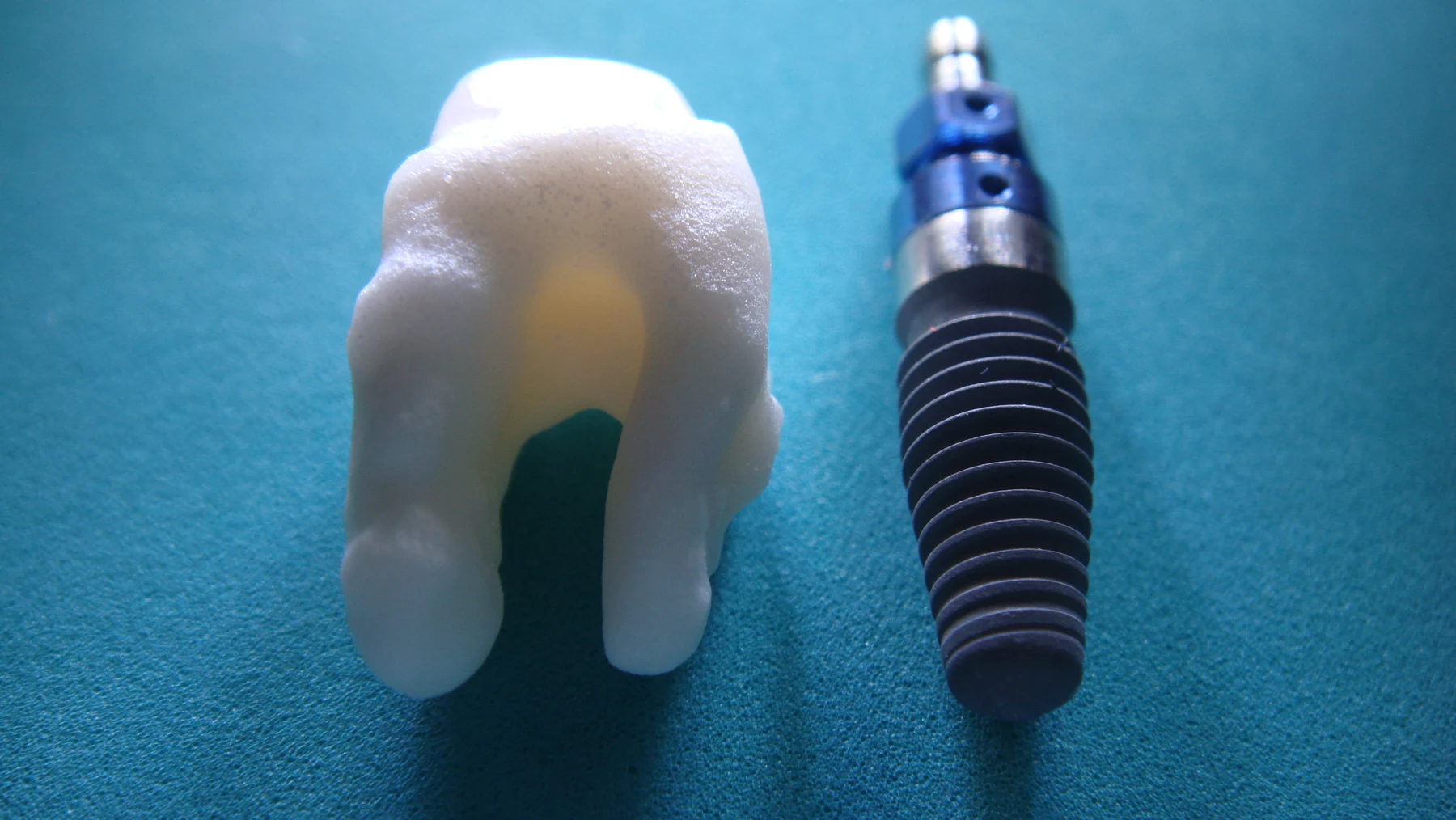 PHOTOGRAPH
Root analogue ceramic dental implant vs titanium screw type implant
Two different dental-implant designs: a root-analogue ceramic form and a titanium screw form. Their shapes illustrate different approaches to anchoring an implant.
Logicwhatelse / Wikimedia Commons. Original source ↗
CC BY-SA 4.0. Resized from 3648 × 2056 to 1800 × 1014 pixels, EXIF orientation applied, and encoded as WebP. Original framing retained. Display layout may crop the image.
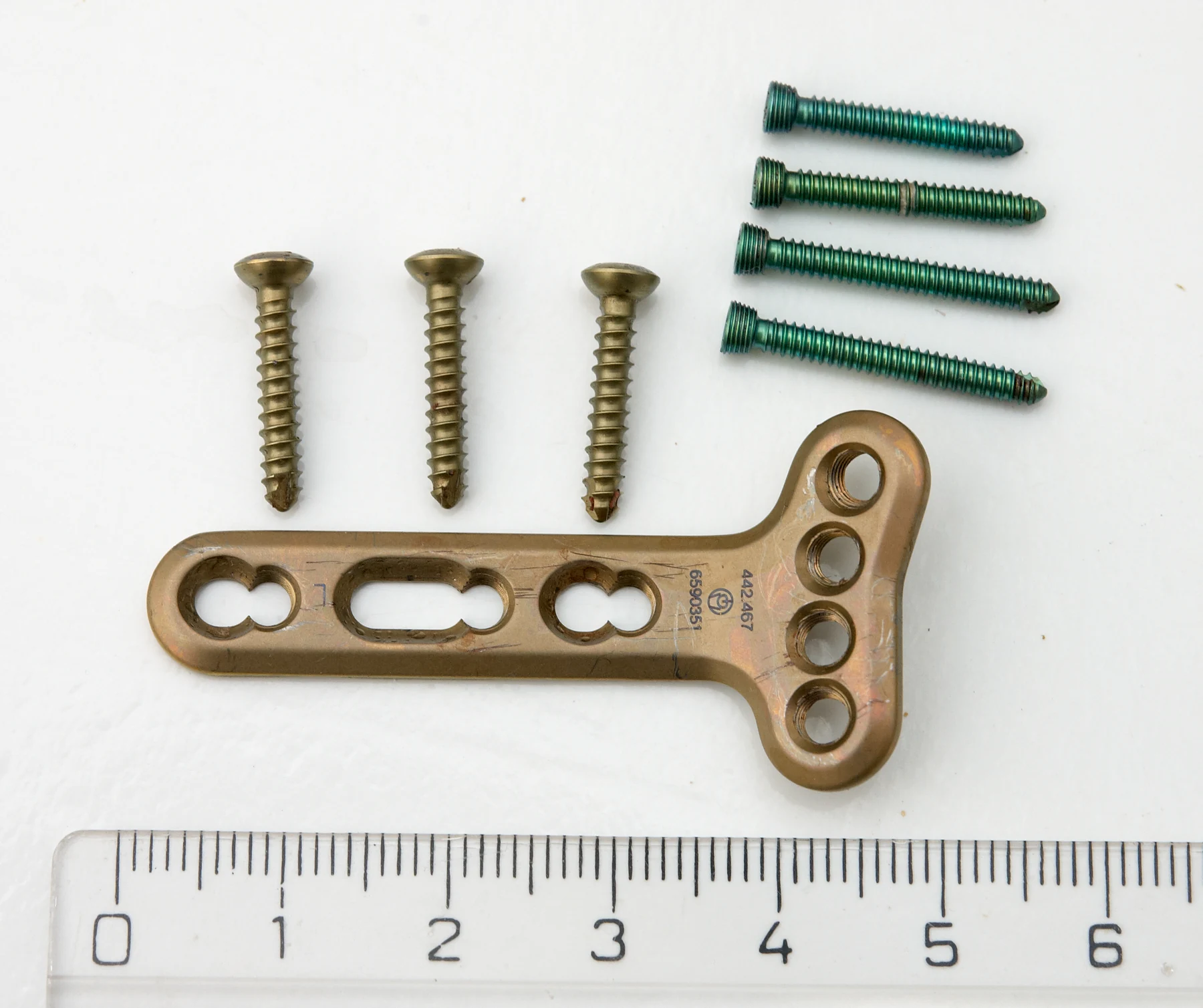 PHOTOGRAPH
Titanium plaatje voor pols
A titanium plate and screws used for wrist-fracture fixation. The ruler provides a scale for the hardware.
Wouter Hagens / Wikimedia Commons. Original source ↗
CC BY-SA 3.0. Resized from 2230 × 1870 to 1800 × 1509 pixels, EXIF orientation applied, and encoded as WebP. Original framing retained. Display layout may crop the image.
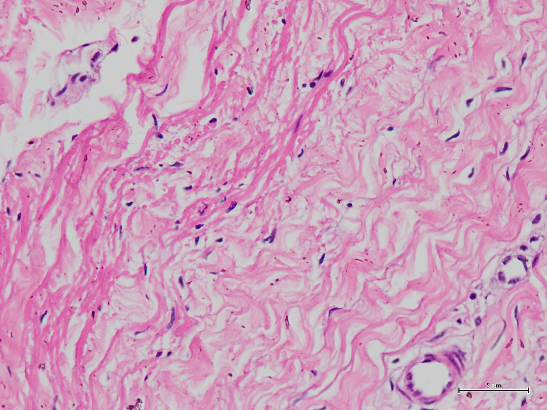 PHOTOGRAPH
'The Living Fabric of the Heart'- Native Bovine Pericardium under the Microscope
Light microscopy of native bovine pericardium, photographed at BUET's Bio-innovation and Research Group. Tissue architecture matters when biological materials are characterized.
Hibiscus Rosa Sinensis / Wikimedia Commons. Original source ↗
CC BY 4.0. Resized from 1920 × 1440 to 1800 × 1350 pixels, EXIF orientation applied, and encoded as WebP. Original framing retained. Display layout may crop the image.
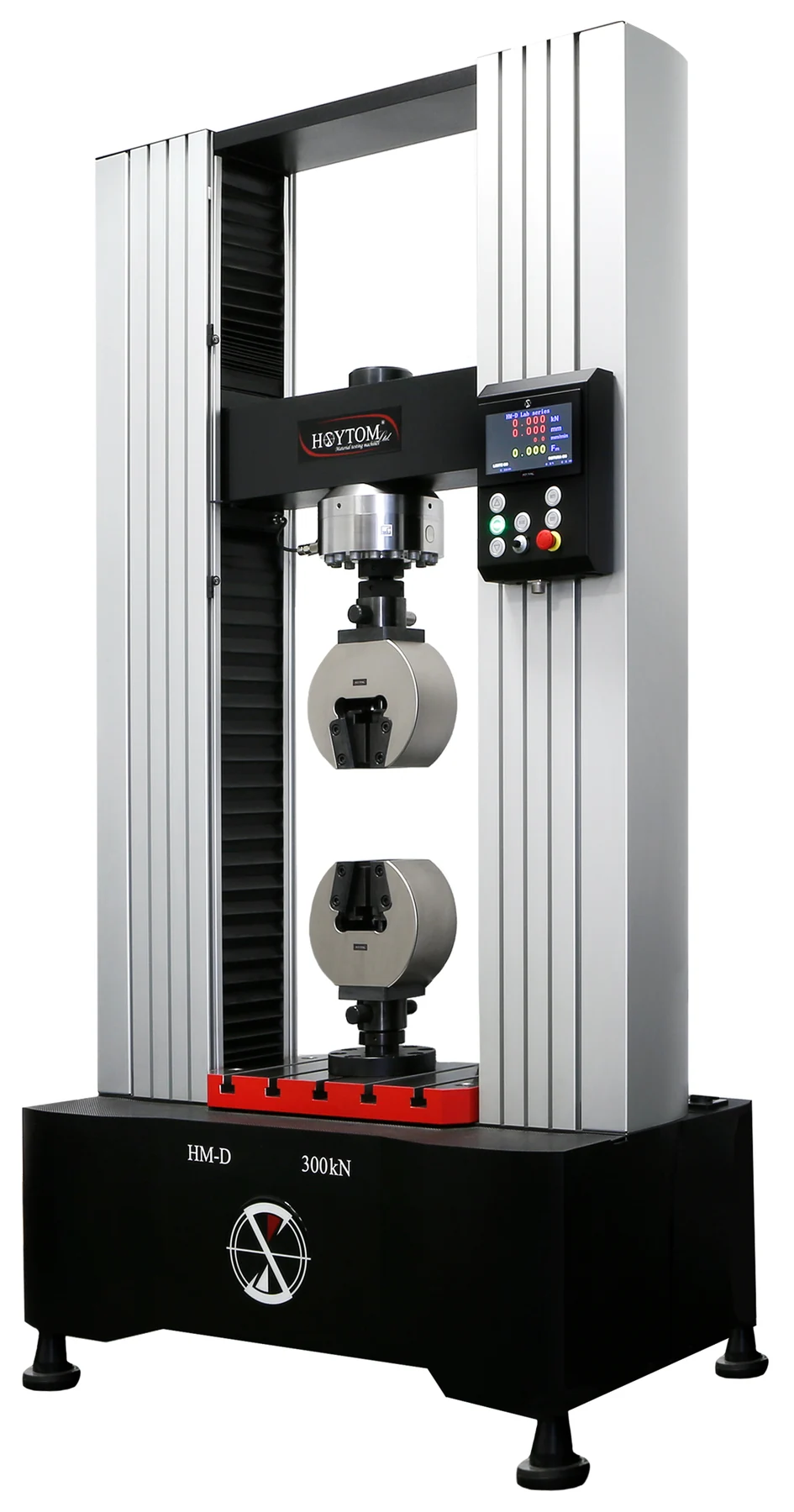 PHOTOGRAPH
Universal testing machine
A universal materials-testing machine. Controlled forces and measured deformation help characterize strength and stiffness.
Ganitz / Wikimedia Commons. Original source ↗
CC BY-SA 4.0. Resized from 2880 × 5488 to 945 × 1800 pixels, EXIF orientation applied, and encoded as WebP. Original framing retained. Display layout may crop the image.
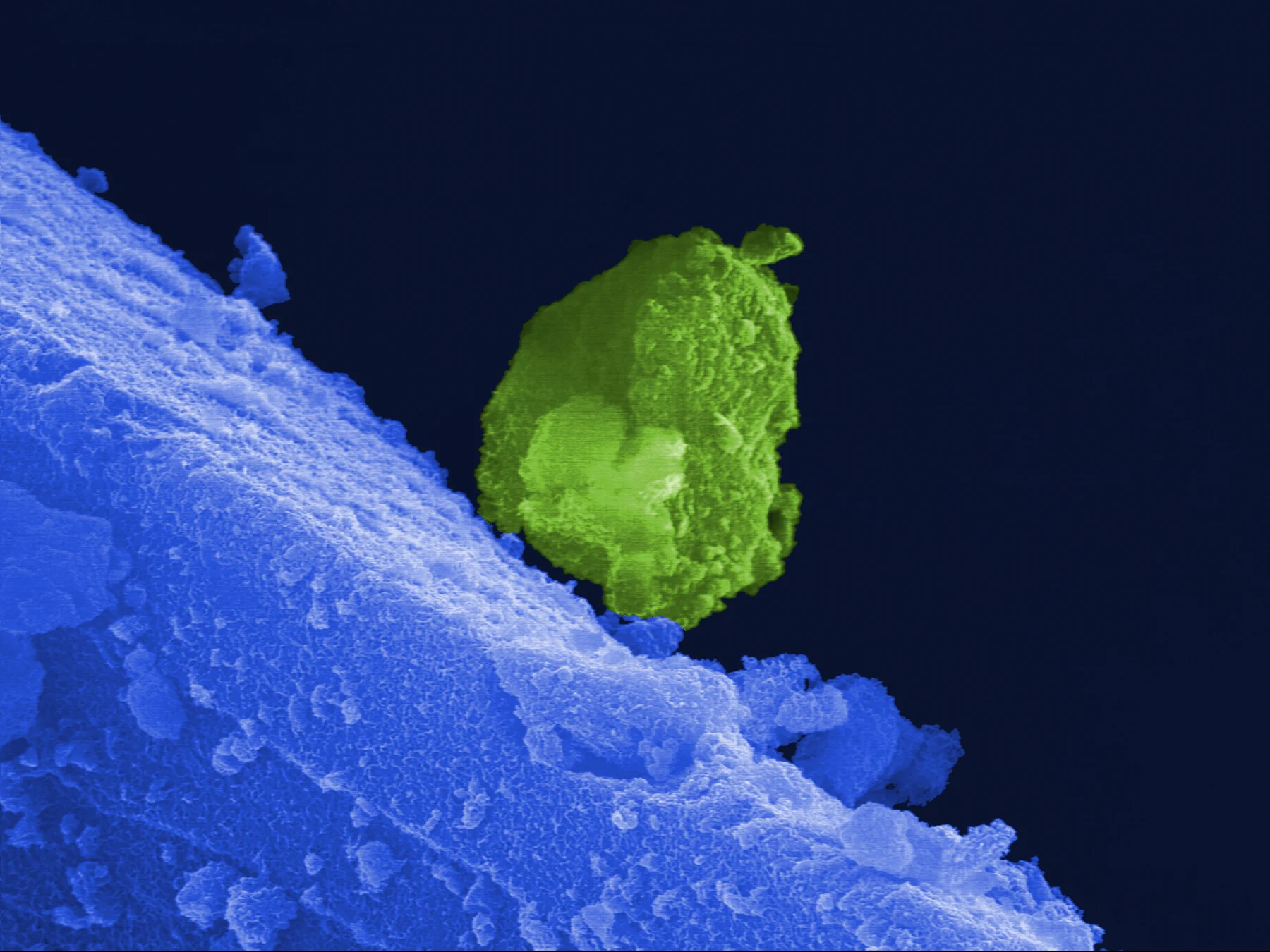 PHOTOGRAPH
Micropear
False-colour scanning electron microscopy shows hydroxyapatite crystallization on bioglass after immersion in simulated body fluid. The colour was added in the source image.
Rob Pullar / Wikimedia Commons. Original source ↗
CC BY 4.0. Resized from 3233 × 2425 to 1800 × 1350 pixels, EXIF orientation applied, and encoded as WebP. Original framing retained. Display layout may crop the image.
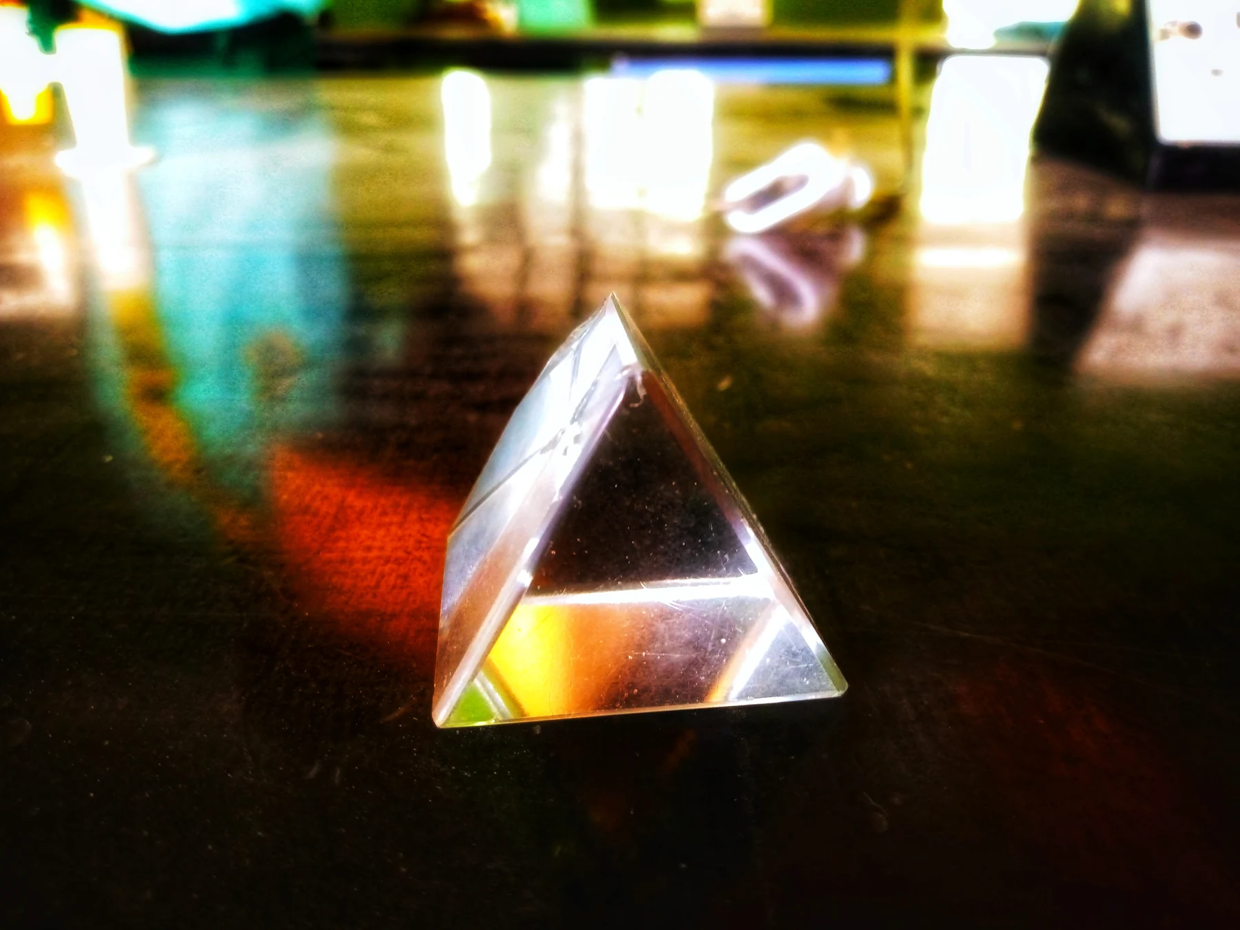 PHOTOGRAPH
Triangular Glass Prism
A triangular glass prism illustrates how the direction of light changes at material boundaries.
Jeremyida002 / Wikimedia Commons. Original source ↗
CC BY-SA 4.0. Resized from 4160 × 3120 to 1800 × 1350 pixels, EXIF orientation applied, and encoded as WebP. Original framing retained. Display layout may crop the image.
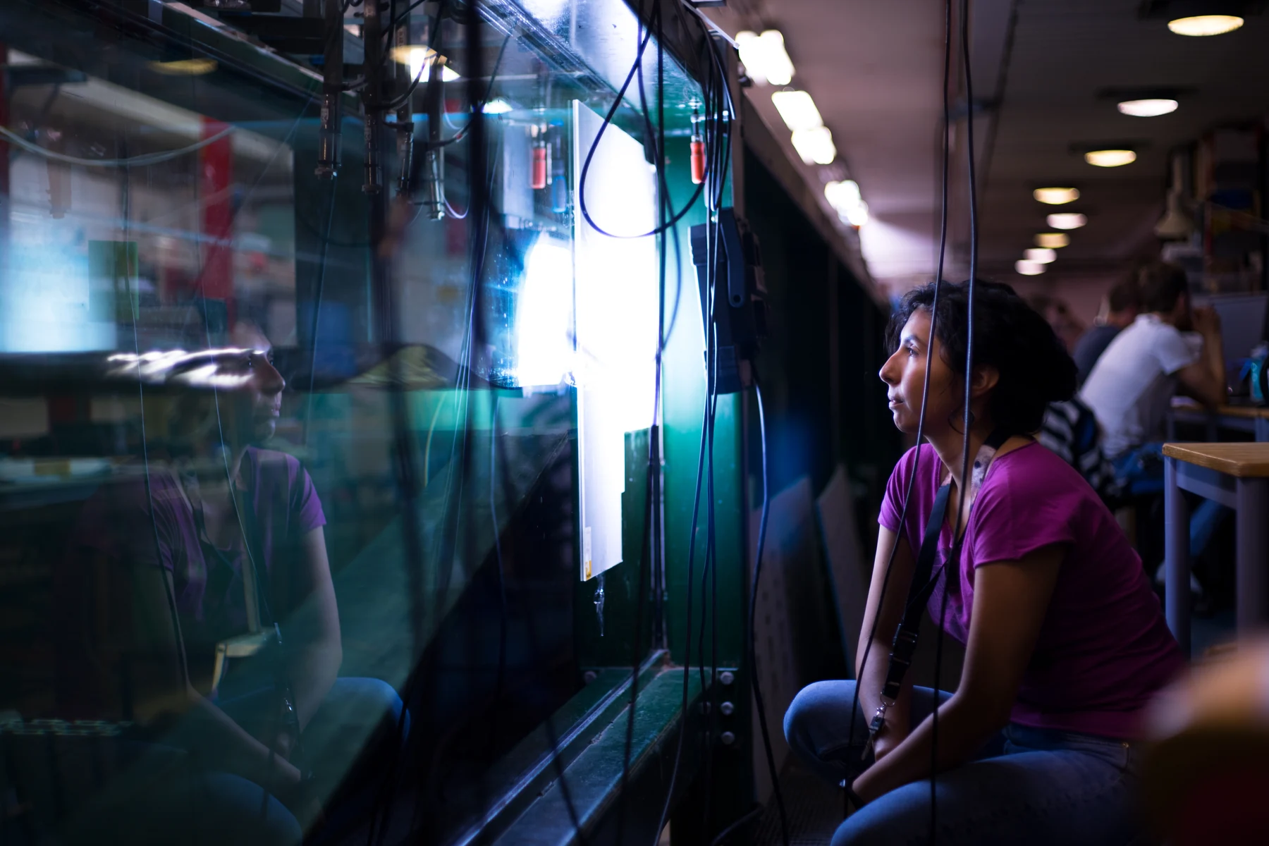 PHOTOGRAPH
Mesmerizing wave
A wave demonstration viewed through a transparent tank. Physical models help make propagation and fluid motion visible.
Michele Giordano / Wikimedia Commons. Original source ↗
CC BY 4.0. Resized from 5630 × 3753 to 1800 × 1200 pixels, EXIF orientation applied, and encoded as WebP. Original framing retained. Display layout may crop the image.
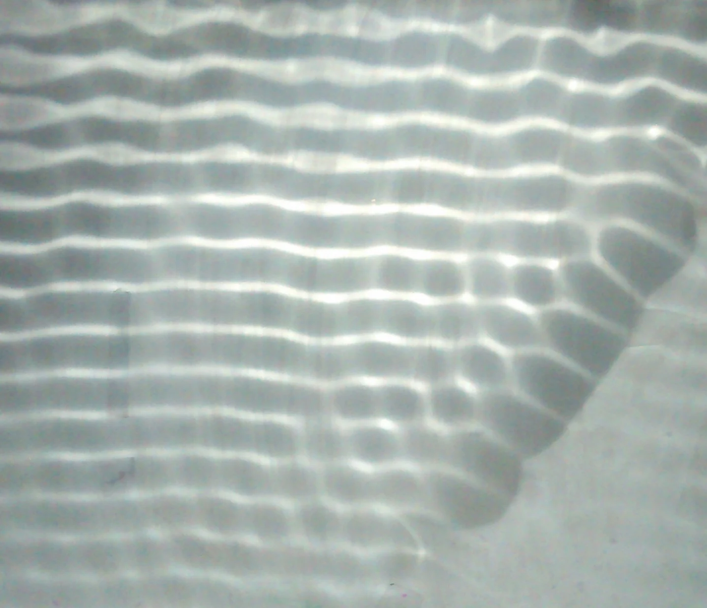 PHOTOGRAPH
Reflection of waves observed using a ripple tank.
A ripple-tank experiment makes wave reflection visible through patterns of light and dark.
Psgs123xyz ( talk ) ( Uploads ). / Wikimedia Commons. Original source ↗
CC0 1.0. Resized from 1437 × 1235 to 1437 × 1235 pixels, EXIF orientation applied, and encoded as WebP. Original framing retained. Display layout may crop the image.
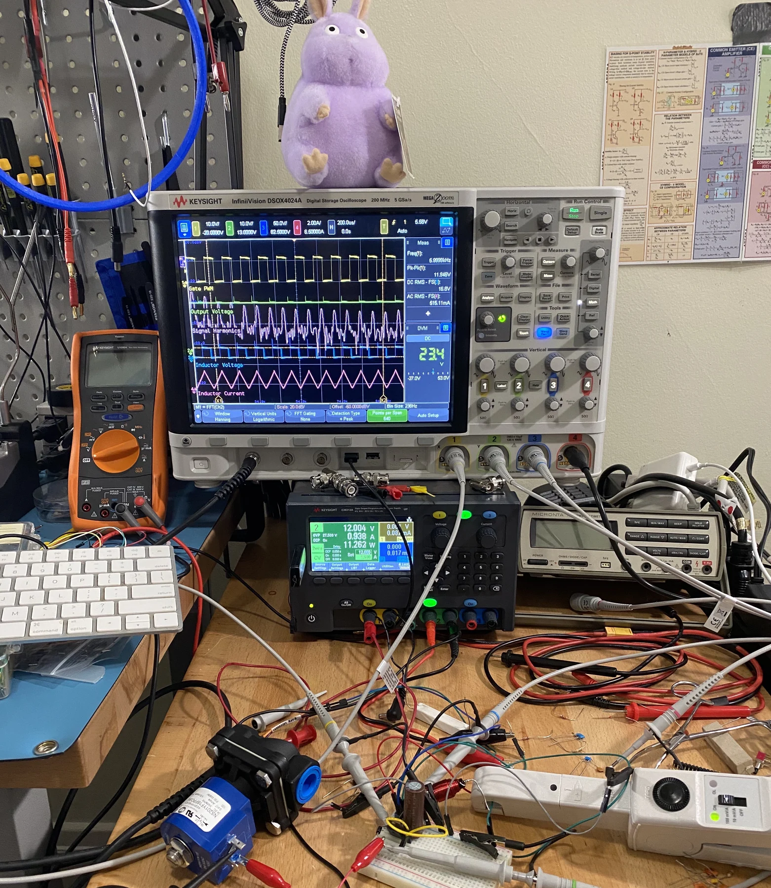 PHOTOGRAPH
Digital oscilloscope in use
An oscilloscope in use at a laboratory bench. The instrument displays how electrical signals change with time.
Radarvector / Wikimedia Commons. Original source ↗
CC BY-SA 4.0. Resized from 3024 × 3484 to 1562 × 1800 pixels, EXIF orientation applied, and encoded as WebP. Original framing retained. Display layout may crop the image.
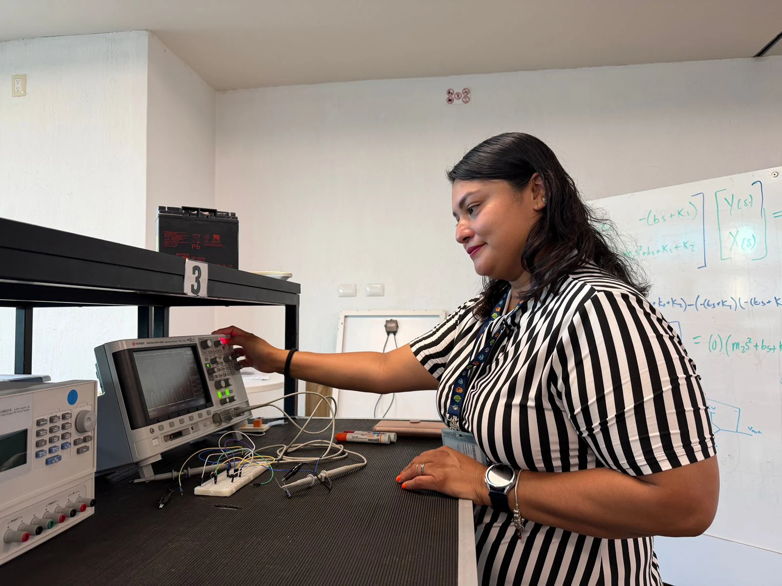 PHOTOGRAPH
Hands that Calibrate Dreams
Hands-on electronic measurement in a teaching laboratory. Calibration and careful connections are part of turning a signal into a useful measurement.
AmmyLucio / Wikimedia Commons. Original source ↗
CC BY 4.0. Resized from 1600 × 1200 to 1600 × 1200 pixels, EXIF orientation applied, and encoded as WebP. Original framing retained. Display layout may crop the image.
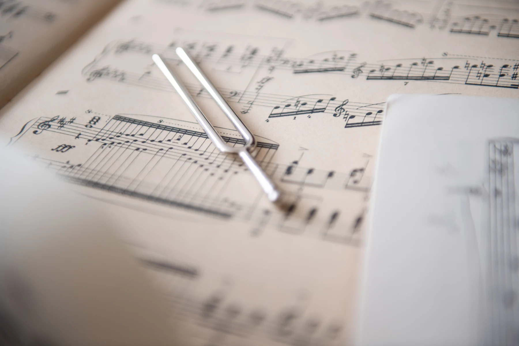 PHOTOGRAPH
A tuning fork placed on sheet music 2
A tuning fork provides a simple example of mechanical resonance and sound generation.
Lukasz Kobus, European Commission · © European Union, 2026 / Wikimedia Commons. Original source ↗
CC BY 4.0. Resized from 6000 × 4000 to 1800 × 1200 pixels, EXIF orientation applied, and encoded as WebP. Original framing retained. Display layout may crop the image.
 PHOTOGRAPH
020220209 134149 pulse oximeter
A fingertip pulse oximeter estimates blood oxygen saturation from light transmitted through tissue. A displayed reading is an example measurement.
Silar / Wikimedia Commons. Original source ↗
CC BY-SA 4.0. Resized from 3143 × 2056 to 1800 × 1177 pixels, EXIF orientation applied, and encoded as WebP. Original framing retained. Display layout may crop the image.
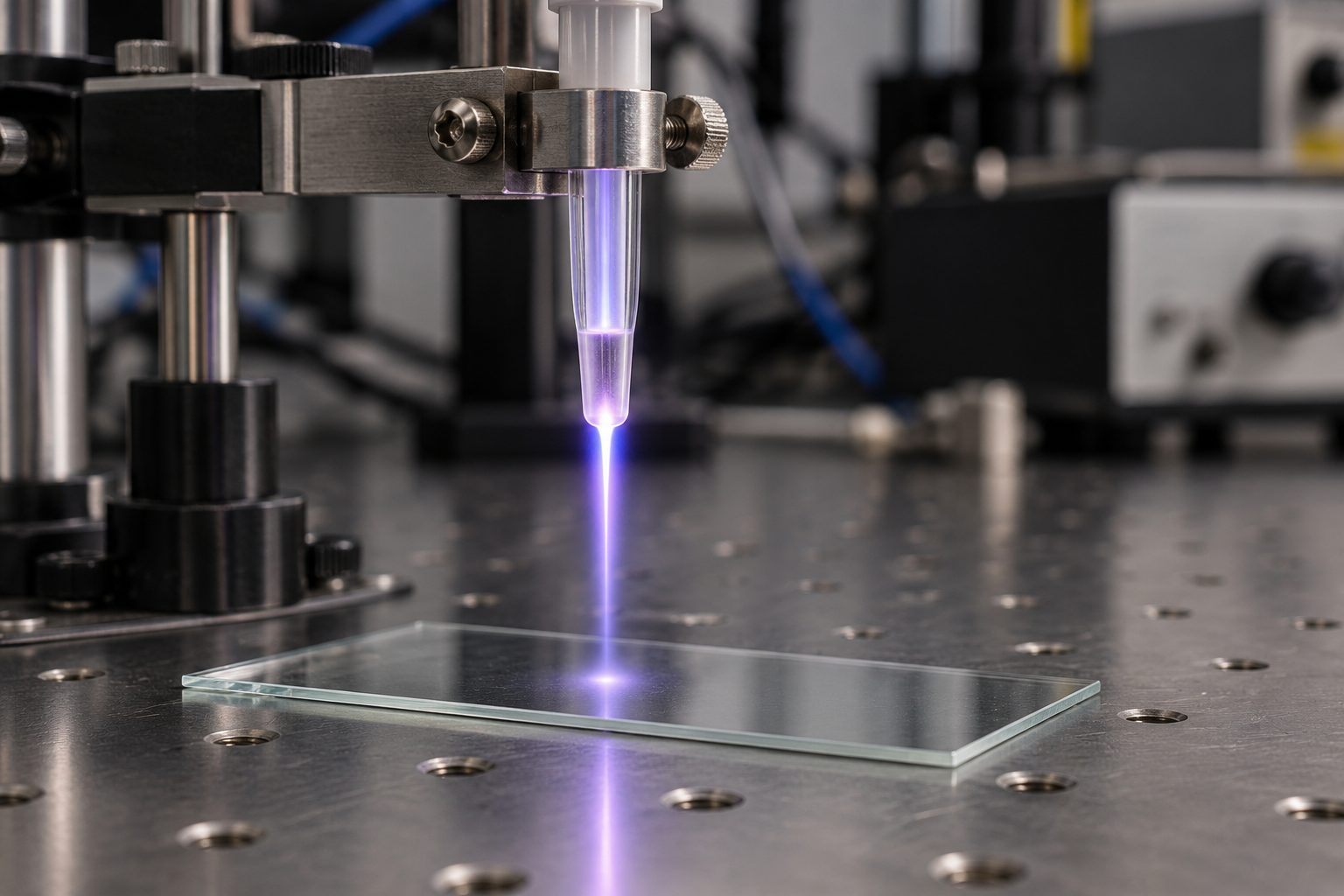 AI-GENERATED EDITORIAL ILLUSTRATION
Generated Cold Plasma Jet
A conceptual laboratory plasma jet above a glass slide.
Created for PSI Center.
Original AI-generated asset. Original generated pixels retained.
AI-generated editorial illustration. This constructed scene conveys a scientific idea; it is not photographic evidence of a clinical treatment or a specific instrument.
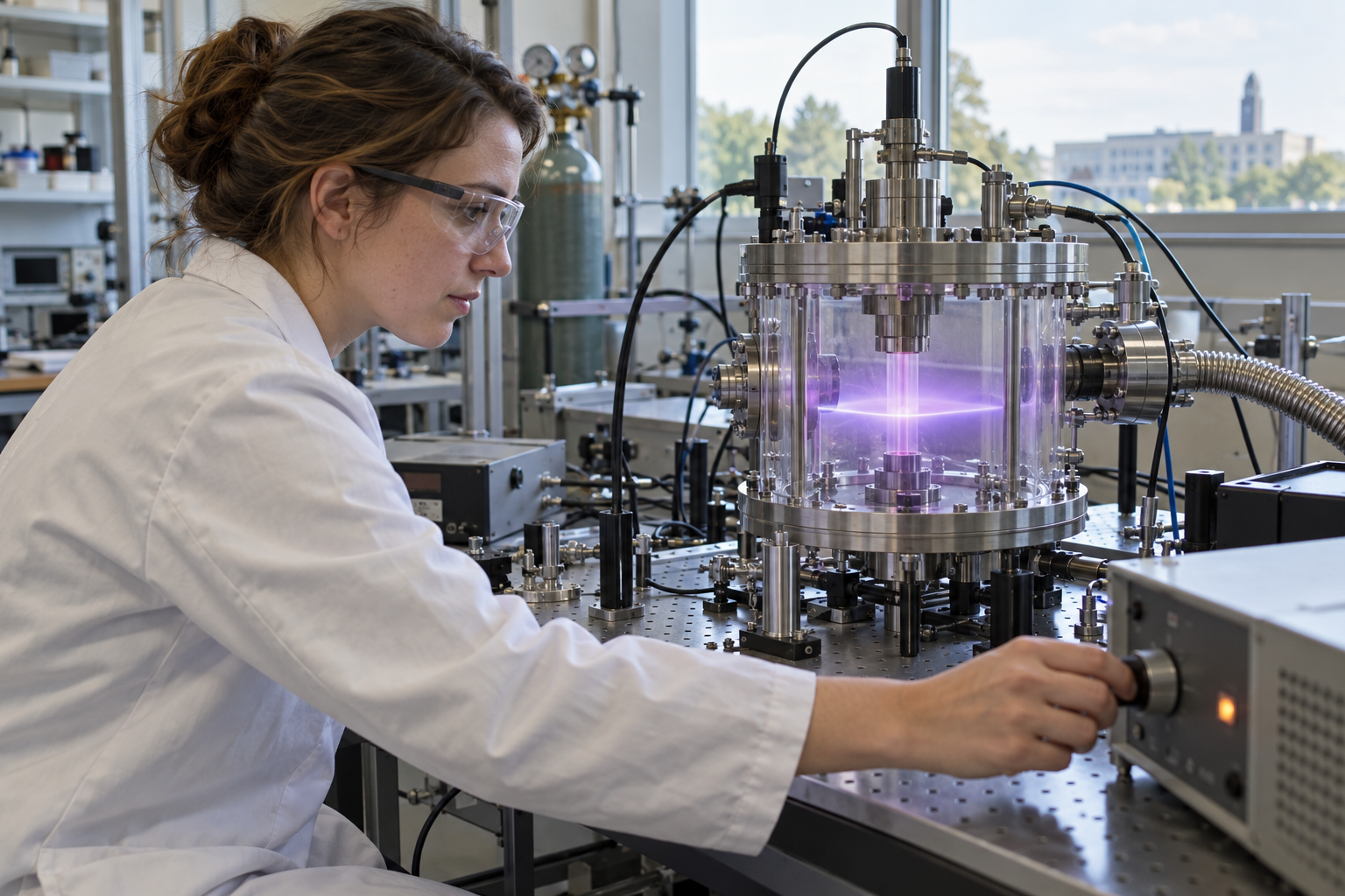 AI-GENERATED EDITORIAL ILLUSTRATION
Generated Plasma Lab Researcher
A conceptual laboratory scene showing a researcher beside a plasma chamber.
Created for PSI Center.
Original AI-generated asset. Original generated pixels retained.
AI-generated editorial illustration. This constructed scene conveys a scientific idea; it is not photographic evidence of a clinical treatment or a specific instrument.
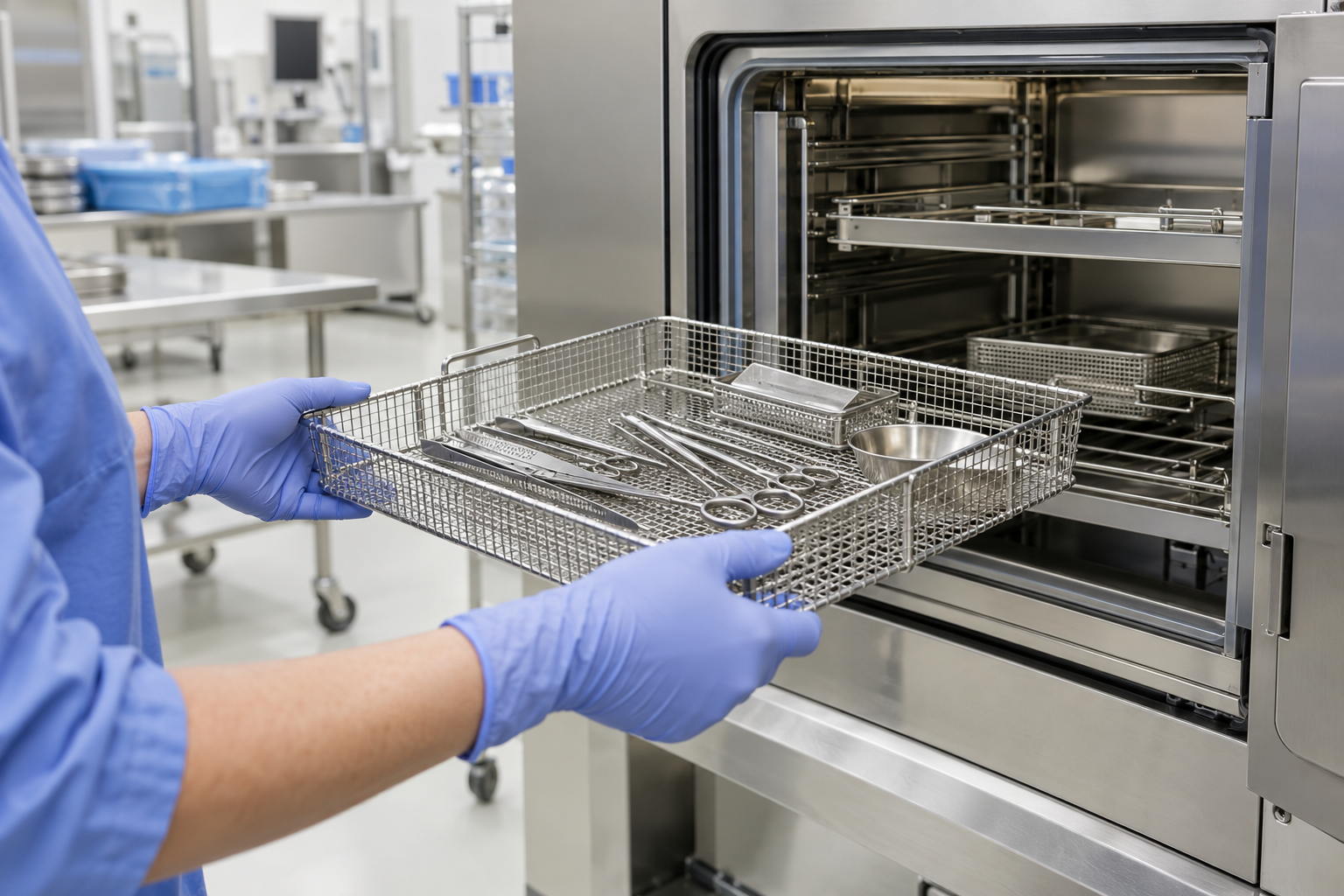 AI-GENERATED EDITORIAL ILLUSTRATION
Generated Sterile Instrument Tray
A conceptual instrument-processing scene beside a sterilization cabinet.
Created for PSI Center.
Original AI-generated asset. Original generated pixels retained.
AI-generated editorial illustration. This constructed scene conveys a scientific idea; it is not photographic evidence of a clinical treatment or a specific instrument.
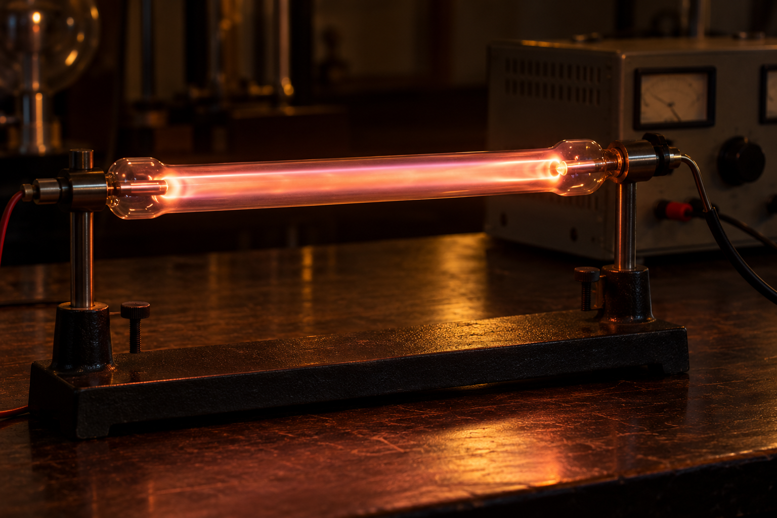 AI-GENERATED EDITORIAL ILLUSTRATION
Generated Discharge Tube
A conceptual electrically excited gas-discharge tube.
Created for PSI Center.
Original AI-generated asset. Original generated pixels retained.
AI-generated editorial illustration. This constructed scene conveys a scientific idea; it is not photographic evidence of a clinical treatment or a specific instrument.
 AI-GENERATED EDITORIAL ILLUSTRATION
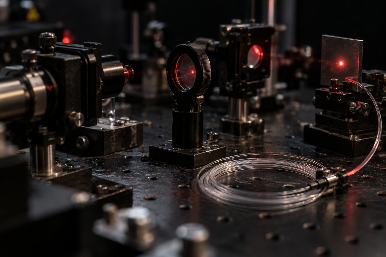
Generated Fiber Optics Bench
A conceptual optical bench with fiber and laser-alignment components.
Created for PSI Center.
Original AI-generated asset. Original generated pixels retained.
AI-generated editorial illustration. This constructed scene conveys a scientific idea; it is not photographic evidence of a clinical treatment or a specific instrument.
 AI-GENERATED EDITORIAL ILLUSTRATION
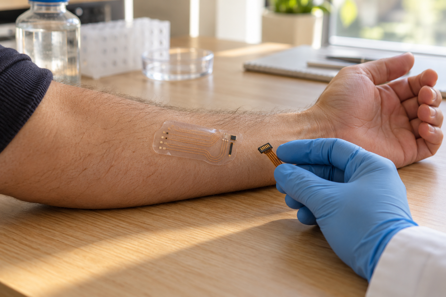
Generated Wearable Sensor
A conceptual flexible sensing patch on a forearm; not a particular approved clinical device.
Created for PSI Center.
Original AI-generated asset. Original generated pixels retained.
AI-generated editorial illustration. This constructed scene conveys a scientific idea; it is not photographic evidence of a clinical treatment or a specific instrument.
 AI-GENERATED EDITORIAL ILLUSTRATION
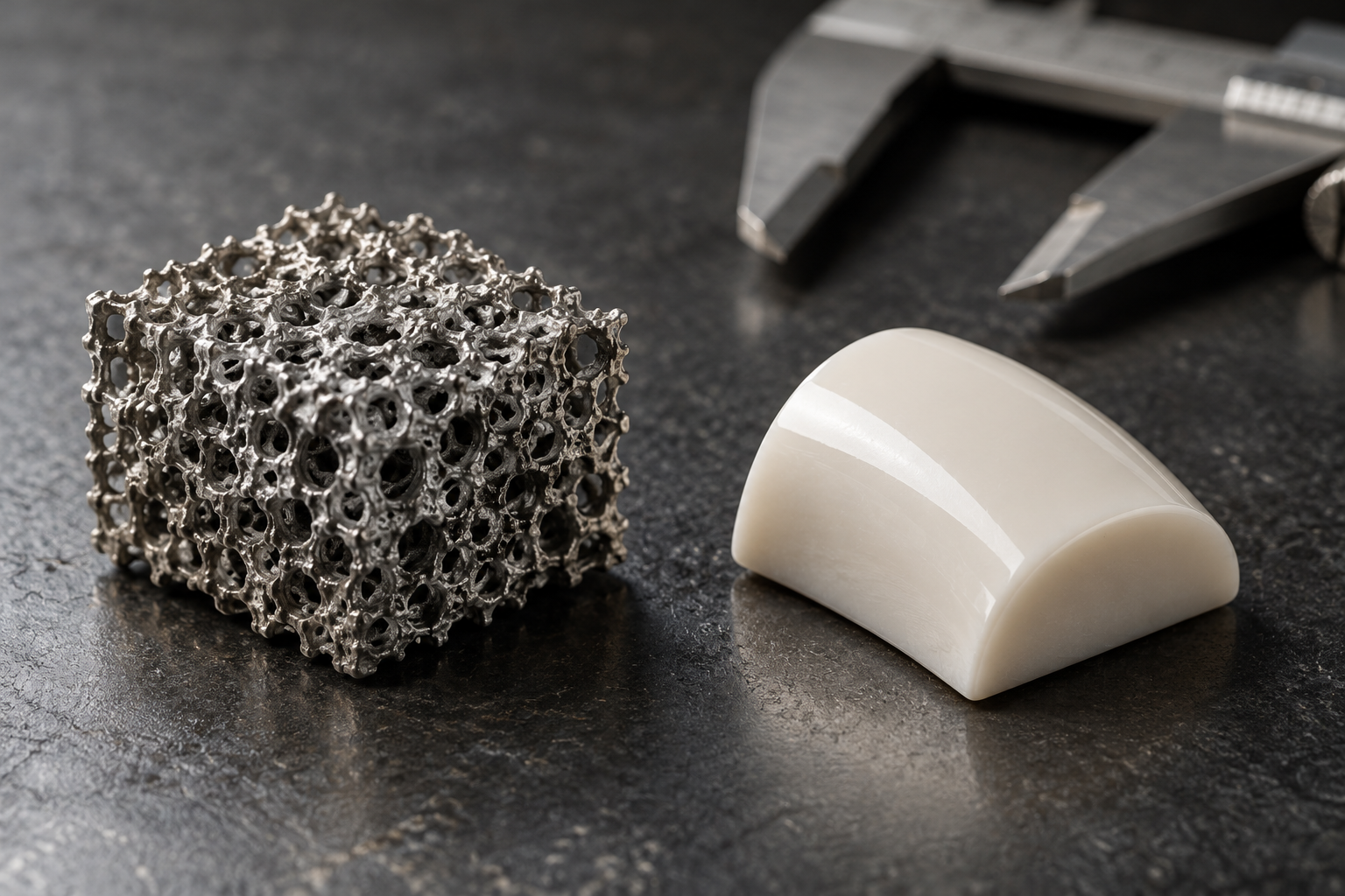
Generated Porous Implant Material
Conceptual porous metal and ceramic biomaterial specimens, not a particular approved implant.
Created for PSI Center.
Original AI-generated asset. Original generated pixels retained.
AI-generated editorial illustration. This constructed scene conveys a scientific idea; it is not photographic evidence of a clinical treatment or a specific instrument.
 AI-GENERATED EDITORIAL ILLUSTRATION
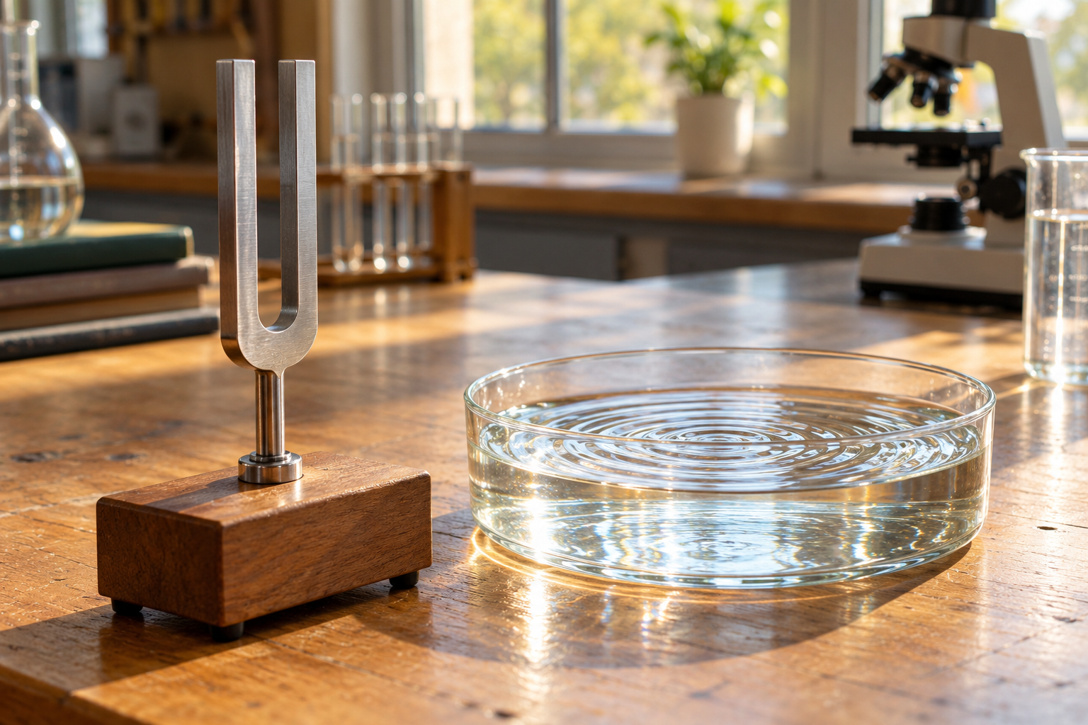
Generated Tuning Fork Water
A conceptual tuning-fork and water-wave demonstration.
Created for PSI Center.
Original AI-generated asset. Original generated pixels retained.
AI-generated editorial illustration. This constructed scene conveys a scientific idea; it is not photographic evidence of a clinical treatment or a specific instrument.
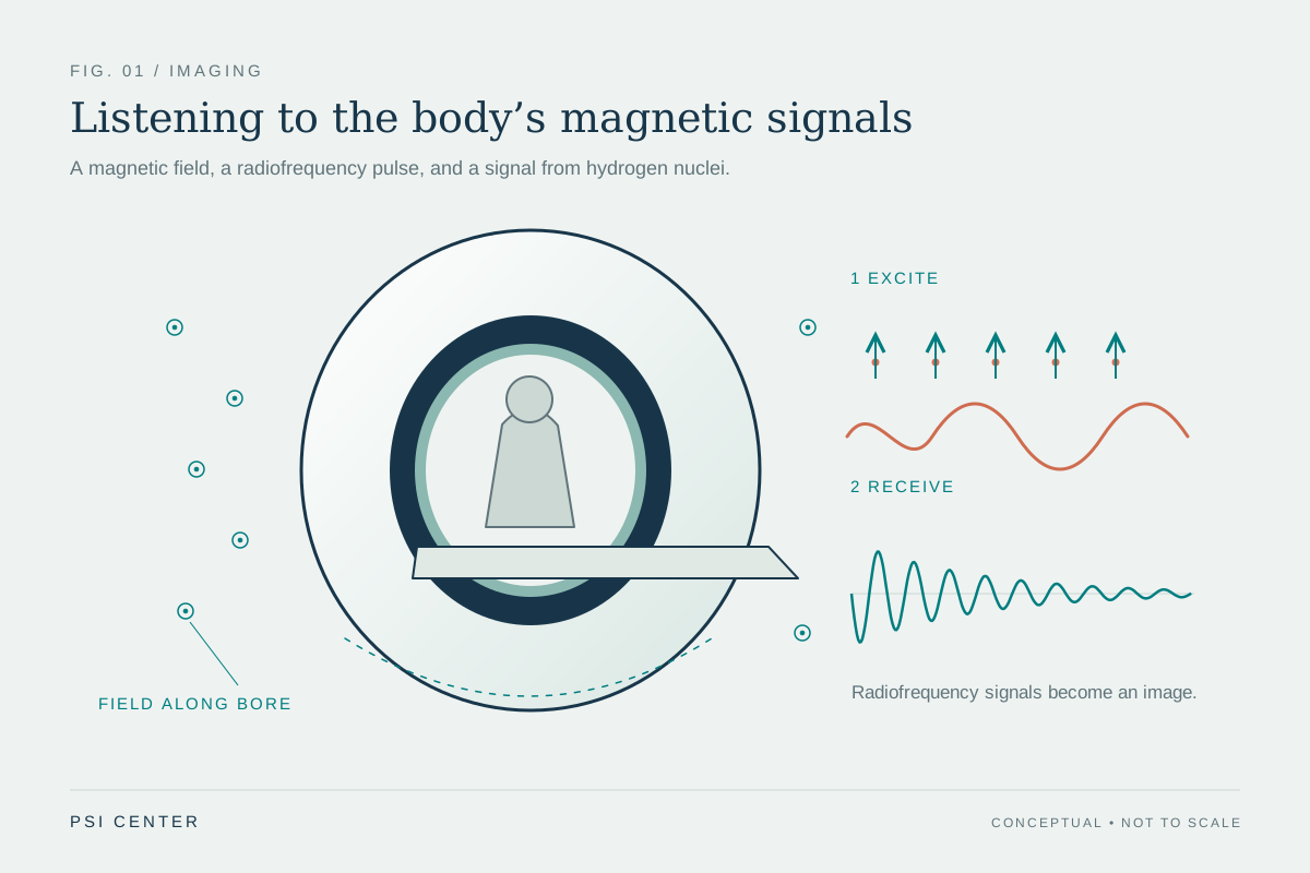 ORIGINAL SCIENTIFIC ILLUSTRATION
Listening to the body’s magnetic signals
MRI uses a strong magnetic field and radiofrequency pulses to measure signals from hydrogen nuclei. Gradient fields help encode where the signals originate. Dot symbols indicate the main field along the bore toward the viewer; the scanner does not require a plasma.
Original illustration by PSI Center. All diagrams are conceptual illustrations; none is a patient record or a measured dataset.
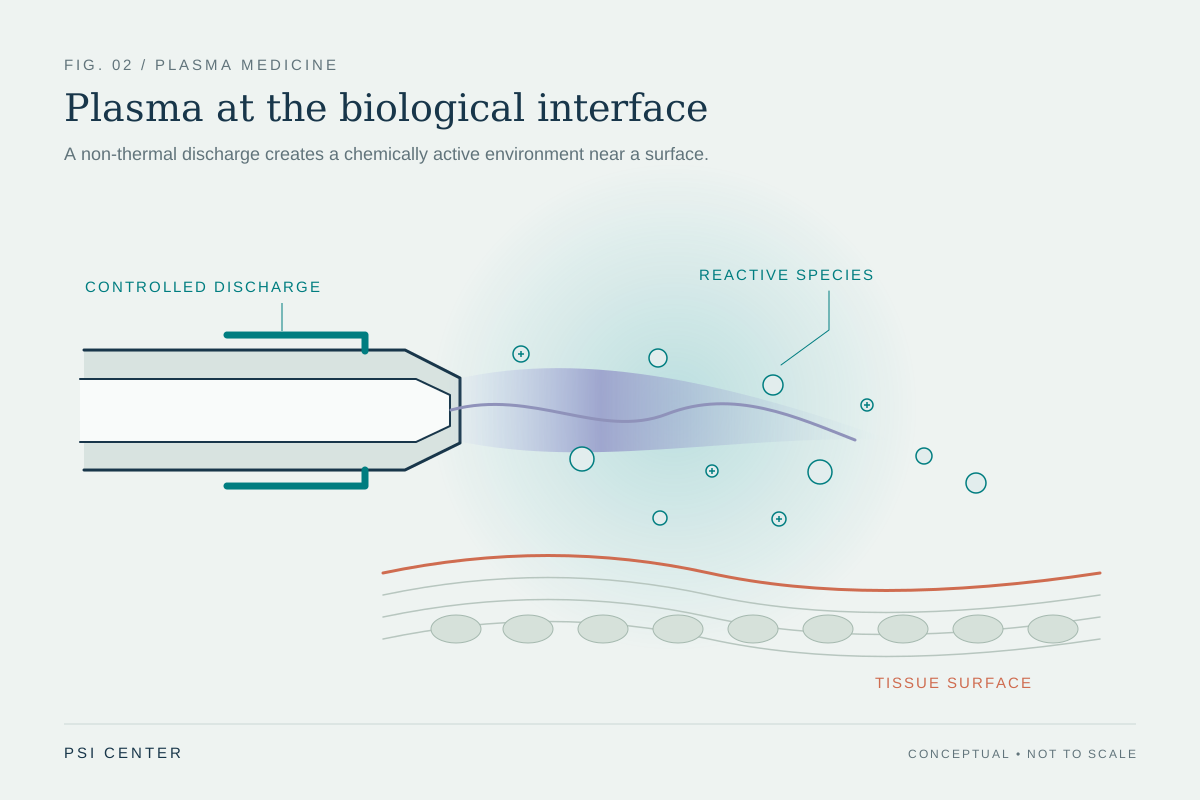 ORIGINAL SCIENTIFIC ILLUSTRATION
Plasma at the biological interface
A cold atmospheric plasma discharge can produce reactive oxygen and nitrogen species, charged particles, and light. Biological effects depend on the device, gas, exposure, and tissue; this diagram does not imply a proven cancer treatment.
Original illustration by PSI Center. All diagrams are conceptual illustrations; none is a patient record or a measured dataset.
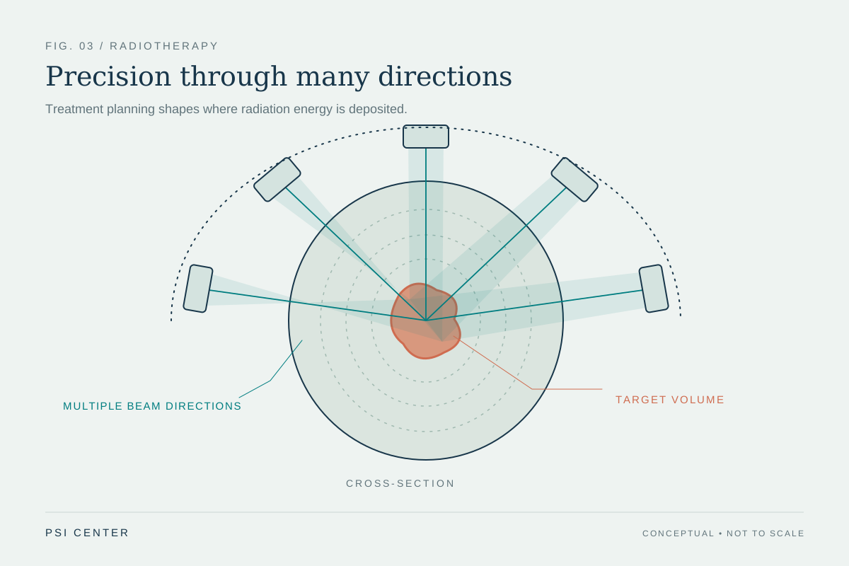 ORIGINAL SCIENTIFIC ILLUSTRATION
Precision through many directions
External-beam radiotherapy can combine beams from multiple directions to concentrate dose in a target while limiting exposure to surrounding tissue. Actual treatment requires an individualized plan, imaging, and verification; the illustrated beams and contours are schematic.
Original illustration by PSI Center. All diagrams are conceptual illustrations; none is a patient record or a measured dataset.
 ORIGINAL SCIENTIFIC ILLUSTRATION
An image carried by echoes
An ultrasound transducer sends sound into tissue and detects returning echoes. Differences in acoustic impedance help determine reflection strength, while echo travel time provides depth information. The focused beam and boundaries are conceptual.
Original illustration by PSI Center. All diagrams are conceptual illustrations; none is a patient record or a measured dataset.
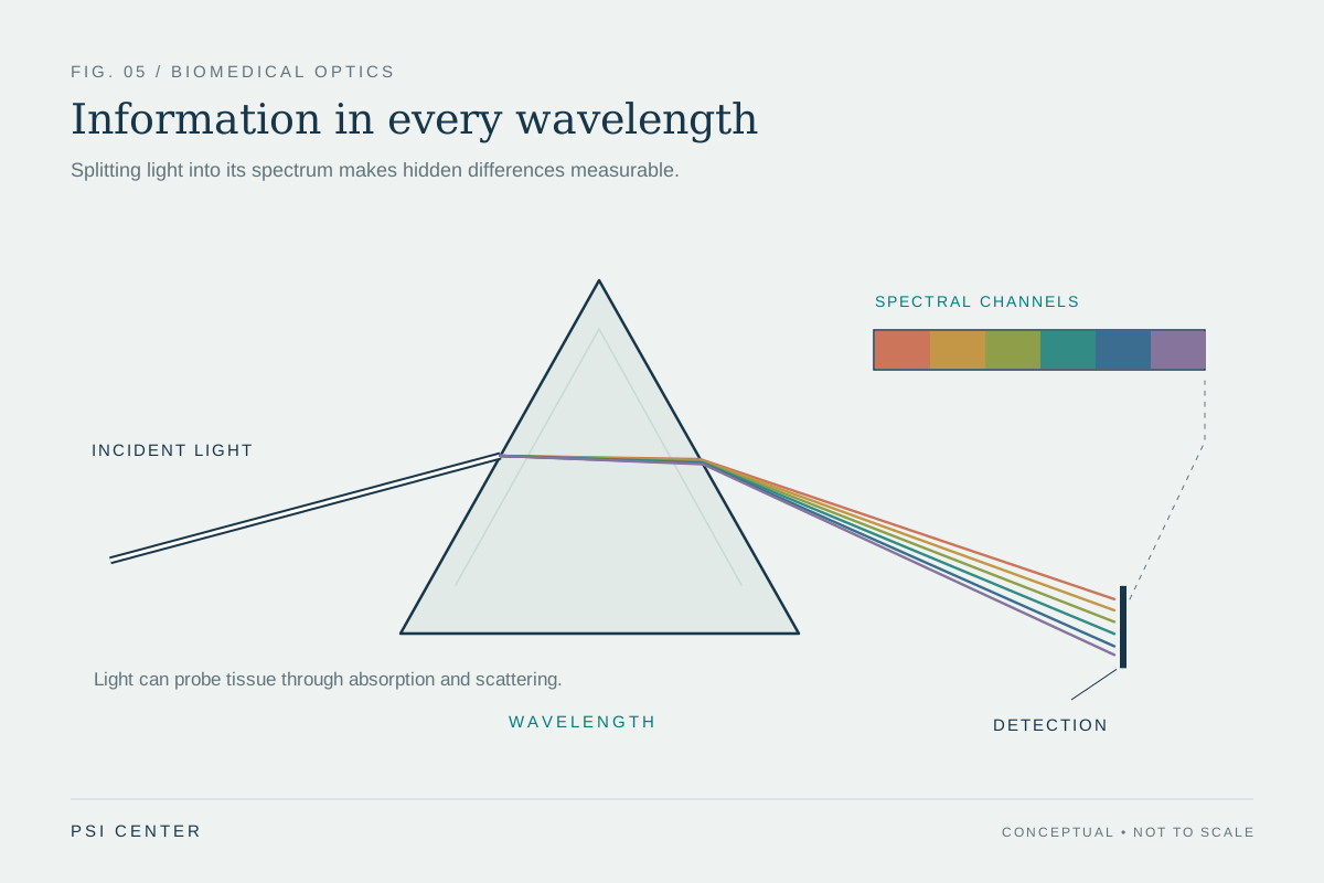 ORIGINAL SCIENTIFIC ILLUSTRATION
Information in every wavelength
Different wavelengths interact differently with molecules and tissue. Spectroscopy uses wavelength-dependent signals to investigate composition and physiology. This idealized prism diagram illustrates spectral separation rather than a complete medical instrument.
Original illustration by PSI Center. All diagrams are conceptual illustrations; none is a patient record or a measured dataset.
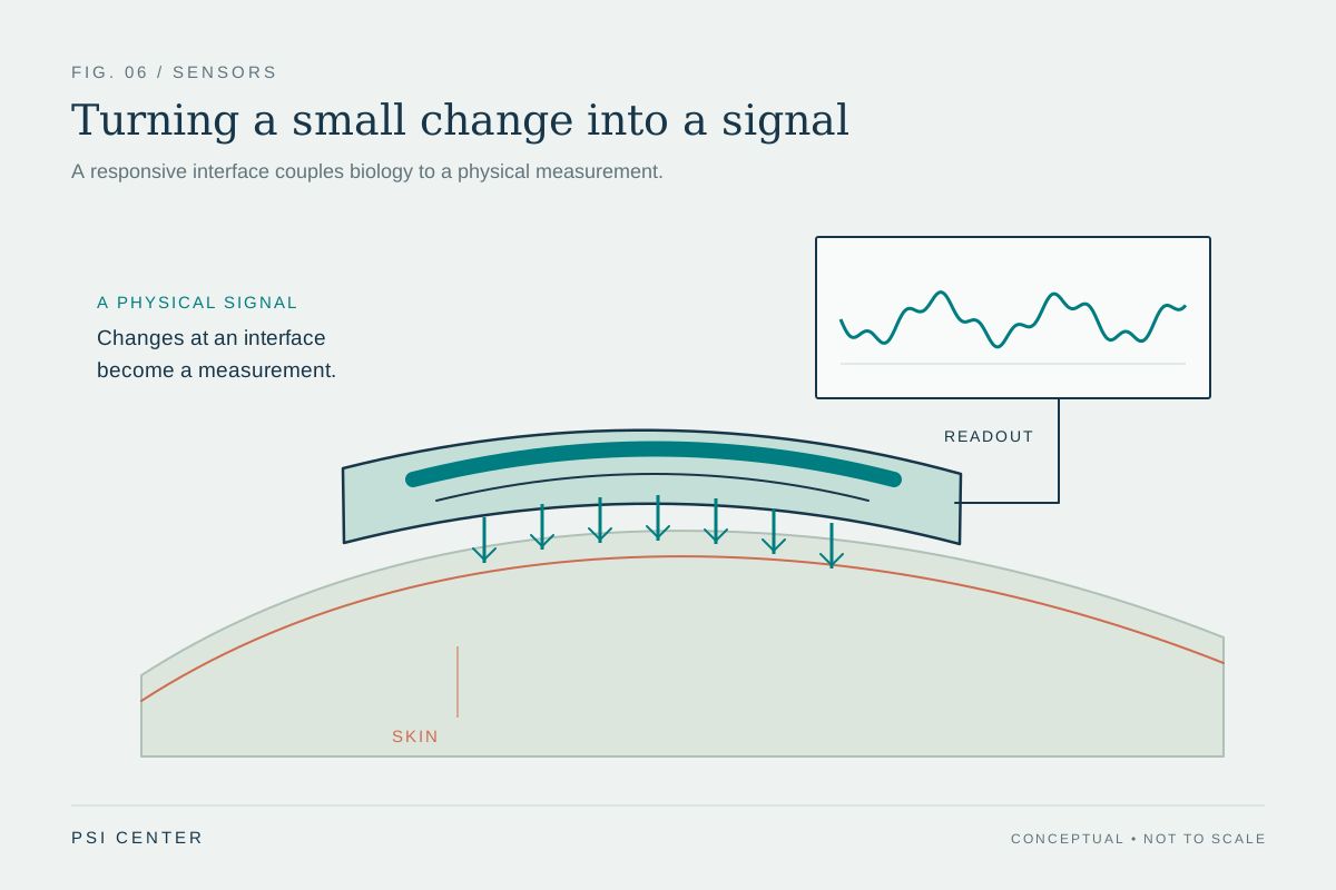 ORIGINAL SCIENTIFIC ILLUSTRATION
Turning a small change into a signal
Wearable sensors convert physical or chemical changes into measurable signals. The interface, electronics, calibration, and contact with skin all affect the result. This conceptual patch and waveform do not represent a specific device or patient measurement.
Original illustration by PSI Center. All diagrams are conceptual illustrations; none is a patient record or a measured dataset.
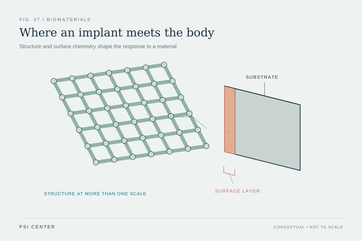 ORIGINAL SCIENTIFIC ILLUSTRATION
Where an implant meets the body
A biomaterial’s pore structure, mechanical properties, and surface chemistry influence how it interacts with cells and tissue. Plasma processing can modify surfaces or deposit coatings. The scaffold and magnified coating are illustrative, not a particular implant design.
Original illustration by PSI Center. All diagrams are conceptual illustrations; none is a patient record or a measured dataset.
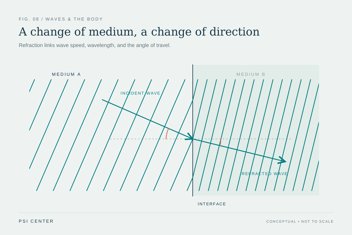 ORIGINAL SCIENTIFIC ILLUSTRATION
A change of medium, a change of direction
At an interface, a transmitted wave can change direction when its speed changes. Frequency remains the same across a stationary boundary while wavelength changes. Refraction matters in optical instruments and ultrasound; these ray and wavefront geometries are conceptual.
Original illustration by PSI Center. All diagrams are conceptual illustrations; none is a patient record or a measured dataset.
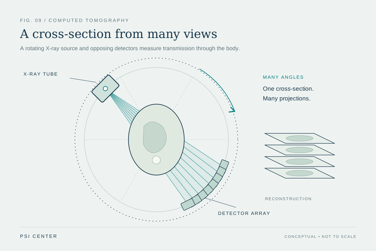 ORIGINAL SCIENTIFIC ILLUSTRATION
A cross-section from many views
CT collects X-ray transmission measurements from many directions using a rotating source and opposing detectors. A computer reconstructs cross-sectional images from the measurements. The body contours, projections, and slice stack are conceptual, not patient data.
Original illustration by PSI Center. All diagrams are conceptual illustrations; none is a patient record or a measured dataset.
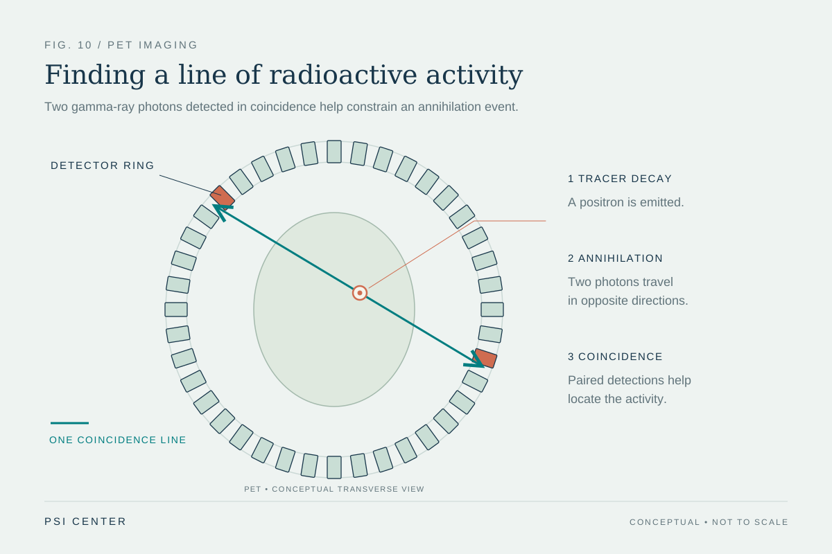 ORIGINAL SCIENTIFIC ILLUSTRATION
Finding a line of radioactive activity
In PET, a radiotracer emits a positron, which can annihilate with an electron to produce two gamma-ray photons traveling in approximately opposite directions. Detecting the pair within a coincidence window identifies a line of response; many such detections support image reconstruction. The example is schematic and contains no patient measurements.
Original illustration by PSI Center. All diagrams are conceptual illustrations; none is a patient record or a measured dataset.
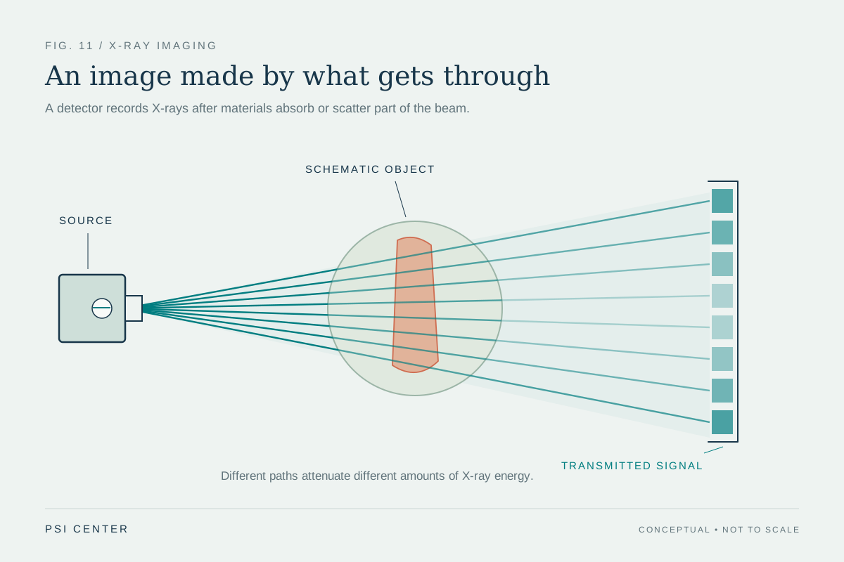 ORIGINAL SCIENTIFIC ILLUSTRATION
An image made by what gets through
X-rays are attenuated by absorption and scattering as they pass through matter. A detector measures the transmitted signal, which varies with the materials and thicknesses along each path. The ray strengths and detector shading are illustrative; this is not a measured radiograph or dose comparison.
Original illustration by PSI Center. All diagrams are conceptual illustrations; none is a patient record or a measured dataset.
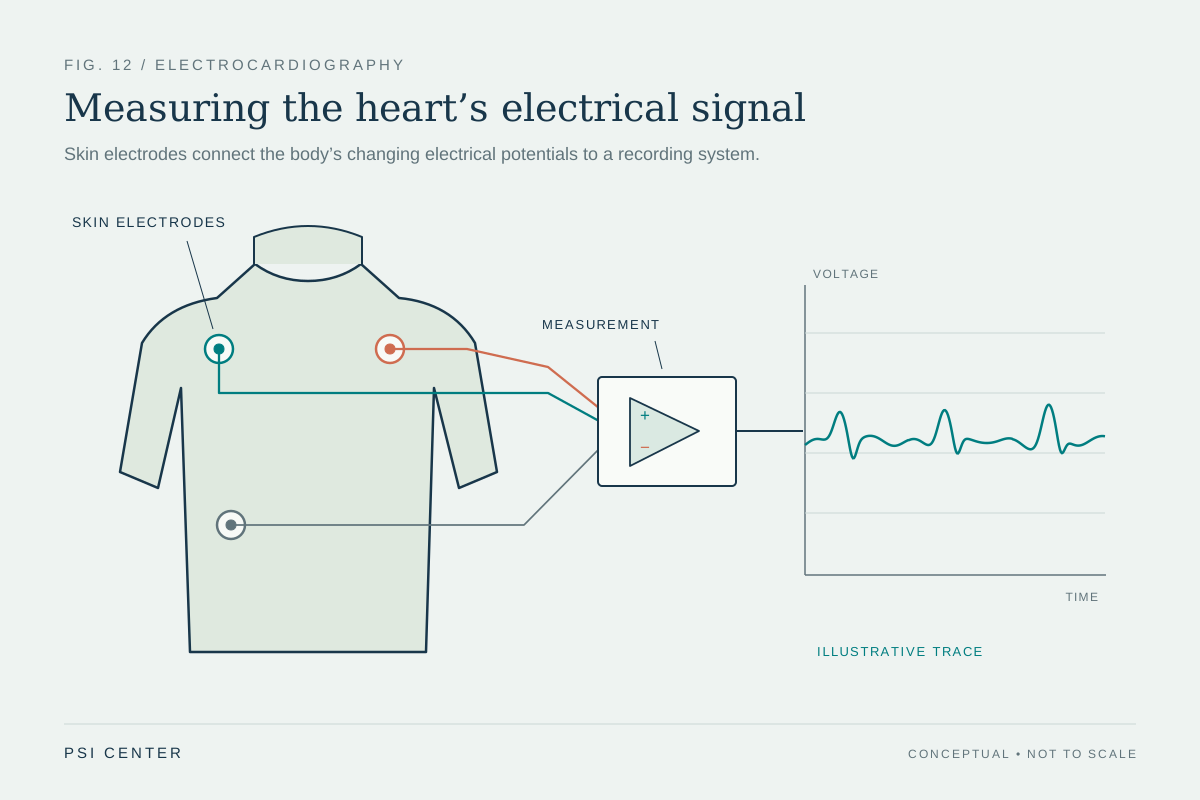 ORIGINAL SCIENTIFIC ILLUSTRATION
Measuring the heart’s electrical signal
An ECG records differences in electrical potential using electrodes placed on the skin. It measures electrical activity rather than sending therapeutic current into the heart. Electrode positions, the circuit, and the unscaled waveform shown here are conceptual and do not represent a clinical lead arrangement, patient tracing, or diagnosis.
Original illustration by PSI Center. All diagrams are conceptual illustrations; none is a patient record or a measured dataset.