Why can a blood vessel be hard to distinguish from the tissue around it in an X-ray image? The detector records how much of an X-ray beam makes it through the body, and neighboring soft tissues may absorb similar amounts. A contrast material changes that pattern. It makes a selected structure easier to see by interacting with X-rays differently from nearby tissue, giving the image a clearer boundary or pathway to follow.
An image is a pattern of transmitted rays
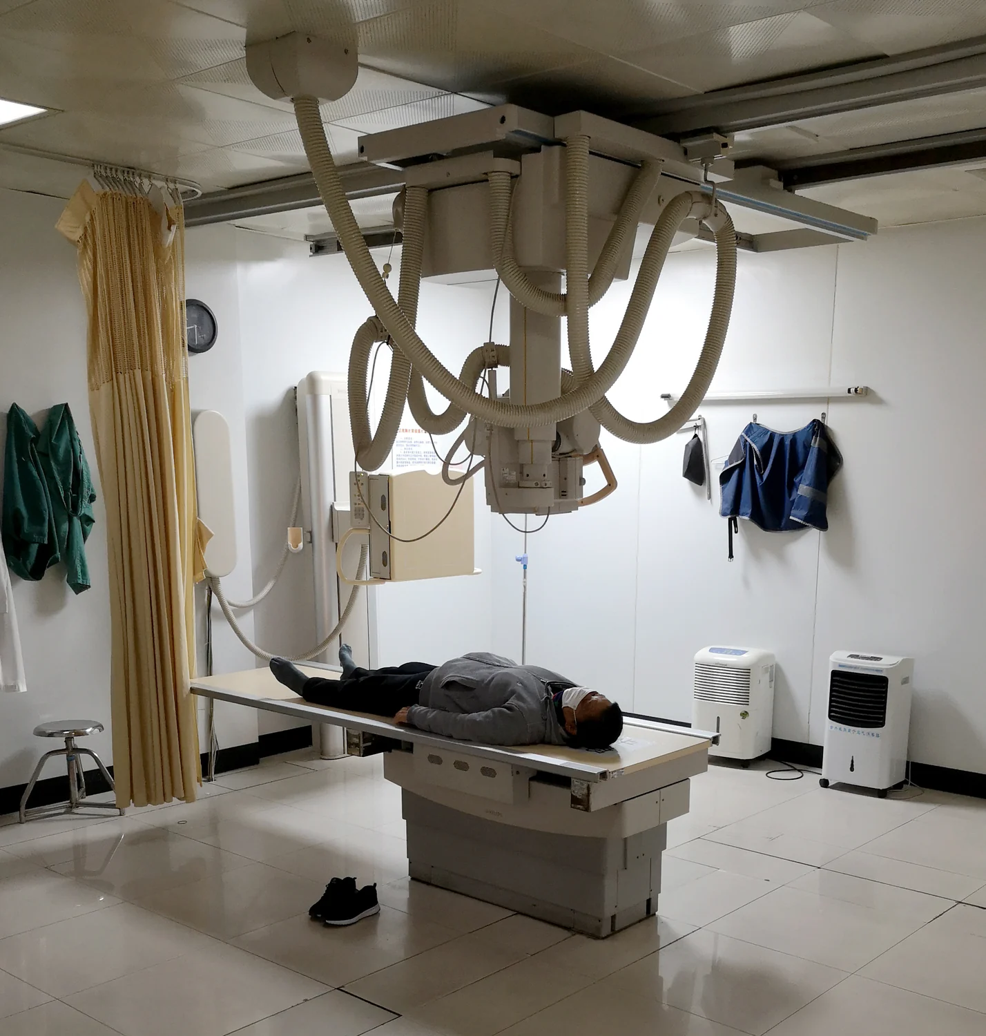
X-rays pass through the body, but not all of them reach the detector. Some are absorbed or scattered by internal structures. The detector records the remaining pattern, which appears as differences in brightness. Bone is relatively easy to see because it is dense, while soft tissues can be faint or similar in appearance on a conventional X-ray [2].
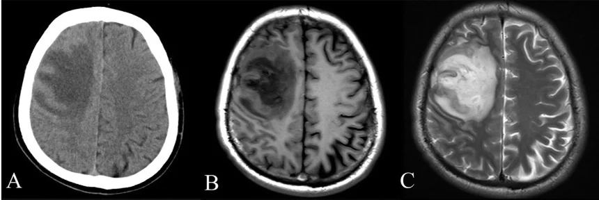
CT uses the same underlying interaction, but collects many X-ray projections as the source rotates around the body. A computer reconstructs those measurements into slices, reducing the problem of overlapping structures that can obscure detail in a single projection [1]. Contrast material adds another way to distinguish a target within those slices.
Changing how a structure stands out
A contrast agent contains substances that stop X-rays more strongly than the surrounding area, so it shows up clearly on an X-ray or CT image [1]. For example, iodine-based contrast may be injected into the bloodstream when clinicians need to examine blood vessels. As the material travels with the blood, the vessel’s path becomes easier to identify against adjacent tissue.
Oral contrast serves a different anatomical task. Barium-based compounds can help visualize the digestive tract, including the esophagus, stomach, and intestines [1]. These examples illustrate a general principle: the material is chosen to make a particular space or pathway more visible. The image does not become clearer everywhere in the same way; contrast changes the pattern where the agent is present.
Following flow and separating structures
Imagine a branching river hidden beneath a map of similar-colored terrain. If a visible tracer moves through the river, its route becomes easier to trace. In a CT angiographic image, an intravenous contrast agent can act in a comparable way: it travels through the circulation, and the X-ray detector registers a stronger signal difference along the blood vessels. That can help reveal possible obstructions or clarify the arrangement of vessels [1].
Contrast can also help distinguish compartments and organs that would otherwise blend together. Its usefulness depends on where the material goes and when images are obtained. The physical idea remains simple—change the attenuation, or weakening, of X-rays through a region—but the interpretation depends on anatomy and timing. The material supports the image; it does not by itself establish what a visible finding means.
Visibility comes with practical limits
Contrast agents are not required for every X-ray or CT examination. They are used when they help answer the imaging question. The FDA notes that injected contrast can be associated with reactions, and that X-ray imaging involves ionizing radiation; for a clinically appropriate exam, benefits and risks are considered together [2]. The NIBIB description of CT likewise notes possible allergic reactions and, rarely, temporary kidney effects for some contrast agents [1].
The word “dye” is often used informally, but contrast agents work through physical properties, not ordinary color. They are selected for the imaging method and body region, and their presence changes the measurements that a computer turns into an image. A more visible vessel or digestive tract is therefore the result of a chain: material moves to the target, X-rays interact with it, detectors measure the altered pattern, and reconstruction presents anatomy with greater separation from its surroundings.
Sources & further reading
- Computed Tomography NIBIB
- Medical X-rays NIBIB
Prepared for the independent PSI Center reading library. About edition dates and source checking.
This explainer describes scientific principles. It does not assess individual treatment choices.