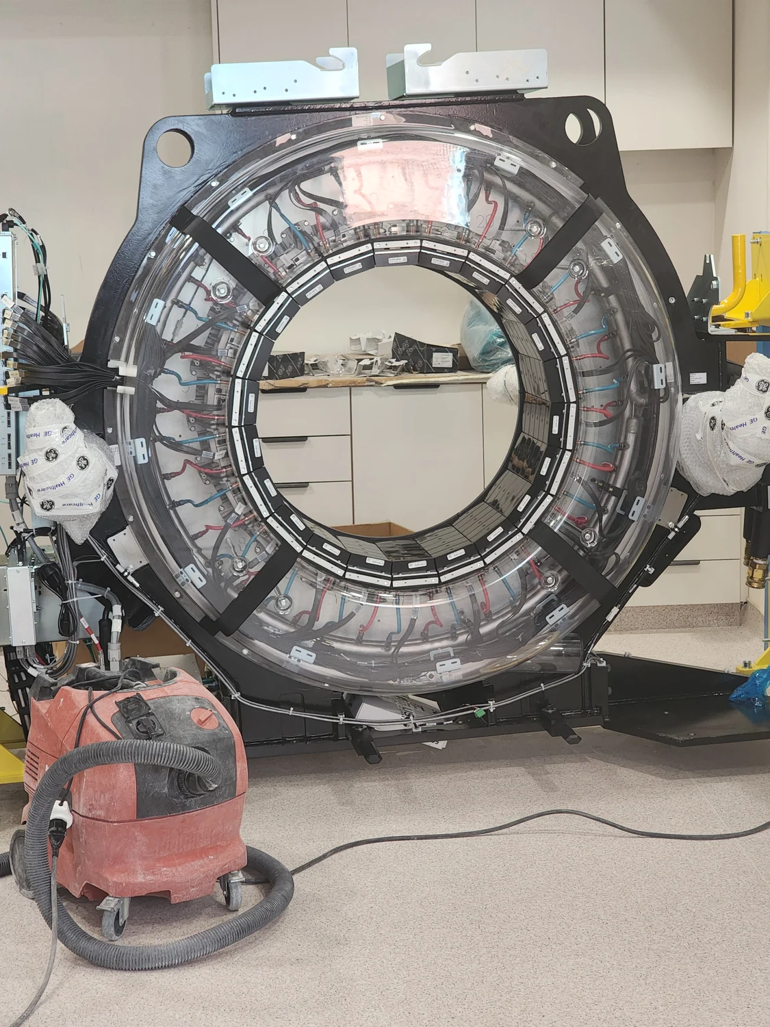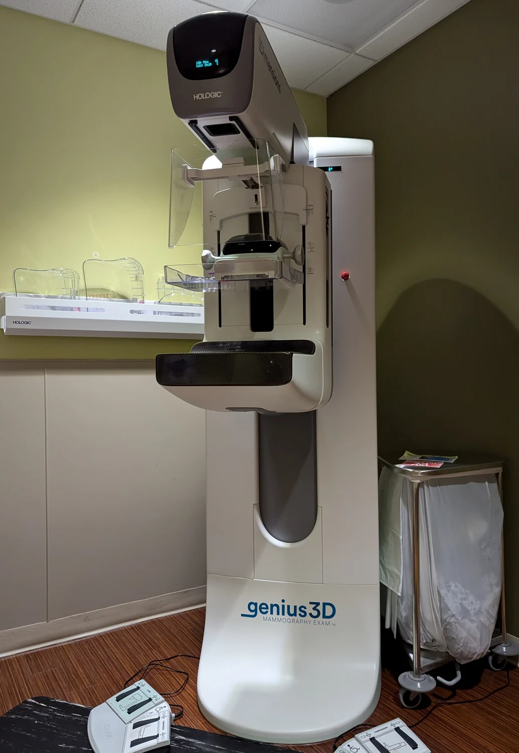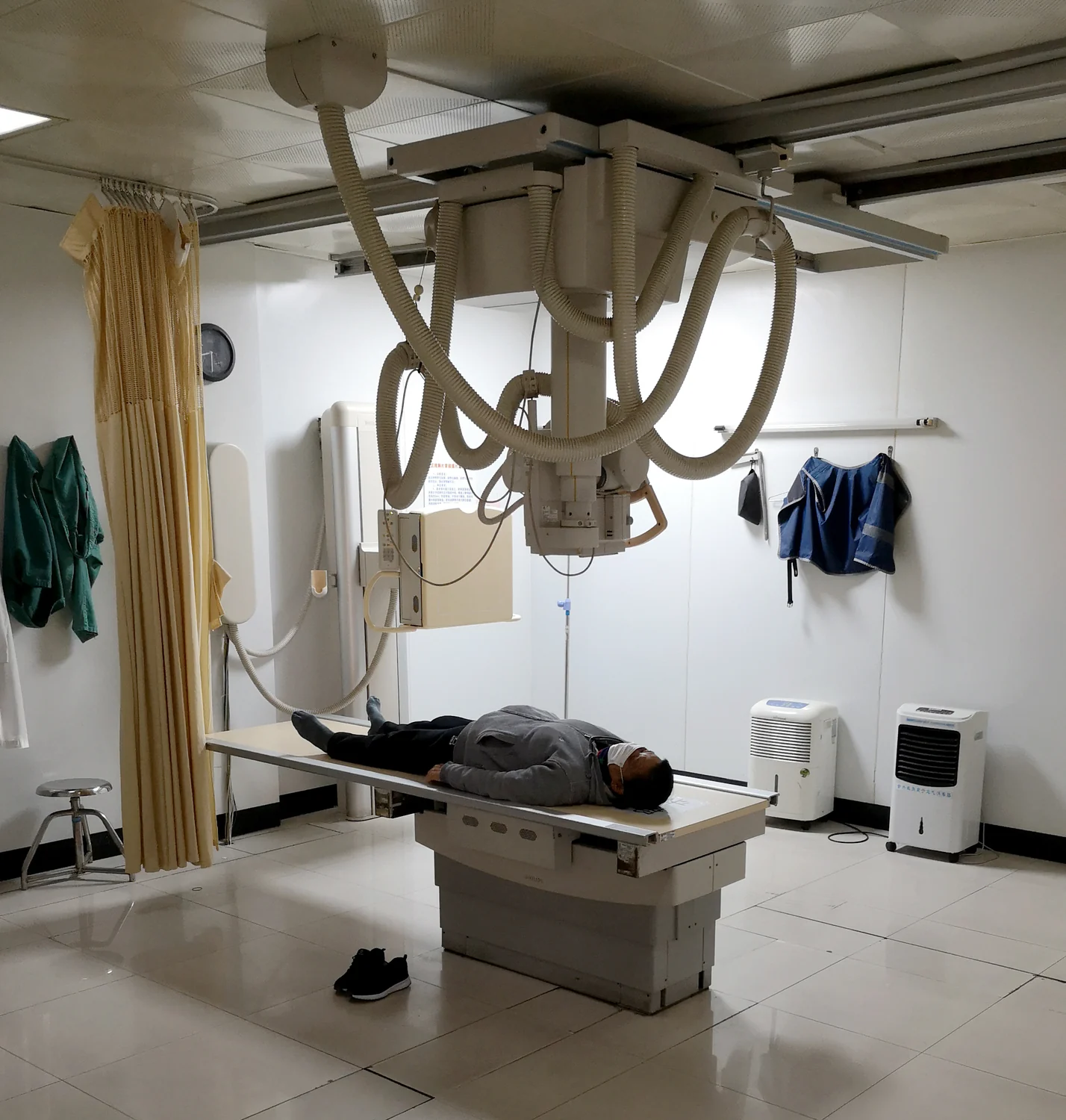
What changes when an x-ray camera moves around you instead of taking one picture from a fixed direction? A plain x-ray records a projection: structures along the beam’s path overlap on the detector. Computed tomography, or CT, gathers many measurements from different angles and calculates cross-sectional images. The physics of x-ray absorption is shared, but the extra viewpoints help separate anatomy that a single shadow-like image can hide.
One projection versus many angles
X-rays are high-energy electromagnetic radiation that can pass through the body. As they travel, different tissues attenuate them by different amounts; a detector records what emerges. A conventional radiograph uses a fixed source and produces a two-dimensional projection, so bone, soft tissue, and other structures can be superimposed [2].
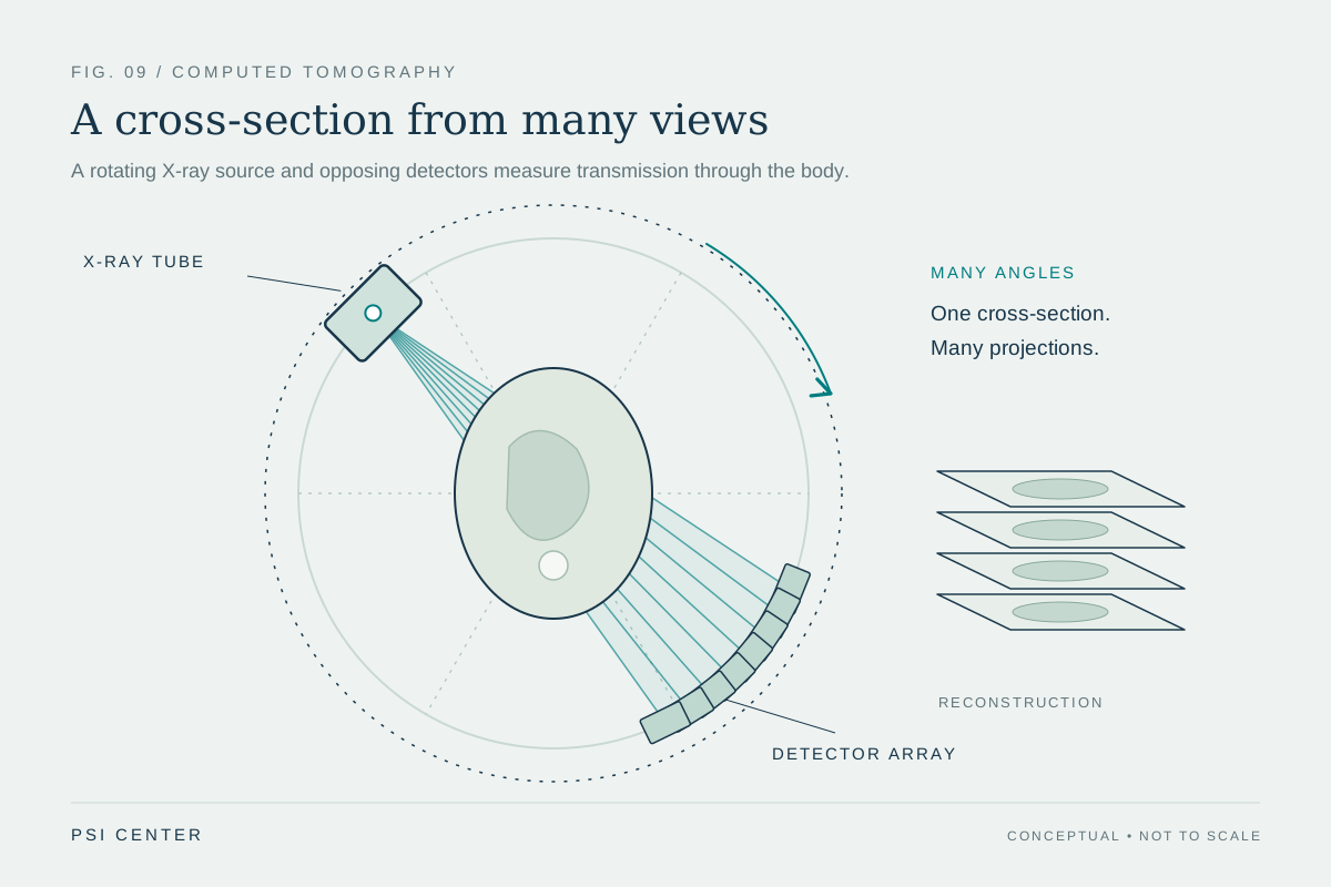
Imagine photographing a closed book with a coin hidden behind it: from one angle, the coin may disappear behind the cover. Move around the book and the coin’s position becomes easier to infer. CT applies a related idea to anatomy. Its x-ray source rotates around the patient while detectors opposite it measure transmitted beams from many directions [1].
How a scanner builds slices
During a CT scan, the patient lies on a bed that moves gradually through a ring-shaped gantry as the x-ray source rotates. For each rotation, a computer uses the detector signals and mathematical reconstruction to estimate the distribution of x-ray attenuation in a cross-section. The resulting slices are commonly a few millimeters thick, though thickness varies by machine [1].
The computer can display slices individually or stack them into a three-dimensional representation. A clinician can scroll through the stack or rotate the model to inspect a structure from different directions. This added spatial context can help locate a complex fracture or distinguish a finding’s position in relation to nearby anatomy [1].
The advantage depends on the question
A plain x-ray is often useful when the question concerns a broad outline, such as whether a bone is broken or whether the lungs show a visible abnormality. It is quick and provides a projection that can answer many focused questions. But overlapping tissues can obscure details: a structure in front of another is laid over it in the final image [2].
CT’s multiple views reduce that overlap by reconstructing slices. It can be particularly useful for complex bone fractures and for examining organs or regions such as the head, chest, and abdomen [1]. More detail does not mean CT is automatically the better choice for every question; the useful view depends on what needs to be seen and on the balance of information and exposure.
Shared radiation, different dose context
Both plain x-rays and CT use ionizing radiation, which has enough energy to alter atoms and can affect living tissue. The amount of exposure depends on the examination and its settings. The NIBIB notes that risk from diagnostic x-ray exposure is generally small, while accumulated exposure over a lifetime and greater sensitivity in children are considerations [2].
CT may also use contrast material when vessels or certain tissues need clearer distinction. Iodine-based contrast in the bloodstream can make blood vessels more visible because it absorbs x-rays; contrast is not required for every scan [1]. The core distinction remains straightforward: a plain radiograph records one projection, while CT combines measurements around the body into slices. Both are tools whose value rests on matching the image to the clinical question.
Explore the physics: an explanatory diagram
Sources & further reading
- Computed Tomography NIBIB
- Medical X-rays NIBIB
Prepared for the independent PSI Center reading library. About edition dates and source checking.
This explainer describes scientific principles. It does not assess individual treatment choices.