How can a scanner make a cross-sectional image without cutting through the body? It measures how x-rays pass through the body from many directions, then uses mathematics to reconstruct a slice. As the scanner gathers successive slices, a computer can stack them into a three-dimensional view. The picture is therefore built from measurements and computation, not captured in one single snapshot.
Many angles create useful clues
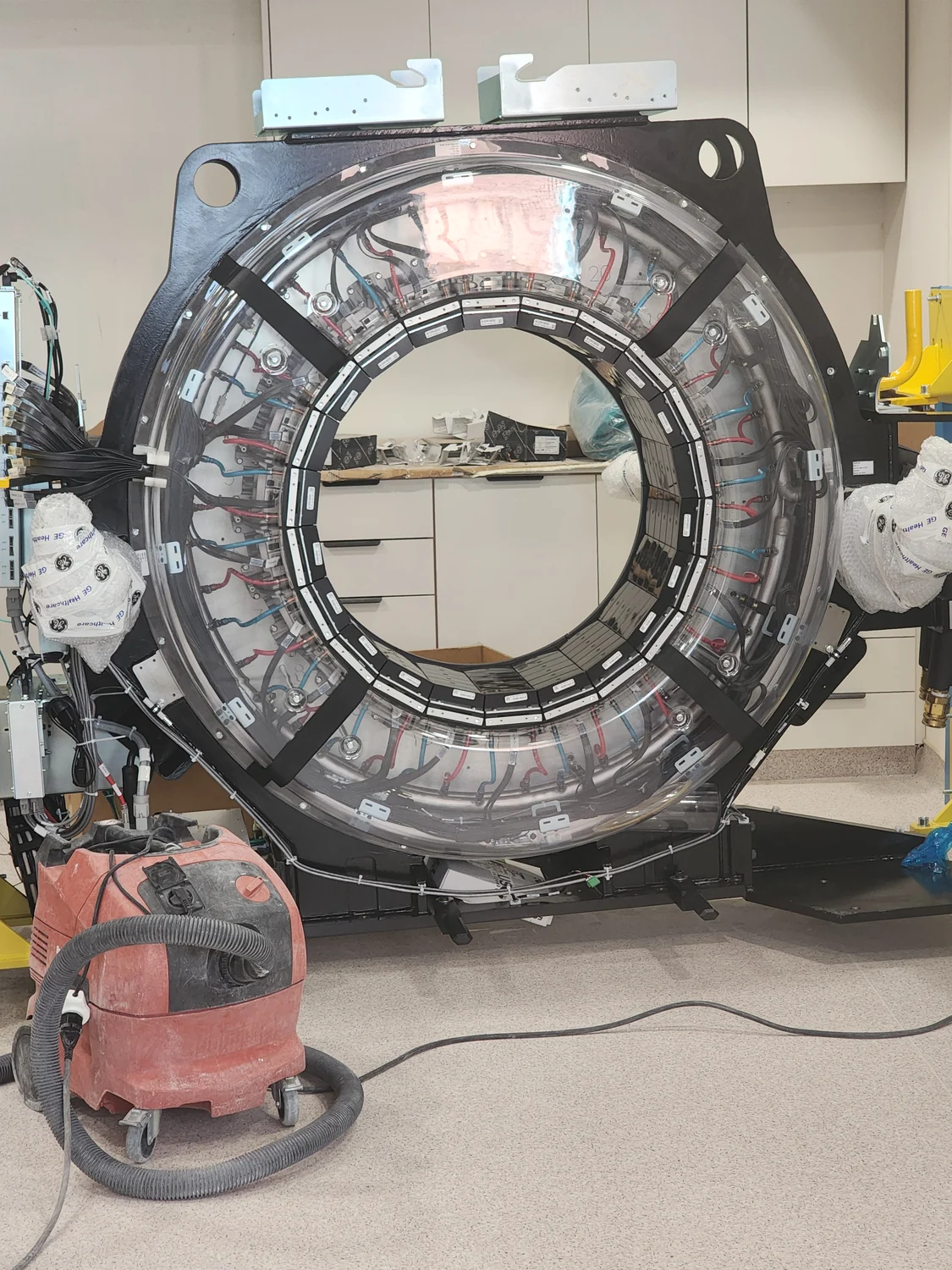
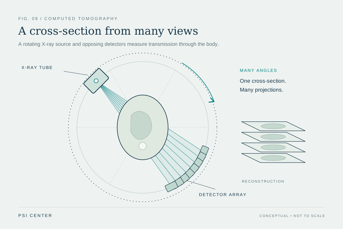
A computed tomography (CT) scanner has an x-ray source and digital detectors mounted opposite each other on a rotating structure called a gantry. The patient lies on a bed that moves through its circular opening while the source rotates around the body. Each beam is narrowed, and detectors register how much of it emerges after passing through tissue. [1]
A single projection cannot show exactly where along the beam a particular structure sits. But measurements from many angles constrain the possibilities. Bone, for example, stops more x-rays than many soft tissues, so it tends to appear clearly. CT’s strength comes from combining these views into a cross-section rather than relying on one fixed direction as a conventional x-ray does. [1]
The computer reconstructs a slice
After the source completes a rotation, the computer applies mathematical methods to the detector measurements to calculate a two-dimensional slice. The reconstruction estimates how x-ray attenuation—the reduction in beam intensity as it passes through material—varies across the scanned region. The output is not a raw detector image; it is a calculated map based on measurements taken around the patient. [1]
Slice thickness can vary by scanner and examination; the NIBIB overview gives a typical range of 1 to 10 millimeters. Once one slice is stored, the bed advances incrementally and another rotation supplies data for the next. Repeating the sequence creates a set of neighboring cross-sections through the body. [1]
From slices to a volume
Imagine a loaf of bread cut into thin slices. Each slice shows a cross-section, and the ordered stack preserves how features shift from one level to the next. CT software can similarly display slices in sequence or combine them into a three-dimensional representation of bones, organs, and other tissues. A clinician can rotate that rendering or inspect individual levels to locate a feature more precisely. [1]
Computational modeling provides a useful distinction here. A model simulates a system using mathematical or physical rules and variables, whereas CT reconstruction calculates an image from measured x-ray data. Both rely on computation, but CT is not simply a prediction of what anatomy might look like. Its reconstructed image is constrained by signals recorded during the scan. [2]
The image has strengths and limits
CT can be useful for complex fractures and for examining the head, lungs, abdomen, and heart. Contrast agents can make selected structures more visible by stopping x-rays more strongly; iodine-based material may help show blood vessels, while barium-based compounds are used for parts of the digestive tract. Contrast changes the measured signal, helping distinguish structures that would otherwise be harder to separate. [1]
The measurement method also sets limits. CT uses x-rays, which are ionizing radiation, and exposure accumulates across imaging encounters; the risk from an individual scan is generally small but depends on context. MRI uses magnetic fields and radio waves instead of x-rays, and may be preferable for some questions, while CT can provide answers when speed or the detail needed makes it useful. The two techniques build images through different physical signals. [1] [2]
Explore the physics: an explanatory diagram
Sources & further reading
- Computed Tomography NIBIB
- Computational Modeling NIBIB
Prepared for the independent PSI Center reading library. About edition dates and source checking.
This explainer describes scientific principles. It does not assess individual treatment choices.