Why rotate an X-ray source around a patient instead of taking one picture from the front? A single projection stacks structures along the beam's path, hiding their depth. Computed tomography, or CT, collects measurements from many directions so a computer can estimate how the X-rays were absorbed at different locations inside the body.
A projection collapses depth
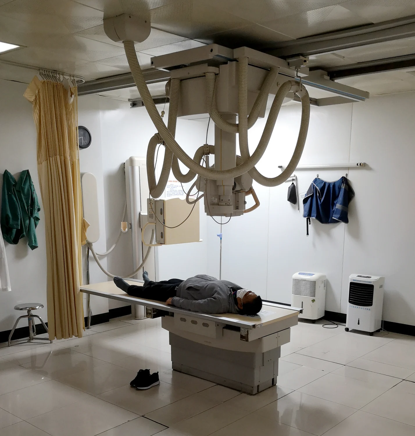
In a conventional X-ray image, each point on the detector records the combined effect of structures that lie along the beam. A rib in front of a lung and another structure behind it may overlap in the final projection. The image can be informative, but it does not independently say how far along the beam each feature sits.
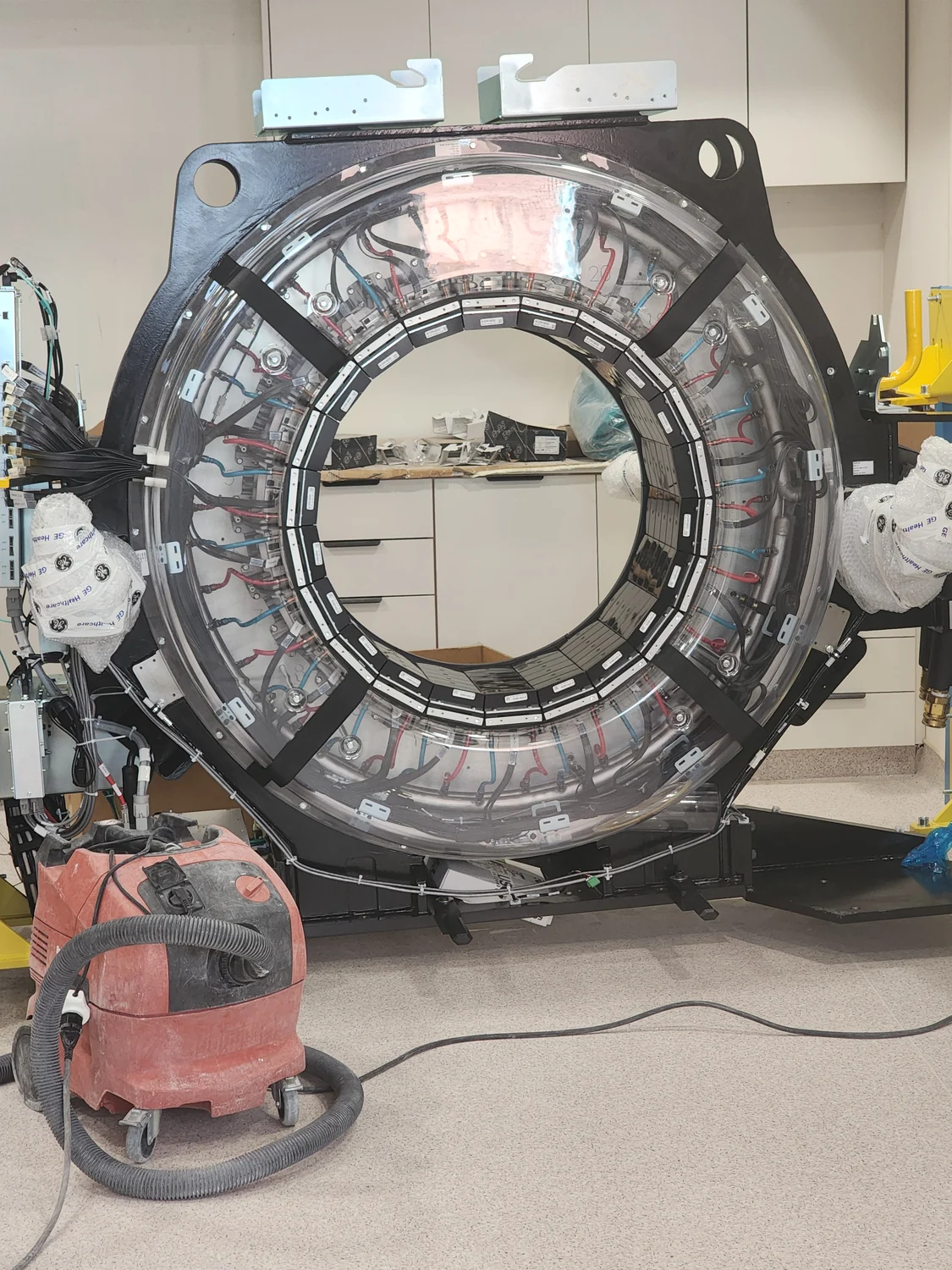
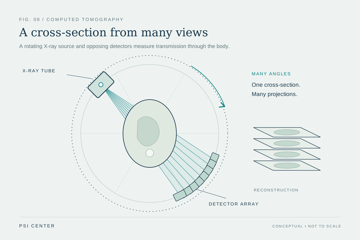
CT changes the geometry. An X-ray source and detector rotate around the body while the patient moves through the scanner's circular opening. The detectors record snapshots of X-rays that pass through the body at many angles during a rotation [2]. Each snapshot is still a projection, but the collection contains more spatial clues than a single view.
Angles supply different clues
Imagine trying to locate a small object hidden in a loaf of bread by looking at one cut surface. Many positions along the viewing direction could explain the same mark. If you inspect many slices from different orientations, the possibilities shrink. CT's angular measurements work on a related principle: an object that lies in one path through the body will affect projections differently as the viewing direction changes.
The X-ray source emits a narrow beam, and detectors measure the radiation that emerges after crossing tissue [1]. Dense materials such as bone stop more X-rays and are readily visible; soft tissues vary in how much they stop, so they can be harder to distinguish without contrast [1]. Across many angles, the computer uses these varying measurements to reconstruct cross-sectional images [2].
Reconstruction turns data into slices
The computer's task is an inverse problem: infer the internal structure that could have produced the observed projections. It does not simply paste the snapshots together. Mathematical reconstruction assigns estimated X-ray attenuation to locations in a slice, using the many paths through those locations. The result is a cross-section, a map of how strongly different parts of the body reduce the beam.
As the table advances through the gantry, the scanner repeats the collection process at successive positions. The resulting slices can be viewed individually or digitally stacked into a three-dimensional representation [1]. Slices can help identify the location of a finding, while a 3D view can make the relationships among structures easier to inspect. These are different ways of displaying reconstructed data, not separate measurements.
More views have a cost
CT's many angles improve localization and detail, but the method uses ionizing X-rays. The FDA notes that exposure can produce a small increase in lifetime cancer risk, and that children are more sensitive per unit dose and have a longer lifetime for effects to emerge [2]. The practical aim is to obtain image quality adequate for the clinical question while limiting exposure. The FDA also advises considering whether another exam using less or no radiation can answer the question and avoiding duplicate scans [2].
Contrast agents can make particular structures more visible. For example, iodine-based intravenous contrast can help illuminate blood vessels because it stops X-rays effectively [1]. Contrast does not replace the angular measurements; it changes the physical differences the scanner detects. CT is valuable when its detailed cross-sections can answer a relevant question, but a finer picture is useful only when the information it adds justifies the method's costs and risks.
Explore the physics: an explanatory diagram
Sources & further reading
- Computed Tomography NIBIB
- Computed Tomography FDA
Prepared for the independent PSI Center reading library. About edition dates and source checking.
This explainer describes scientific principles. It does not assess individual treatment choices.