How can a scanner infer where a radioactive tracer is inside the body without opening the body to look? In positron emission tomography, or PET, the key clue is a pair of gamma-ray photons created when a positron meets an electron. Detectors arranged around the patient register photons arriving at nearly the same time. Each pair points to a line across the scanner, and many such lines help reconstruct where tracer activity is concentrated.
The tracer sets the event in motion
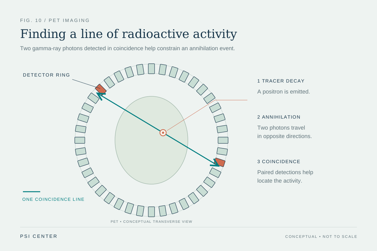
A PET scan begins with a radiotracer: a carrier molecule joined to a radioactive atom. The carrier is chosen for the biological process being studied, while the radioactive atom makes the molecule detectable. A tracer may be administered by injection or another route, depending on the question. As the radioactive atom decays, it emits a positron, a particle with the same mass as an electron but opposite electric charge [1].
The positron travels a short distance through tissue and encounters an electron. The two particles annihilate, meaning their mass is converted into energy. In this process, two gamma-ray photons shoot off in nearly opposite directions. Their energy is characteristic of the annihilation process rather than the specific positron-emitting tracer [1].
Coincidence turns a pair into a clue
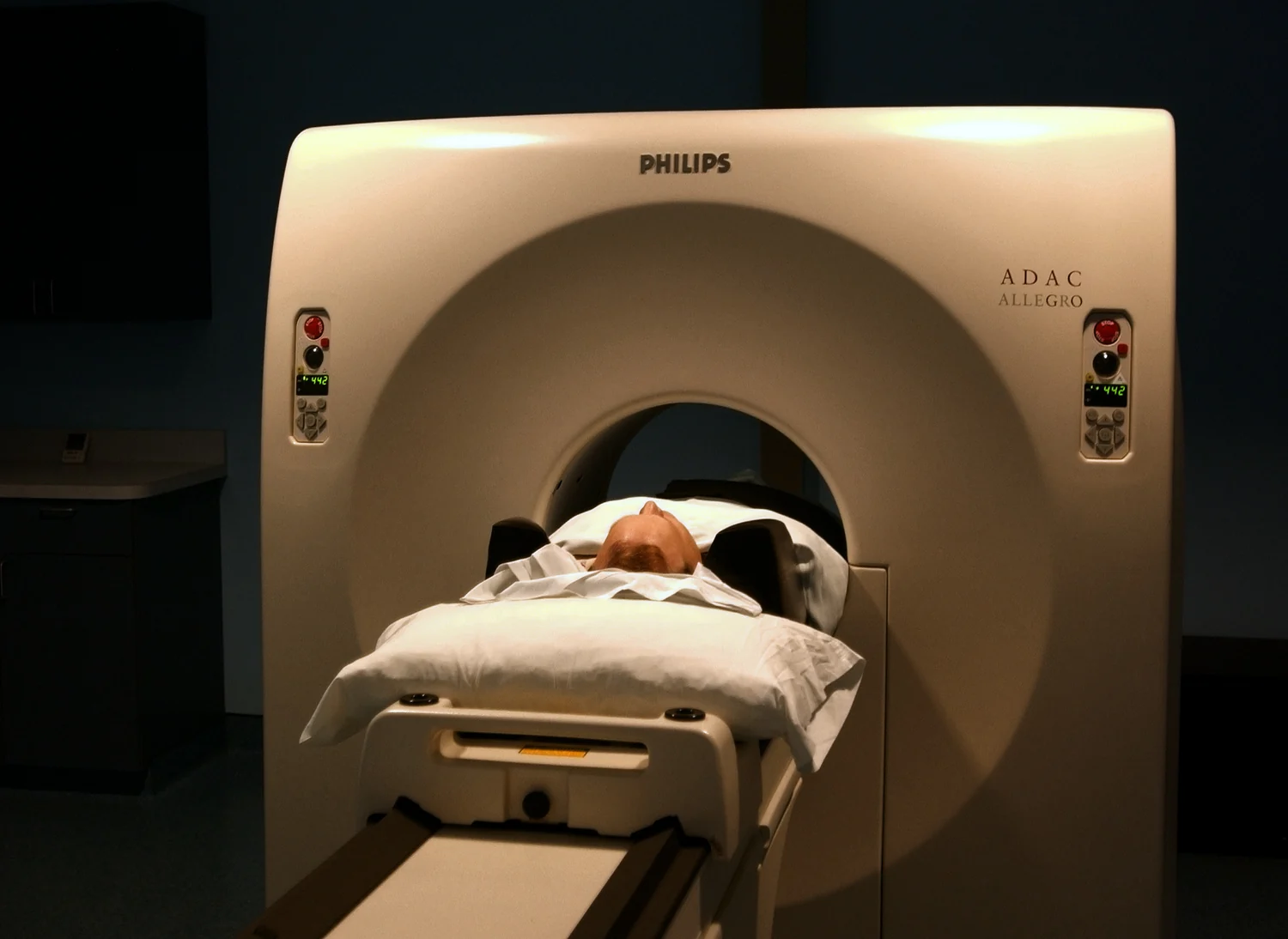
A PET scanner surrounds the body with detectors. If two detectors on opposite sides register photons at nearly the same time, the scanner treats them as a coincident pair. The event indicates that an annihilation likely occurred somewhere along the line connecting those detectors. It does not pinpoint the exact location by itself; it narrows the possibilities to a line.
Imagine drawing a straight line through each detected pair. One line could pass through many possible points, but millions of lines from different directions overlap more densely where emissions are more likely to have occurred. A computer uses the accumulated detections to estimate the three-dimensional distribution of tracer activity. The method is called tomography: constructing an image of internal structure or activity from measurements collected around the body [1].
PET maps function through chemistry
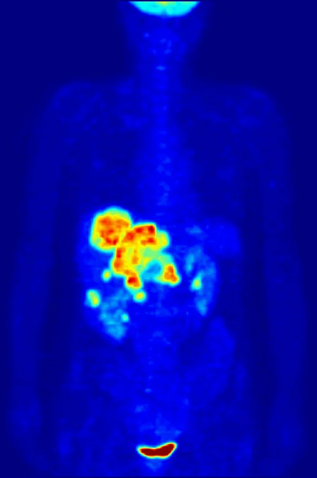
The image reflects where the tracer goes and how much is detected, not simply the outline of an organ. For example, the radiolabeled glucose molecule FDG is used because cells take up glucose for energy, and cellular activity can affect that uptake. PET can therefore help assess functional molecular processes, including patterns used in cancer imaging and brain or cardiac studies [1].
This functional information differs from the kind of picture produced by X-ray imaging. In radiography or CT, an external X-ray beam passes through the body, and detectors record how much is absorbed or scattered by internal structures. Those techniques use ionizing radiation too, but their physical signal and image formation are different from PET’s detection of photons originating inside the body [2].
A line is not a diagnosis
PET findings need interpretation in context. A bright region means the tracer signal is relatively concentrated there; it does not, on its own, identify the cause. The tracer’s biological target, the scan protocol, and other clinical information shape what the image can show. Nuclear medicine uses different radiopharmaceuticals for different questions, and the chosen tracer determines what process is being examined [1].
PET is also often paired with CT, which provides anatomical information alongside the functional map. The combination can help locate a signal within the body’s structures, but it does not erase the distinction between the two methods. CT uses X-rays; PET detects coincident gamma photons from positron annihilation. Each imaging procedure has its own benefits and risks, and X-ray imaging is used when it is medically appropriate to answer a clinical question [2].
Explore the physics: an explanatory diagram
Sources & further reading
- Nuclear Medicine NIBIB
- Medical X-ray Imaging FDA
Prepared for the independent PSI Center reading library. About edition dates and source checking.
This explainer describes scientific principles. It does not assess individual treatment choices.