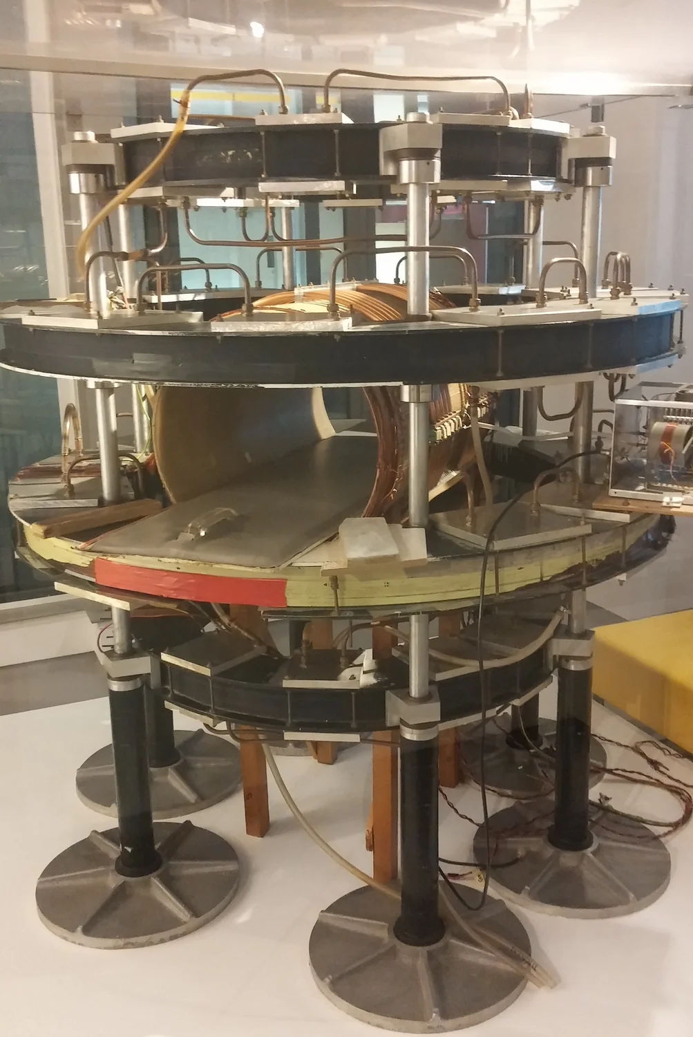
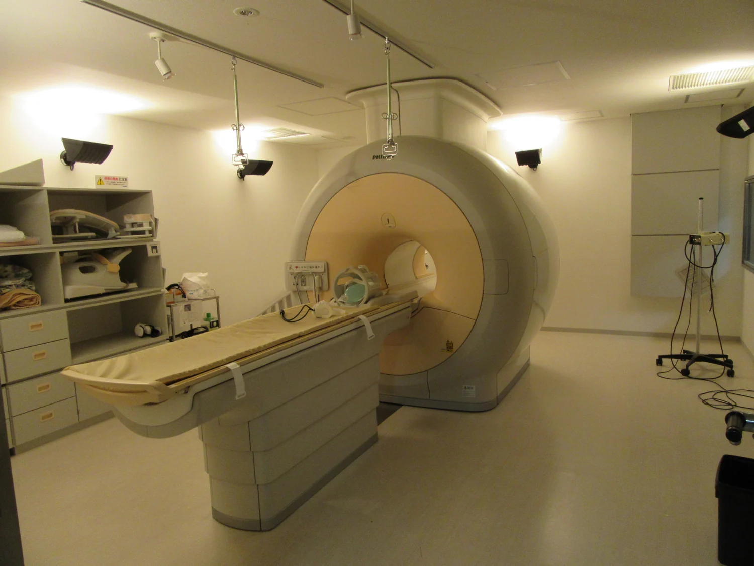
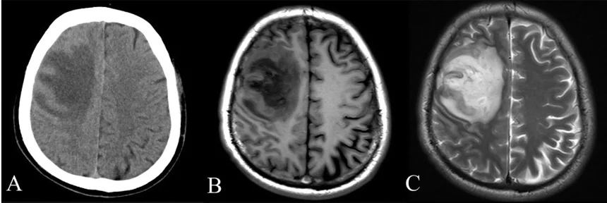
Why can two MRI images of the same body region make tissue look strikingly different? The scanner is not simply taking a photograph: it measures signals from protons and turns them into an image using chosen settings. Different tissues return those signals on different schedules, and the scan sequence determines which differences stand out. The result is a contrast map shaped by physics and by the question the scan is designed to answer.
Protons respond to a magnetic field
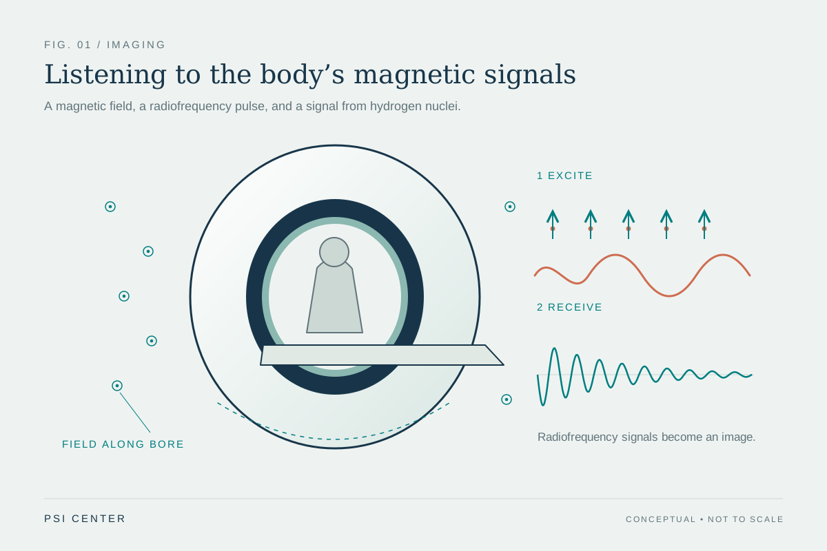
The MRI scanner’s strong magnetic field nudges many hydrogen protons in the body, especially those in water, into alignment. A radiofrequency pulse disturbs that alignment. When the pulse ends, the protons move back toward equilibrium and release energy that sensors detect. Their recovery depends on the surrounding molecular environment, so water-rich, fatty, and other tissues can produce different signals. MRI uses magnetic fields and radio waves, not plasma or ionizing x-rays. [1]
A proton is a positively charged particle found in a hydrogen nucleus. In a simplified mental picture, it behaves like a tiny magnetic compass, though the actual quantum physics is more subtle. The scanner’s signal comes from many protons acting together. Their response is affected by local chemistry and structure, which gives MRI a way to distinguish soft tissues that may look similar in an ordinary x-ray. [1]
Settings decide which differences appear
MRI sequences choose how pulses and measurements are timed. Those choices emphasize different aspects of proton behavior, making a tissue bright in one image and darker in another. The scanner is therefore not assigning one permanent brightness to each tissue; it is measuring under a particular set of conditions. Comparing images requires knowing that sequence, because apparent contrast depends on the acquisition as well as the anatomy.
Contrast agents can change the recovery behavior of protons and make some structures more conspicuous. NIBIB describes agents often containing gadolinium that may be given before or during a scan to increase the speed at which protons realign; this can alter image brightness. The decision to use contrast and the interpretation of resulting images belong within clinical imaging practice, where the scan’s purpose and patient context matter. [1]
MRI and optical imaging answer different questions
Optical imaging uses light that may be absorbed, scattered, reflected, or emitted by tissue. Its techniques span scales: microscopy can examine cells, while optical coherence tomography uses infrared light to form cross-sectional views beneath surfaces, including in the retina. Diffuse optical methods can measure properties such as hemoglobin concentration and blood oxygen saturation in soft tissue. This is a different physical signal from MRI’s proton response. [2]
The methods can complement each other because they reveal different properties and operate at different scales. Optical approaches can be sensitive to light interactions in accessible tissue, while MRI can form detailed three-dimensional anatomical images and distinguish soft tissues throughout the body. Neither modality is a universal camera. The useful choice depends on what needs to be measured, how deep it is, and what kind of contrast can reveal it. [1] [2]
Reading an image means reading its conditions
Imagine photographing a landscape at dawn and at noon: the objects remain, but the lighting changes which edges and colors are prominent. MRI sequences are not literally lighting, yet the analogy captures why contrast shifts. A sequence tuned to one tissue property can make boundaries easier to see; another may emphasize a different contrast. Motion can blur the measurement, so patients are asked to remain still while the scan is acquired. [1]
MRI’s physical advantages and constraints both matter. It does not use x-rays, and it is particularly useful for soft tissues such as brain, spinal cord, muscles, ligaments, and tendons. It does use a powerful magnetic field, so implanted devices and magnetizable objects require careful consideration. Optical imaging can also be combined with MRI to provide complementary information. In each case, the visible image is evidence produced by a specific measurement process, not a transparent view of everything happening in the body. [1] [2]
Explore the physics: an explanatory diagram
Sources & further reading
- Magnetic Resonance Imaging NIBIB
- Optical Imaging NIBIB
Prepared for the independent PSI Center reading library. About edition dates and source checking.
This explainer describes scientific principles. It does not assess individual treatment choices.