What can a scan taken today tell us that a scan from last month cannot? The answer may lie in the difference between images, not in either image alone. Repeated scans let clinicians compare structure or biological activity across time, but each imaging method measures a particular physical signal. Knowing what that signal represents helps distinguish real change from a change in the way the image was made.
MRI turns proton behavior into contrast
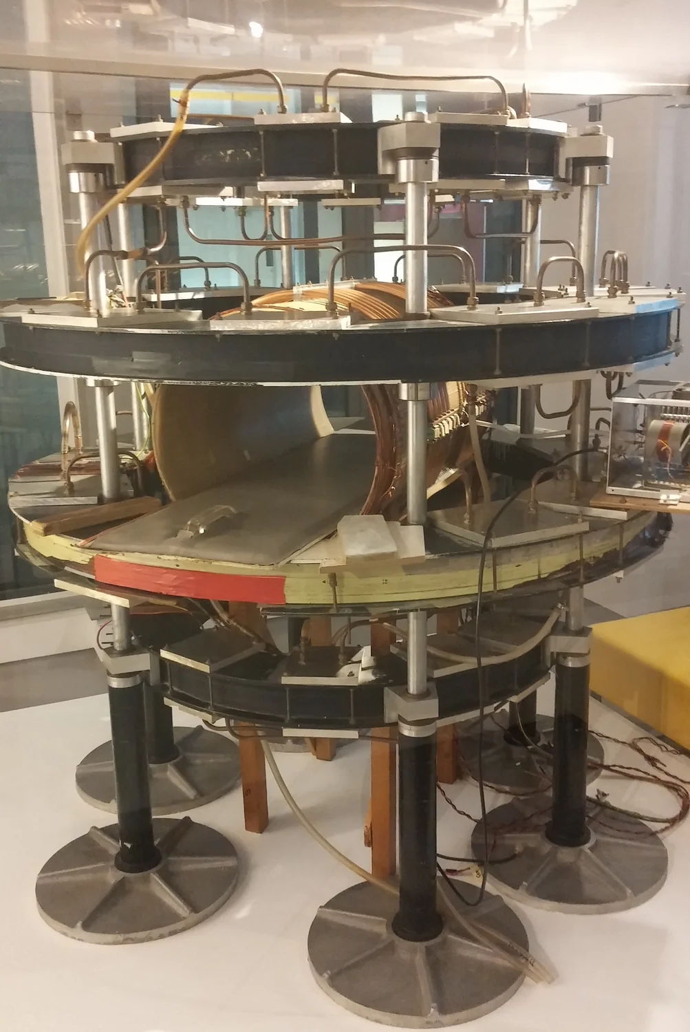
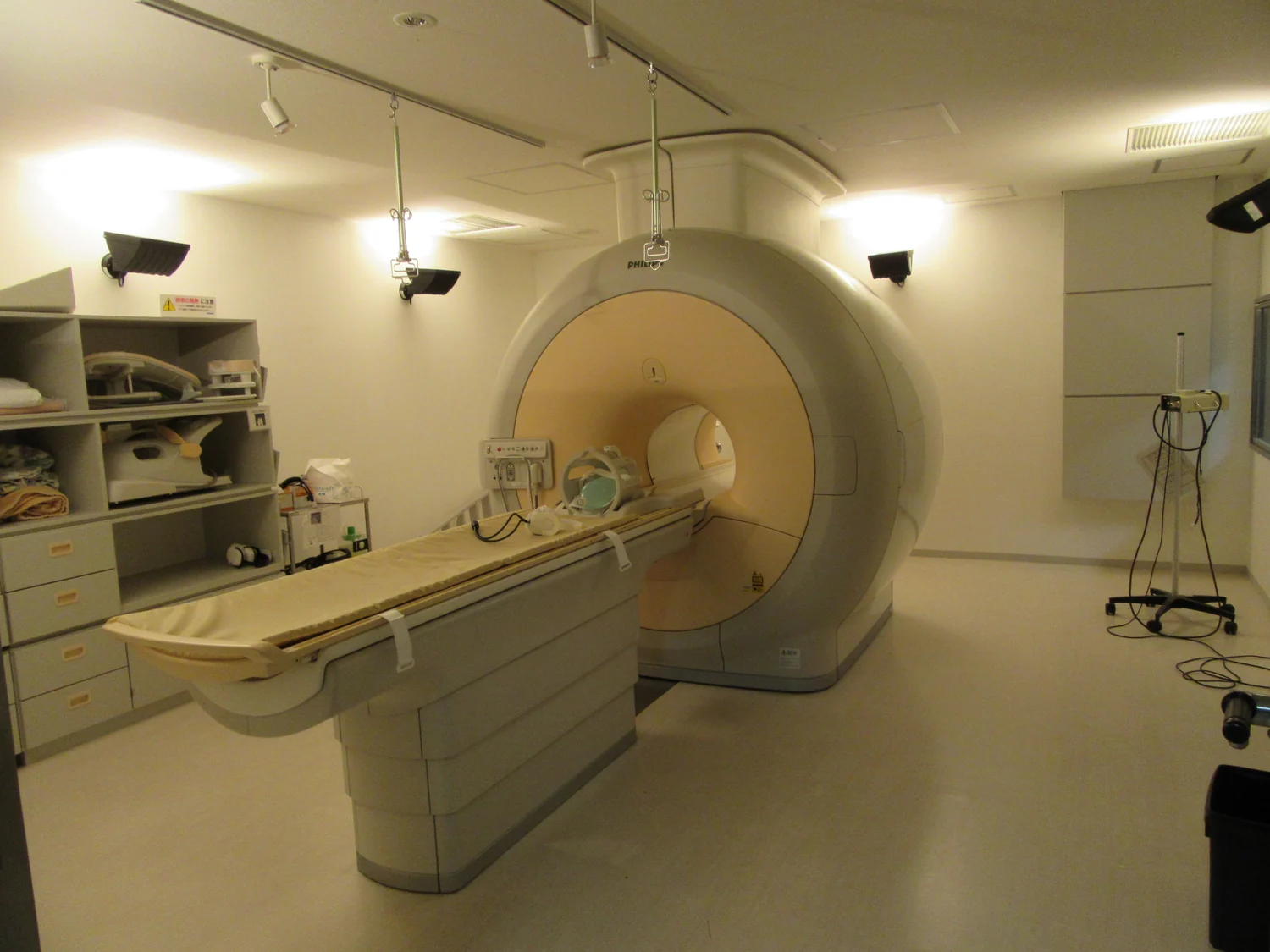
Magnetic resonance imaging (MRI) uses a strong magnetic field and radiofrequency pulses to disturb the alignment of protons in the body’s water. When the pulse stops, sensors detect energy released as the protons realign. The timing and amount of that signal vary with the chemical and physical surroundings, allowing different tissues to appear with contrast [1]. MRI uses magnetic fields and radio waves, not plasma or ionizing x-rays.
Imagine comparing two maps of the same neighborhood taken months apart. To interpret a changed boundary, the maps need to represent the same region in a comparable way. MRI likewise requires the patient to remain still during acquisition; movement can blur the image [1]. When scans are repeated, consistent positioning and image quality help make a difference between them more interpretable. MRI is often used to examine soft tissues, including the brain, spinal cord, muscles, ligaments, and tendons.
Some scans show function as well as form
Nuclear medicine imaging follows radiotracers: radioactive atoms attached to carrier molecules that travel or interact in the body. PET and SPECT cameras detect signals from those tracers and use them to construct three-dimensional images. PET, for example, can use a radiolabeled glucose-like molecule to show patterns of uptake associated with cellular activity; it is used to detect cancer and monitor progression or response to treatment [2]. This is a different kind of information from an MRI’s tissue contrast.
The distinction matters in follow-up. A structural scan may help show the size or location of an abnormality, while a tracer-based scan can show where a particular molecule accumulates. The tracer determines what biological process is visible, so the image does not represent all activity in the body. A comparison over time is meaningful only in relation to what the chosen tracer measures and how the examination was performed [2].
A difference between scans needs an explanation
Suppose a later image looks brighter in one region. That may reflect a change in tissue or tracer uptake, but the brightness is not a free-standing diagnosis. MRI signal depends on proton behavior in the local molecular environment and on how the scan is acquired [1]. In PET or SPECT, the distribution of the selected radiopharmaceutical shapes the image [2]. The same visual change can therefore mean different things in different imaging methods.
Combination systems can pair modalities. PET/CT, for instance, overlays functional tracer information with CT anatomy to help locate and monitor tumors [2]. In research, MRI can also be combined with specialized techniques to investigate tissue properties or metabolism [1]. These examples show why a sequence of images is interpreted in context: the image is a measured signal processed into a map, and its clinical meaning depends on the physics and the question asked.
Monitoring is a comparison, not a guarantee
MRI is often useful when frequent imaging is needed because it does not use ionizing x-rays, though it has its own considerations, including powerful magnetic fields and possible implant-related concerns [1]. Nuclear medicine scans introduce radiopharmaceuticals and involve radiation exposure; the tracer and purpose determine the study, and clinicians aim to use the amount needed for a useful examination [2]. Repeating either type is a decision tied to the information sought, not a rule that every condition requires serial scans.
Follow-up imaging can help show whether a disease is changing or a treatment response is detectable. In nuclear medicine, imaging after a therapeutic radiopharmaceutical may help assess response; MRI is also used for disease detection, diagnosis, and treatment monitoring [2] [1]. Yet a scan is one source of evidence. The most useful comparison asks what physical signal each examination measured, whether the images are comparable, and how the observed difference fits the broader clinical question.
Sources & further reading
- Magnetic Resonance Imaging NIBIB
- Nuclear Medicine NIBIB
Prepared for the independent PSI Center reading library. About edition dates and source checking.
This explainer describes scientific principles. It does not assess individual treatment choices.