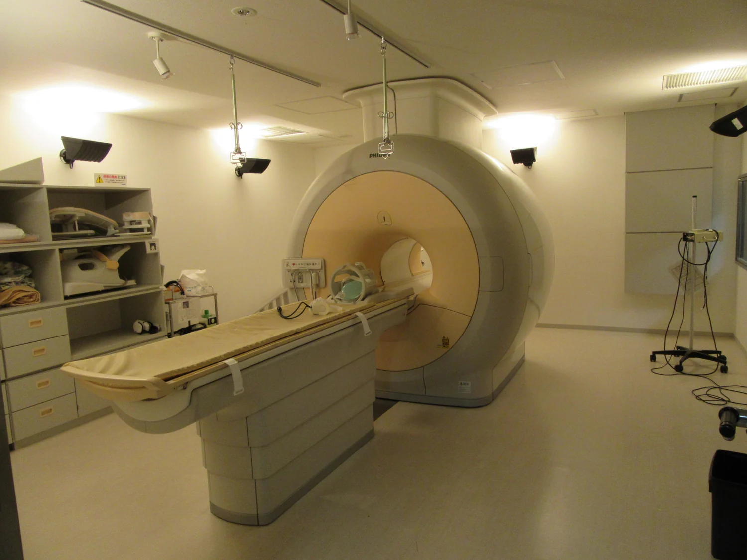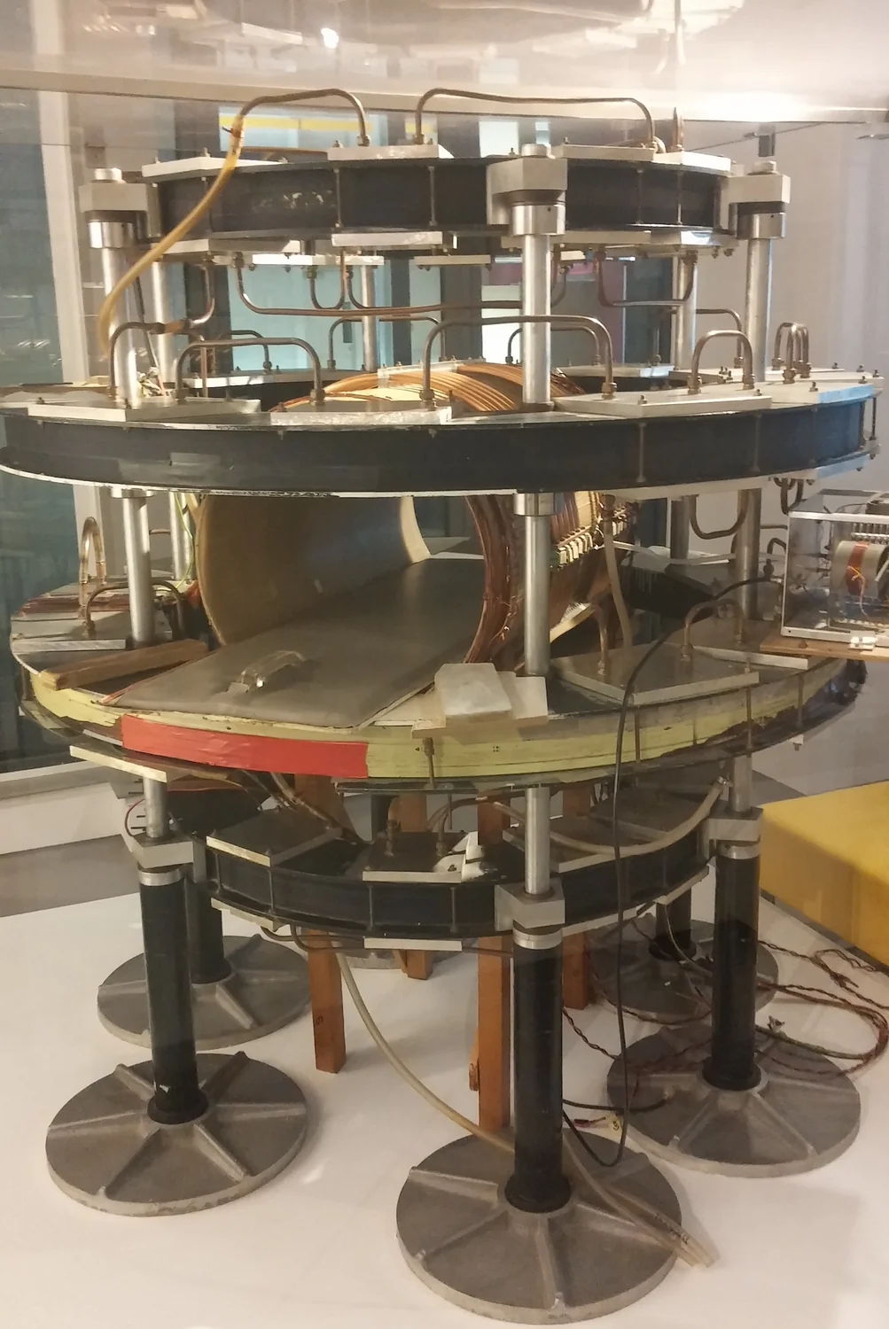
How can a scanner make an image when it never directly photographs the inside of the body? Medical imaging begins with measurements: signals arriving from tissue, often incomplete or noisy, that software must combine into a map. Artificial intelligence can change how that map is reconstructed, speeding some calculations or learning patterns from examples. But the central question remains physical: which features in the image are supported by the measured signal, and which depend on assumptions built into the reconstruction?
From measured signal to image
An imaging system does not usually record a ready-made picture. It collects signals shaped by the interaction of energy with tissue, then uses mathematics to infer where signal sources or boundaries lie. Reconstruction is the computational step that turns those measurements into an image. In MRI, for example, magnetic fields and radio waves produce signals that are processed into images; MRI does not use plasma or ionizing x-rays. Every reconstruction balances limited measurements, noise and the need for a useful representation.
A familiar analogy is assembling a map from scattered location reports. If many reports are missing, a program can interpolate a plausible road network, but plausibility is not proof that every road is present. Image reconstruction has the same tension: a method can fill gaps smoothly while suppressing a small structure or adding a pattern that fits its expectations. The scanner’s measurements constrain the answer, but the algorithm shapes how those constraints become visible.
What a learning algorithm adds
Machine learning is a way of finding patterns in data and using them to make predictions. In medical imaging, a model can be trained on collections of images and corresponding examples, then used to help identify features or process new data. Deep learning uses many layers of computation to learn more complex patterns. Such tools may help clinicians notice subtle changes or help systems assess images more quickly, but the result depends on the examples and labels used to teach the model. [1]
For an image reconstruction task, a learned model may help distinguish likely signal from noise, or infer missing information from patterns encountered during training. That can be useful when measurements are sparse or acquisition is constrained. Yet a model’s output is not a direct view into the body. It is an estimate shaped both by incoming measurements and learned regularities. If an unusual anatomy or disease pattern differs from the training examples, a confident-looking reconstruction may still be wrong.
The physics and the data both matter
Computational models can be built from established physical principles, patterns in datasets, or a combination of both. A mechanistic model encodes an account of how a system works; a data-driven model learns associations without explicitly representing every underlying process. Hybrid models combine the approaches. In imaging, physical knowledge can constrain what reconstructions are plausible, while data-driven methods can capture recurring structures that are difficult to describe with simple equations. [2]
Consider a blurred edge in a scan. A physics-based model may account for how the scanner’s measurement process spreads signal, while a learned component may recognize common tissue boundaries. Both can improve a reconstruction, but only if their assumptions match the case. If the model smooths away a tiny but real feature, an image may look cleaner while losing information. “Sharper” is therefore not synonymous with “truer”; image quality has to be judged against the clinical question and independent evidence.
Validation is part of the image
Training data for medical AI require curation, quality checks, annotation and labels from people with relevant expertise. The distribution of cases matters too: a dataset that underrepresents particular populations or acquisition settings may yield uneven performance. NIBIB notes that AI developers need to assess fairness and performance across groups. These are not administrative details added after the physics; they affect which patterns a model learns to treat as normal. [1]
Validation asks whether a method works beyond the examples used to build it. Researchers can compare reconstructions with known measurements, test them on separate datasets and examine whether performance holds across scanners and patient groups. Computational models can also help identify candidate experiments before laboratory work, but simulated success is not clinical proof. [2] The useful question is not whether AI can make an image look convincing. It is whether the reconstructed features remain reliable when the inputs, bodies and clinical settings change.
Sources & further reading
- Artificial Intelligence NIBIB
- Computational Modeling NIBIB
Prepared for the independent PSI Center reading library. About edition dates and source checking.
This explainer describes scientific principles. It does not assess individual treatment choices.