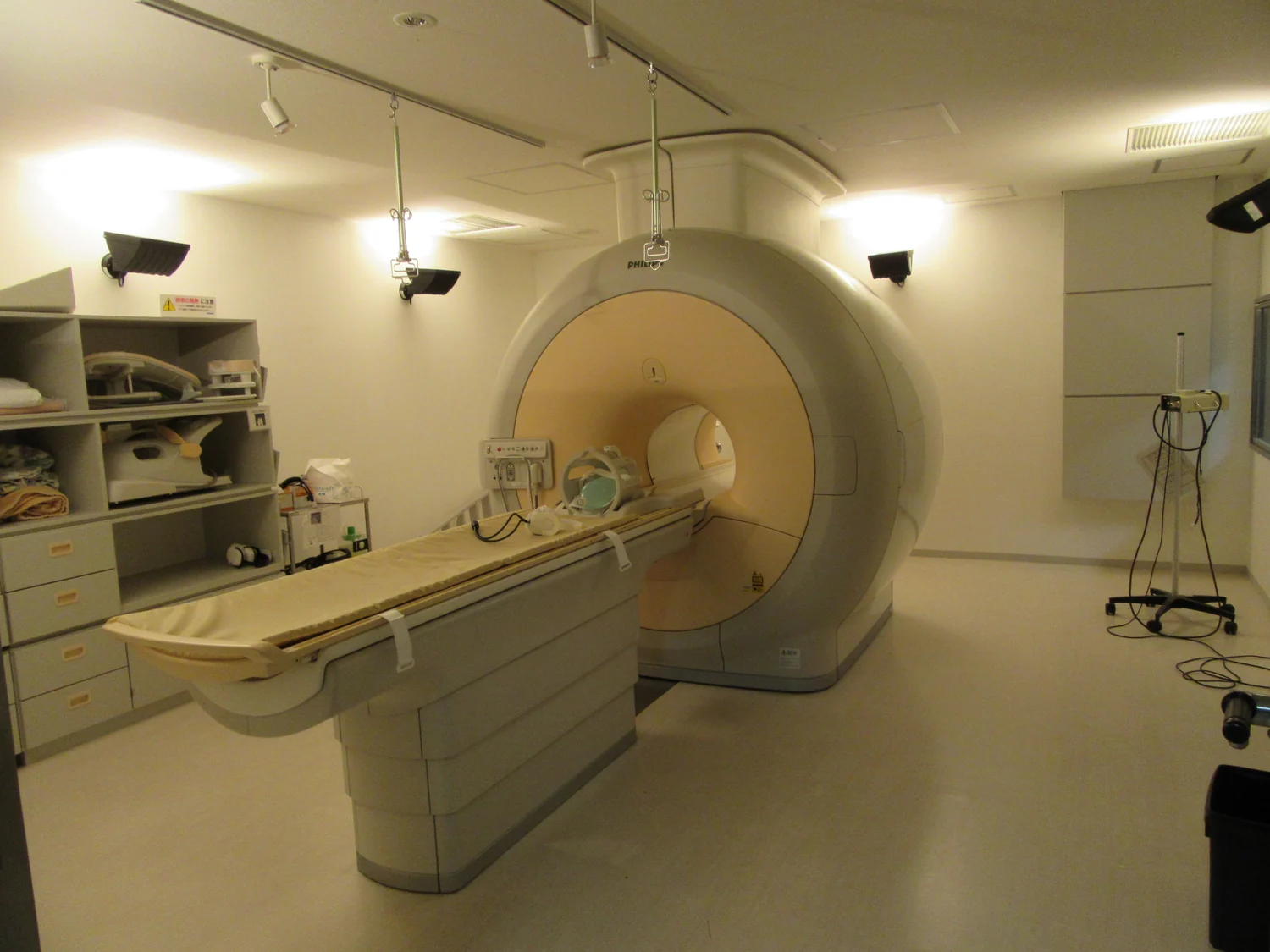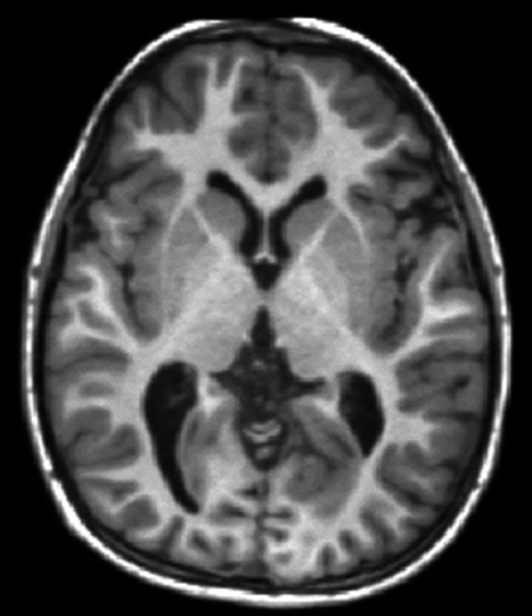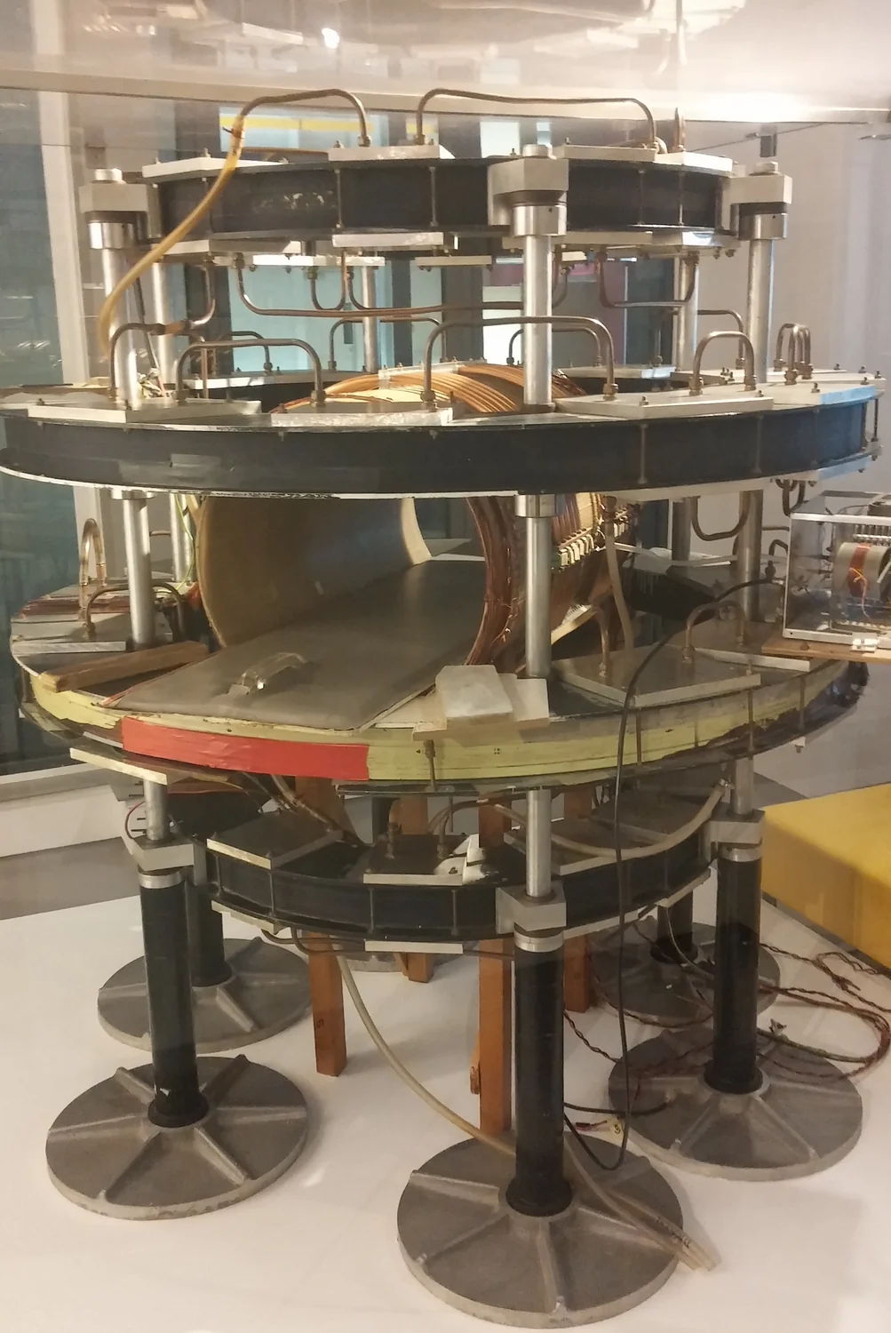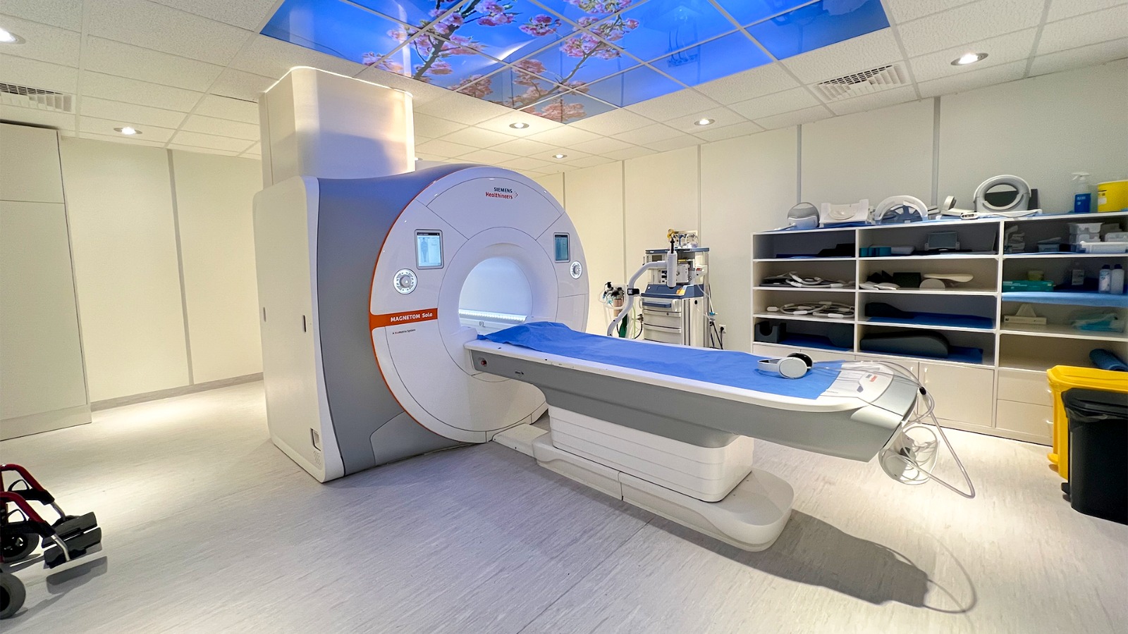
MRI hardware must do more than create a strong magnetic field: its radiofrequency (RF) field also has to tip nuclear spins in a controlled way. At high field strengths, that RF pattern can become uneven across the body. A study published on 9 June 2026 used a physics-constrained learning framework to design a thin metasurface intended to reshape that pattern. The reported demonstration was computational, using an arm phantom in simulations; it was not a scan of a person or a clinical evaluation. [1]
Two magnetic fields serve different roles
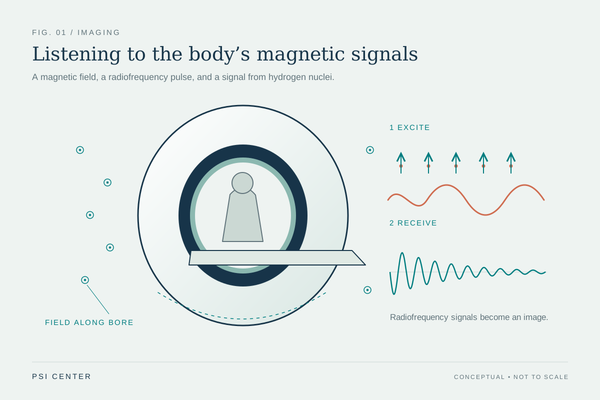
An MRI scanner’s strong static field aligns nuclear spins. A second, much weaker, time-varying radiofrequency field, often described by its transmit component B₁⁺, tips those spins away from alignment. The resulting response contributes to the signal used to form an image. If the transmit field varies substantially over the region being examined, the same pulse can produce different excitation at different locations. This field uniformity problem is especially challenging at ultrahigh field, where RF wavelengths in tissue are short relative to body dimensions. [1] [3]
A metasurface is a deliberately arranged layer of small conducting or dielectric structures. Their interactions with an incoming electromagnetic field can alter its local distribution. In MRI, the design goal is not simply to make the field bigger everywhere: field coverage, efficiency, and the accompanying electric field and power deposition all matter. A useful analogy is a patterned acoustic panel that changes how sound spreads in a room, except MRI structures operate through electromagnetic resonance and must work inside a tightly constrained scanner space.
Why inverse design is difficult
A designer can specify a desired field pattern and ask what arrangement of resonant elements might produce it. That is an inverse problem. Many different structures may create similar field patterns, while small changes in spacing, material properties, loading, or geometry can alter their response. Directly evaluating each candidate with detailed electromagnetic simulation can be computationally demanding. The June study proposed a bidirectional learning framework: a surrogate model estimates electromagnetic behavior, and a second model searches for structures consistent with desired responses while using physics-based constraints. [1]
The physics constraint is important because a network can otherwise return a mathematically plausible pattern that does not behave like a realizable electromagnetic structure. In simplified terms, the method tries to keep a proposed design tied to the equations governing fields, rather than asking a statistical model to guess without that connection. It does not remove the need for simulation or measurement; it changes how candidate designs can be explored.
What the June 2026 result demonstrated
The researchers reported a design tuned to a 63.8 MHz resonance. In their electromagnetic simulations incorporating a human arm phantom, the designed structure produced a more homogeneous field pattern while the modeled specific absorption rate (SAR) remained within the limits used in the study. These are computational findings for the modeled geometry and assumptions. They show that the framework generated a candidate with the intended simulated behavior; they do not establish what a manufactured structure would do beside a person in a scanner. [1]
A resonance frequency is not itself an image-quality result. Nor does a more uniform simulated B₁⁺ map prove faster scans, more accurate diagnoses, or improved care. Those conclusions require a fabricated structure, phantom measurements, heating tests, scanner characterization, and controlled imaging evaluation. The study is a design-method contribution, not a new clinical MRI capability.
RF efficiency and heating must be considered together
MRI uses non-ionizing RF energy, but the FDA notes that this energy can heat the body. The field that helps produce the desired spin excitation is coupled to electric fields and energy absorption. A design that improves one magnetic-field metric can still concentrate electric fields or change local heating, so field maps and SAR estimates need to be considered together. The FDA also identifies special MRI hazards for implants and other devices, including possible heating or malfunction. [2]
SAR estimates RF power absorbed per unit mass over a specified region and averaging period; it is not a direct temperature reading. Temperature also depends on tissue properties, perfusion, geometry, duration, and heat transfer. Simulated SAR within a selected limit describes that model, not safety clearance for a new accessory. [2]
Earlier human work shows a different evidence step
MRI metasurface research is not limited to computation. A 2024 study evaluated a flexible metasurface at 7 T with phantom temperature experiments and in vivo calf imaging in five healthy volunteers. The authors reported higher average signal-to-noise and B₁⁺ measures than with dielectric pads in that experiment. This illustrates a later evidence step—testing hardware in a scanner and acquiring volunteer images—but it was a small technical study, not proof of diagnostic benefit or general safety across scanners, body regions, or patient groups. [3]
The 2026 simulated design is distinct from the 2024 prototype: one develops a computational method, while the other tests hardware in a scanner.
What would move a design toward use
Next, researchers would need to build the structure, compare measured resonance and field maps with simulations, assess heating under relevant conditions, and test image quality and artifacts across hardware and anatomies. Device-specific safety labeling is a separate question from a favorable simulation. [2] [3]
AI-assisted design can search a complex physical design space, but the evidence progresses from computational models to measured prototypes, scanner testing, and appropriately scoped clinical evaluation. The June 2026 study contributes to the first two steps; performance and safety of its specific design remain unestablished.
Explore the physics: an explanatory diagram
Sources & further reading
- Physics-constrained deep-learning framework for MRI metasurfaces Optics Letters (2026)
- Benefits and Risks of MRI U.S. Food and Drug Administration
- In vivo B₁⁺ enhancement of calf MRI at 7 T via optimized flexible metasurfaces Magnetic Resonance in Medicine (2024)
Sources checked: 19 November 2026. The calculations illustrate physical models; they are not clinical settings.
Prepared for the independent PSI Center reading library. About edition dates and source checking.
This explainer describes scientific principles. It does not assess individual treatment choices.