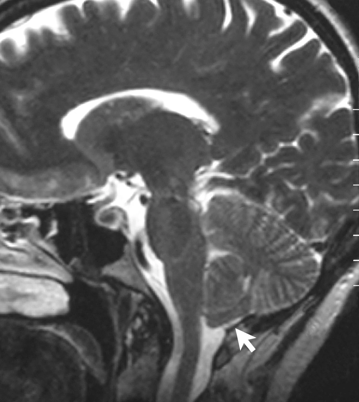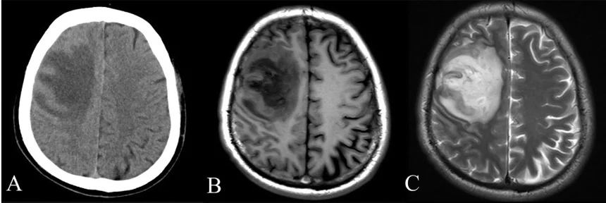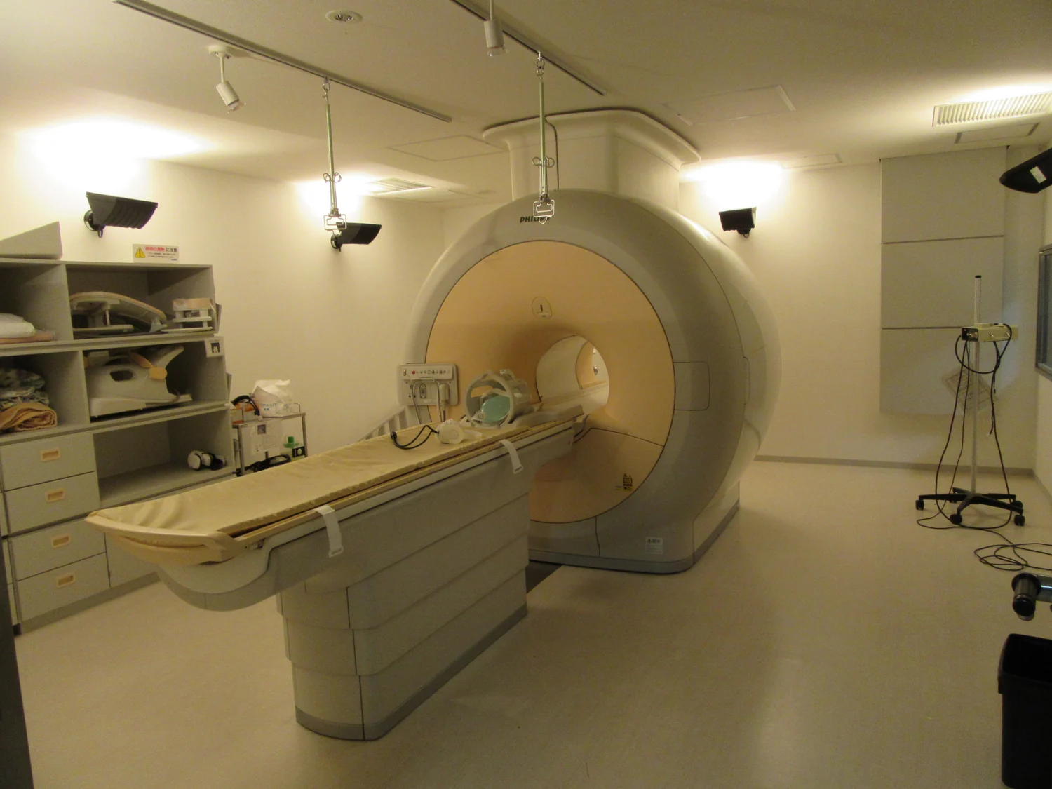
How can a scanner turn radio waves into a picture of a knee or brain? MRI does not take a photograph with radio waves; it perturbs tiny magnetic moments in the body and listens to how they respond. The main field prepares the protons, a radiofrequency pulse nudges them away from their preferred alignment, and sensors pick up signals as they settle back. Differences in that response help distinguish tissues.
The body supplies the signal source
Many hydrogen nuclei in the body are protons, and each behaves like a tiny magnet. Water is abundant in living tissue, so the scanner has a large population of these proton “compasses” to work with. In the strong static magnetic field of an MRI scanner, more protons align with the field than against it, creating a small net magnetization [1].
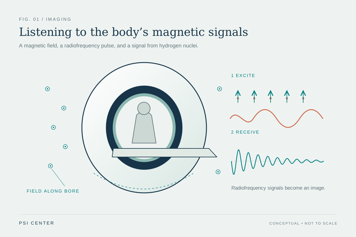
That alignment is not a rigid row of compass needles. The protons also precess, meaning their magnetic directions rotate around the main field at a characteristic rate. The scanner’s radiofrequency pulse is tuned to interact with them. It transfers energy and tips the net magnetization away from the field’s direction, disturbing the equilibrium [1].
Excitation: a carefully timed nudge
Think of a swing that moves most readily when pushed in time with its rhythm. A radiofrequency pulse at the appropriate frequency can similarly transfer energy to the proton system. During the pulse, the protons’ collective magnetization changes direction; when the pulse ends, the system is out of equilibrium and begins to relax toward alignment with the main field.
Radio waves are non-ionizing electromagnetic radiation: their individual photons do not carry the energy associated with x-rays that can ionize atoms. MRI uses magnetic fields and radiofrequency pulses, not plasma or ionizing x-rays [1] [2]. That distinction describes the imaging method; it does not mean every aspect of an MRI visit is risk-free, since the strong magnetic field creates important equipment and implant considerations.
Listening as the protons settle
As the disturbed magnetization changes, it induces a faint voltage in receiver coils. This is the signal the scanner detects. Protons in different molecular environments return toward equilibrium at different rates and release signal differently. The timing and strength of the received signal therefore carry information about the tissue’s local chemical and physical surroundings [1].
A simplified mental experiment: imagine two neighboring regions with different water environments. Give both the same radiofrequency nudge, then listen. If the signals fade or recover at different rates, the regions can have different image brightness under a chosen scan sequence. MRI does not identify tissue from one universal “signature”; pulse timing and signal processing shape which differences become visible.
From signal to image
The scanner also varies magnetic fields across space. These gradient fields make the precession frequency depend slightly on location, allowing the system to encode where signals came from. A computer combines the measured data into slices or three-dimensional images. Remaining still matters because motion can shift or blur the encoded signal, much as movement during a long exposure blurs a photograph.
MRI is especially useful for soft tissues, including brain, muscles, ligaments, and tendons, and can be used for detection, diagnosis, and monitoring [1]. The radiofrequency signal is only one part of the chain: the main magnet establishes alignment, the pulse creates a measurable response, gradients encode location, and reconstruction turns those measurements into anatomy. The scanner is, in effect, both an instrument that excites and one that listens.
Explore the physics: an explanatory diagram
Sources & further reading
- Magnetic Resonance Imaging NIBIB
- Electromagnetic Fields and Cancer National Cancer Institute
Prepared for the independent PSI Center reading library. About edition dates and source checking.
This explainer describes scientific principles. It does not assess individual treatment choices.