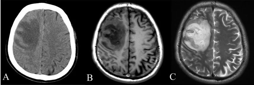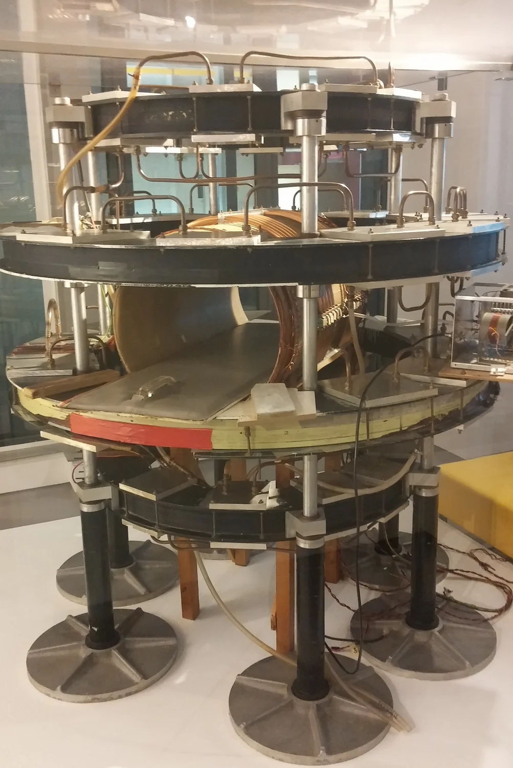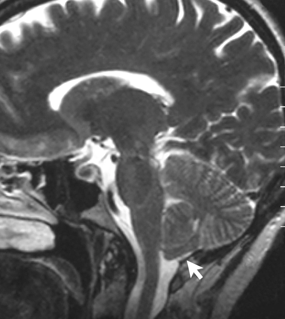
Why can a tiny movement blur a scan that uses no x-rays? MRI does not photograph the body in one instant. It gathers signals from protons in water as they respond to magnetic fields and radio waves, then uses those signals to construct an image. If the body shifts while measurements are being collected, the signals may no longer describe one consistent arrangement of tissue.
From proton response to signal
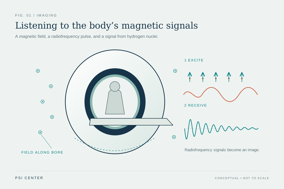
The MRI scanner’s powerful magnetic field encourages protons in the body to align with it. A radiofrequency pulse tips them away from that alignment. When the pulse stops, the protons move back toward the field direction and release energy that sensors detect. The timing and amount of that signal vary with the molecular environment, helping distinguish tissues. MRI uses magnetic fields and radio waves, not ionizing x-rays. [1]
The scanner does not simply capture a complete image at the moment the pulse is sent. Measurements are collected and combined to create the final image. Think of assembling a map from many carefully labeled points: if the map’s landmarks move while the points are recorded, the pieces may not fit together. In MRI, movement during acquisition can produce blur or other artifacts because the signal data correspond to changing positions. [1]
A moving target changes the measurements
A slow shift of the head, a swallow, or ordinary breathing can move tissue relative to the scanner. The signal then comes from a location that may have changed since earlier measurements. When the computer reconstructs the image under the assumption that the anatomy stayed in place, those inconsistent measurements can smear edges or duplicate details. The effect is especially consequential when the feature of interest is small or needs a crisp boundary.
Different kinds of motion create different problems. A brief sudden movement can disrupt part of the acquisition; repeated motion can create patterned artifacts. Some body regions move naturally, so scan design and patient positioning matter. NIBIB notes that people must remain very still during imaging to avoid blur, and describes research into faster imaging and motion compensation, including tracking approaches that adapt MRI pulses to changes in pose. [1]
Why the image is worth protecting
MRI is particularly useful for soft tissues such as the brain, spinal cord, muscles, ligaments, and tendons. A clear image can help clinicians see structures that ordinary x-rays depict less clearly. But image quality is not just a property of the magnet: it depends on signal collection, the patient’s position, and how the acquisition is designed. If motion blurs a region, repeating the scan may be needed to obtain usable information. [1]
Computational models can simulate complex systems by representing variables and calculating what happens under different conditions. In biomedical research, such models can help explore many simulated experiments and identify promising laboratory questions. That broader idea helps explain MRI reconstruction too: computers process measurements according to a mathematical model. A reconstruction can organize data, but it cannot make measurements from incompatible positions equivalent to a perfectly still body. [2]
Stillness is a design challenge, too
Holding still can be difficult for a child, someone in pain, or a person who finds the scanner confining. NIBIB describes research on pediatric coils designed for smaller bodies and on motion correction that could improve image quality for people unable to remain still. These are research efforts aimed at the challenge; they do not mean every scan already compensates fully for motion. [1]
A simple mental experiment is to imagine taking a long exposure of a moving bicycle: the camera records light over time, so the bicycle’s changing position becomes a streak. MRI’s signals are not visible light, but the analogy captures the central issue—measurements gathered across time are easiest to combine when anatomy remains in a consistent position. Stillness supports a sharper reconstruction because it keeps the physical source of the signal aligned with the image being built.
Explore the physics: an explanatory diagram
Sources & further reading
- Magnetic Resonance Imaging NIBIB
- Computational Modeling NIBIB
Prepared for the independent PSI Center reading library. About edition dates and source checking.
This explainer describes scientific principles. It does not assess individual treatment choices.