Why can one scan show a knee ligament clearly while another is better for watching blood move? Imaging methods do not simply take sharper or blurrier pictures of the same thing. Each sends a different kind of signal through the body and records how tissue changes it. Magnetic resonance imaging (MRI) listens to protons responding to magnetic fields and radio waves; ultrasound maps echoes from sound waves. Their physics makes some tissues easier to see than others.
MRI listens to water’s magnetic response
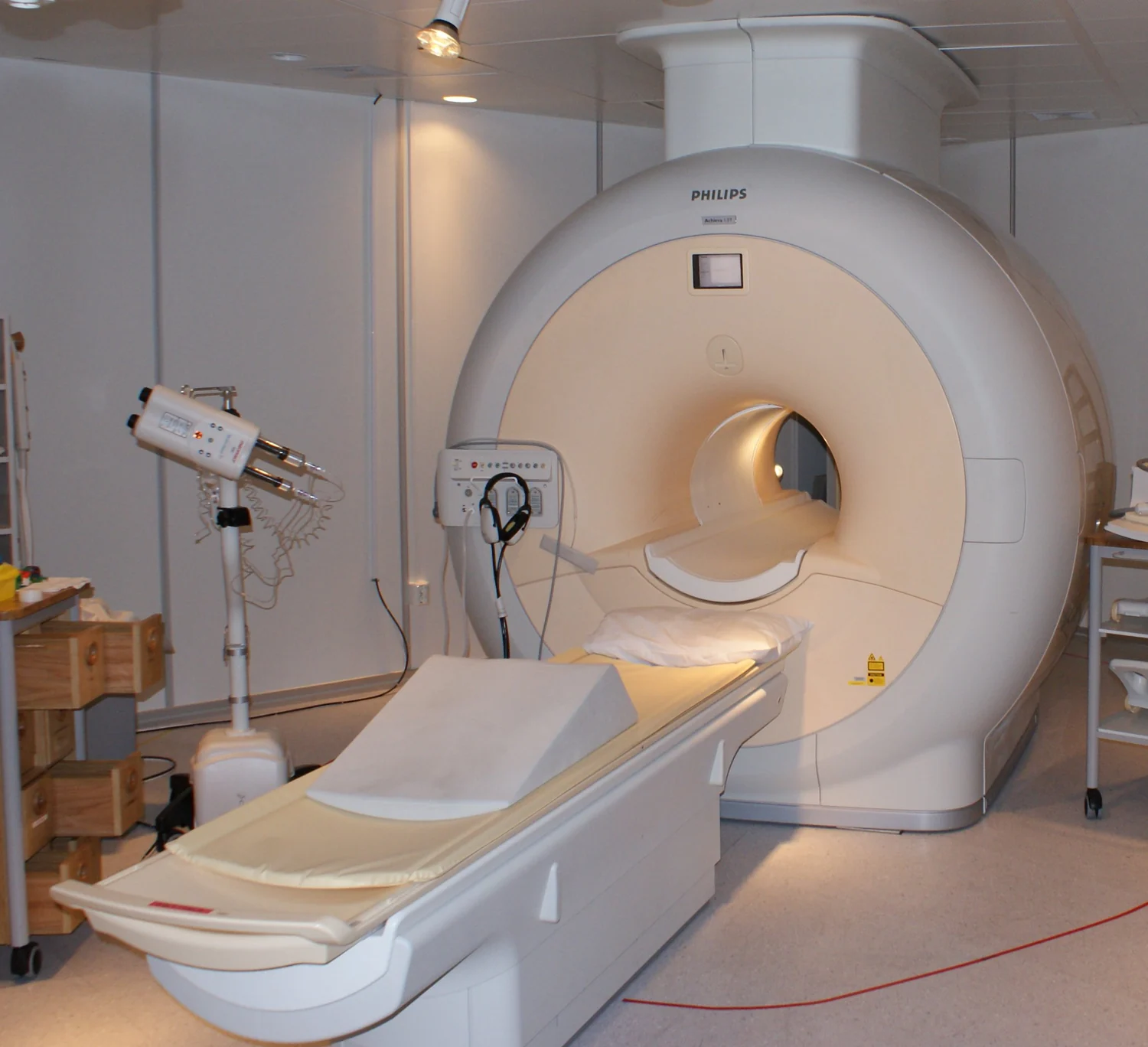
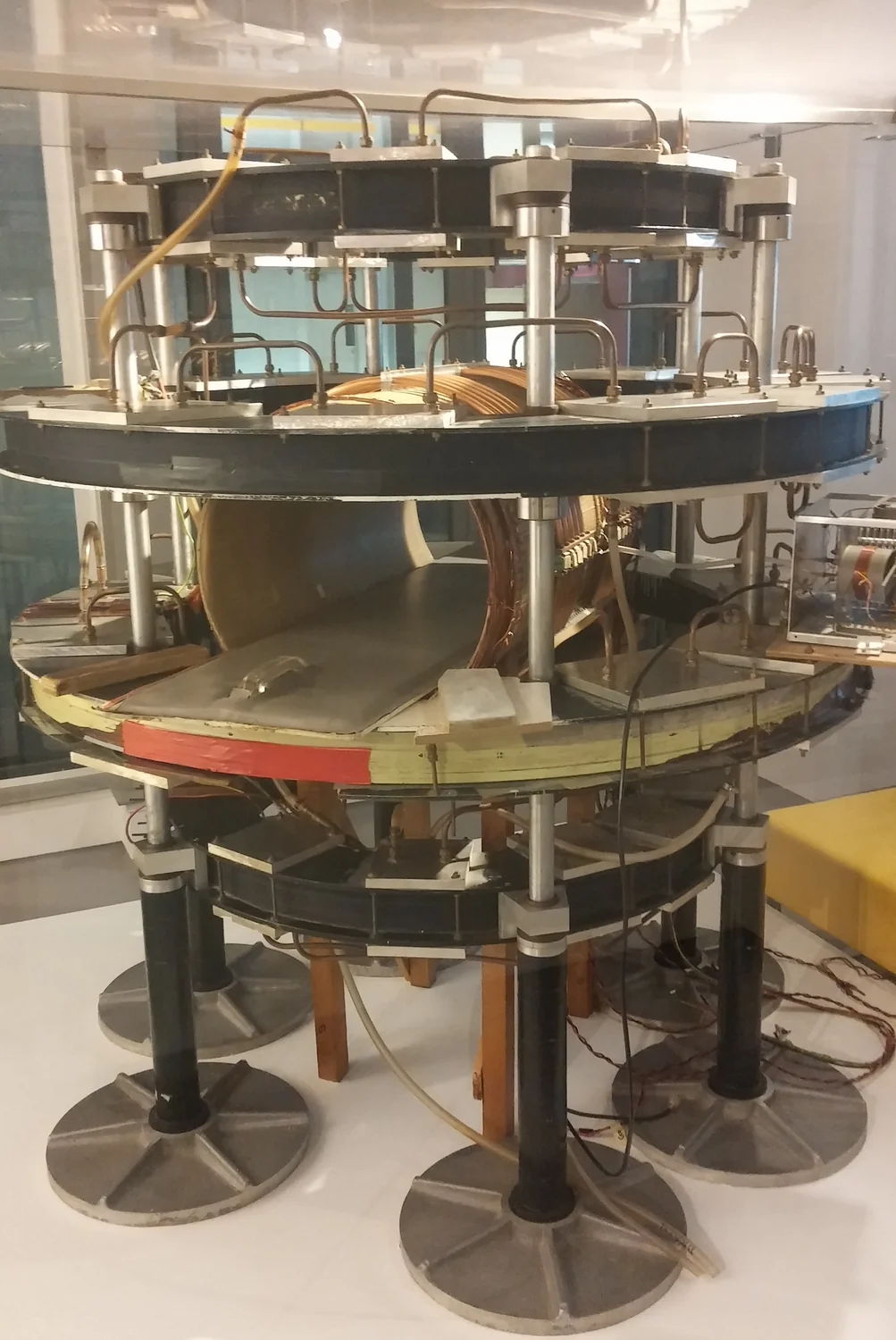
The body contains abundant water, whose hydrogen nuclei are protons. In MRI, a strong magnetic field nudges many proton spins into alignment. A radiofrequency pulse disturbs that alignment; after the pulse ends, protons return toward their prior state and release detectable energy. The timing and strength of that signal vary with the protons’ molecular surroundings, allowing image contrast between tissues [1]. MRI therefore uses magnetic fields and radio waves, not plasma or ionizing x-rays.
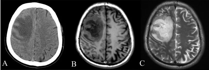
This sensitivity to soft-tissue environments helps explain MRI’s usefulness for structures such as the brain, spinal cord, muscles, ligaments, and tendons. Imagine comparing a knee’s soft ligament with nearby bone: a conventional x-ray is excellent for showing bone structure, while MRI can distinguish soft tissues more clearly. The best method depends on the question, not a single universal scale of image quality [1].
Ultrasound builds a map from echoes
Ultrasound sends high-frequency sound into the body and detects echoes that return from boundaries between materials. A transducer, the probe held against the skin, both emits sound and senses returning vibrations. The scanner estimates depth from how long each echo takes to return, then combines many measurements into an image [2]. Gel helps sound pass from probe to skin by reducing air gaps, since air can block transmission.
A familiar mental picture is calling out in a room and using the echo to estimate where a wall sits. In the body, boundaries between fluid and soft tissue or tissue and bone reflect sound differently. The method can display anatomy, and Doppler ultrasound uses changes in echoes to show blood-flow direction and speed. It can also guide a needle or catheter in real time, which is useful when seeing a moving instrument matters as much as seeing the target [2].
The path through the body changes the answer
Sound does not travel equally well through every material. Ultrasound is limited for imaging bone and air-containing tissues such as lungs, because these strongly impede or reflect the sound path. Under some conditions, fluid around a structure can create an acoustic window. In contrast, MRI is particularly suited to many soft tissues, although it requires a powerful magnet and patients generally need to remain still while signals are collected [2] [1].
MRI’s magnetic field also introduces practical considerations that have no direct equivalent in ordinary ultrasound scanning. Ferromagnetic objects can be pulled forcefully toward the magnet, so implants and metal objects require careful assessment before a scan. MRI can be noisy and long, and some people find the enclosed scanner difficult to tolerate. Ultrasound avoids ionizing radiation too, but it is not simply a universal substitute: the target, access route, and needed information determine which tool fits.
Images answer different questions
Consider a clinician trying to assess a suspected tendon injury. MRI can provide detailed soft-tissue anatomy; ultrasound may offer a view while the joint moves, showing changing relationships in real time. For a blood vessel, Doppler ultrasound can map flow. MRI can instead provide detailed anatomy of surrounding structures. Neither result automatically answers every clinical question, and imaging findings must be interpreted in context [1] [2].
The distinction also prevents a common mix-up: MRI does not use x-rays, while ultrasound uses sound rather than light or magnetic resonance. Both methods can be used diagnostically, but their physical signals create different tradeoffs in access, contrast, motion, and detail. Choosing between them is less like selecting the best camera and more like choosing whether to listen for an echo or measure how water molecules respond to a magnetic field.
Sources & further reading
- Magnetic Resonance Imaging NIBIB
- Ultrasound NIBIB
Prepared for the independent PSI Center reading library. About edition dates and source checking.
This explainer describes scientific principles. It does not assess individual treatment choices.