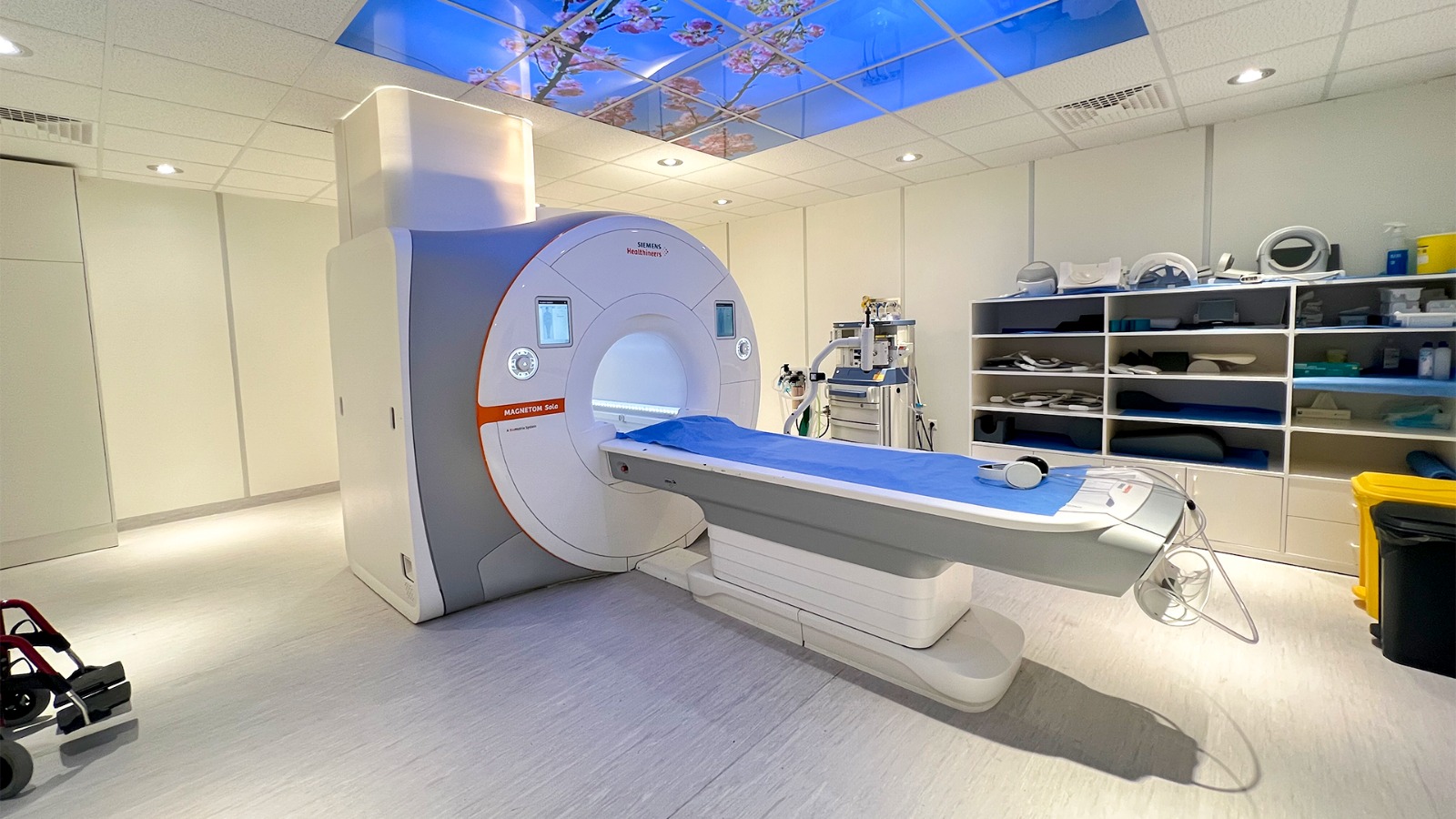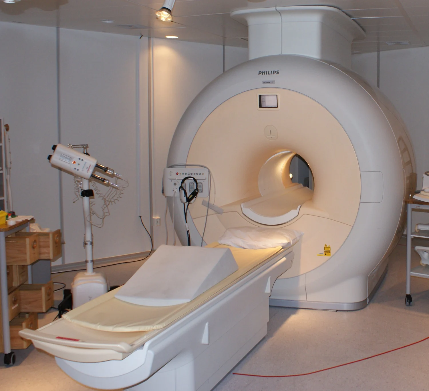
Why put a person inside a powerful magnet to make an image? MRI takes advantage of a tiny magnetic property carried by many hydrogen nuclei in the body’s abundant water. A strong field nudges their orientations into a more organized state; radio waves then disturb that alignment, and sensors listen as the nuclei respond. The signal depends on the surrounding molecular environment, giving the scanner information that can distinguish soft tissues.
Hydrogen nuclei respond to the field
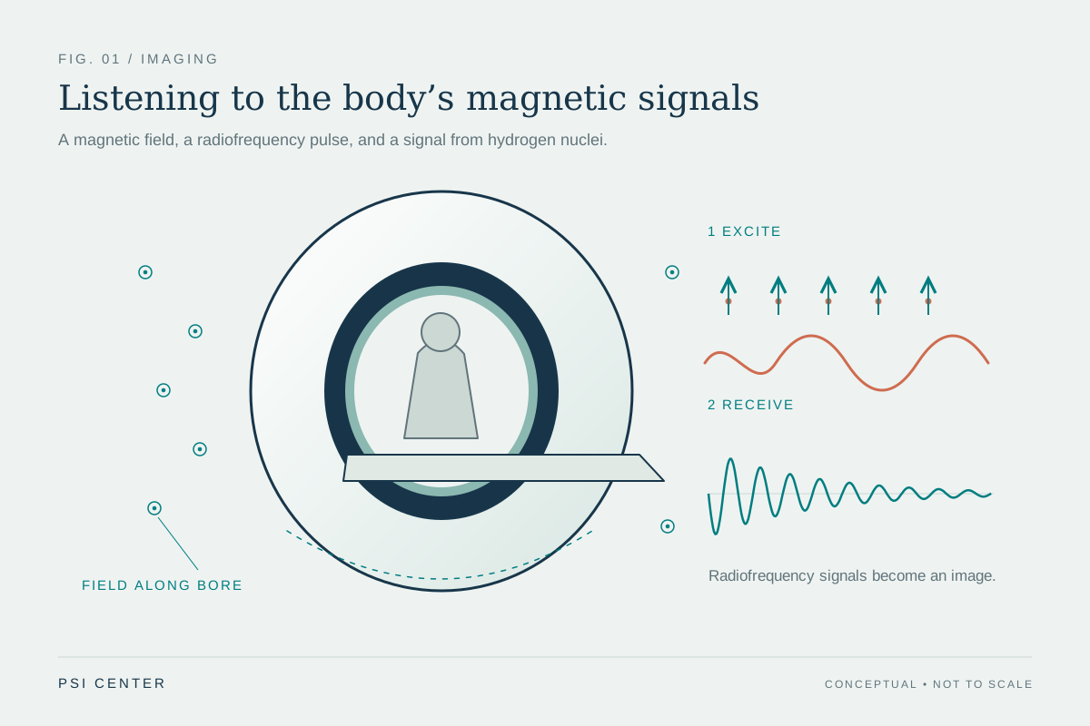
A hydrogen atom’s nucleus is a proton, and protons have a property called spin that gives them a tiny magnetic moment. In the absence of a strong external field, these moments point in many directions. Inside an MRI scanner, the strong magnetic field makes their orientations preferentially align with it, creating a small net magnetization in the body’s tissues [1].
The alignment is not a row of miniature bar magnets locking rigidly into place. Rather, the field changes the balance of possible orientations, and the protons collectively produce a measurable effect. Hydrogen is useful because water is abundant in living tissue. The scanner is therefore listening to a response from many protons, not photographing individual atoms.
Radio waves tip the alignment
A radiofrequency pulse supplies energy at a frequency that interacts with the protons in the magnetic field. This pulse tips the net magnetization away from its preferred alignment. When the pulse stops, the protons move back toward equilibrium and release energy that receiver coils can detect [1]. That returning signal is the raw material from which the scanner constructs an image.
Different tissues produce signals that vary because the protons’ surroundings differ. The time it takes for them to realign and the amount of signal they release depend on the molecular environment. By collecting signals in a carefully controlled sequence, the scanner maps these differences into image contrast. In this way, MRI can distinguish many soft tissues that look similar on a conventional X-ray image.
Gradients provide position information
The main magnet organizes the spins, but the scanner also uses magnetic fields that vary slightly across space, called gradients. These gradients make the proton response depend on location. The scanner can then separate signal from one region from signal elsewhere and assemble a spatial image. Repeating the process with different pulse timings emphasizes different tissue properties.
The need for stillness follows from this spatial encoding. If the body moves while signals are being collected, the scanner may assign returning signal to the wrong position, blurring the image. MRI is therefore a measurement process that combines magnetic fields, radiofrequency energy, timing, and computation. The large magnet is essential, but it does not create a picture by itself.
Magnetic strength brings safety considerations
MRI does not use ionizing X-rays, unlike CT, but that does not mean the environment has no hazards. The static magnetic field can pull on ferromagnetic objects, while changing fields can cause loud knocking sounds and sometimes nerve or muscle stimulation. Radiofrequency energy can heat the body, particularly during longer examinations [2]. Screening of people, implants, and objects is therefore part of safe scanning.
Implanted devices need particular attention because magnetic forces, heating, or interference can affect a device or surrounding tissue. The FDA distinguishes devices that are MR Safe from those that are MR Conditional, which may be used only under specified conditions [2]. MRI’s strong magnet is not a decorative feature: it creates the physical conditions that make the signal measurable, while careful control of the environment allows that measurement to be used for imaging.
Explore the physics: an explanatory diagram
Sources & further reading
Prepared for the independent PSI Center reading library. About edition dates and source checking.
This explainer describes scientific principles. It does not assess individual treatment choices.