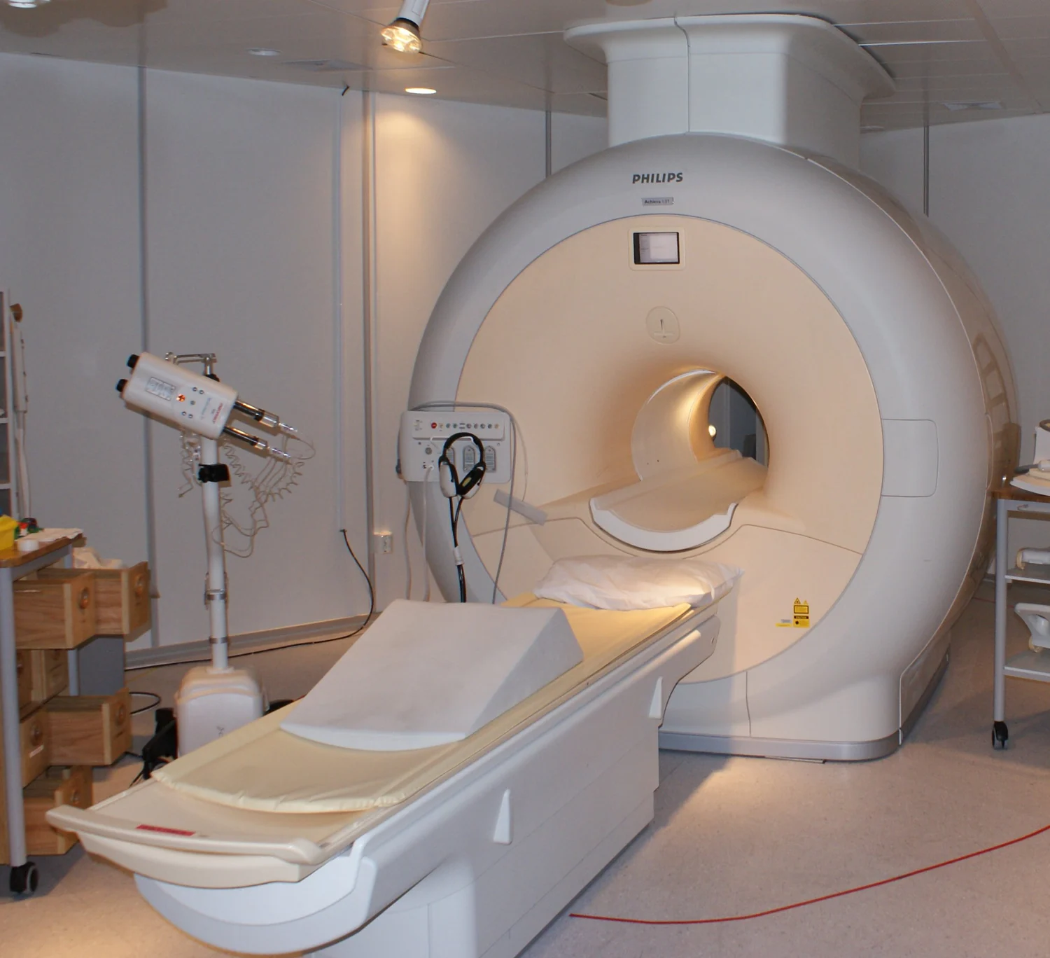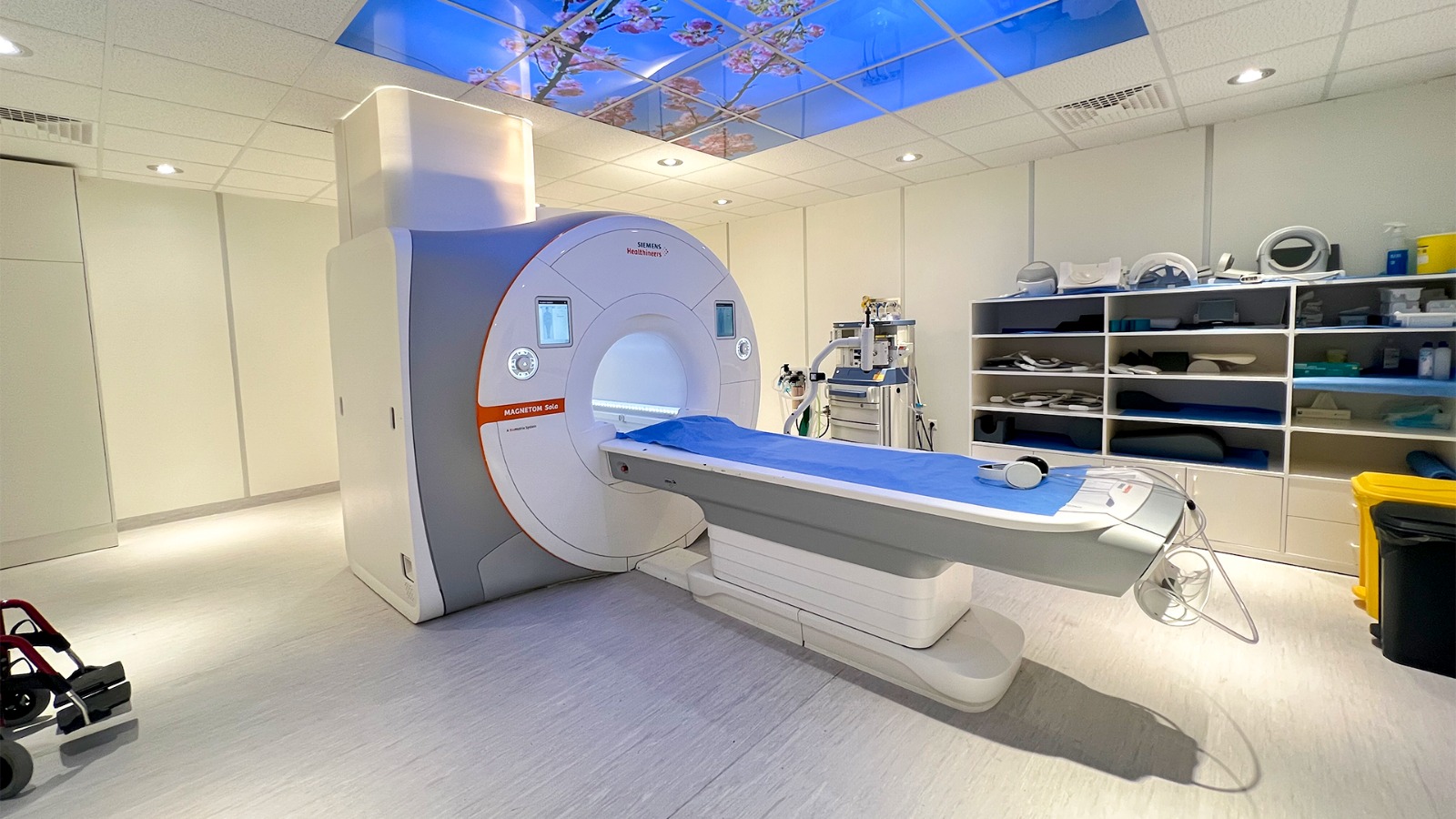
How can a scanner make a detailed picture from hydrogen atoms in your body? It does not photograph them or send x-rays through you. Instead, MRI uses a strong magnetic field and radiofrequency pulses to disturb and detect the behavior of hydrogen nuclei, especially abundant in water. Their signals vary with the molecular surroundings, and the scanner encodes where those differences came from. The image is a map reconstructed from this coordinated physical response.
Hydrogen as a tiny magnetic signal
A hydrogen nucleus is a proton. Protons have a property called spin, which gives them a small magnetic character. In the scanner’s strong magnetic field, many proton magnetic axes tend to align with the field, creating a net signal. They are not little planets literally spinning like tops, but the spinning-top picture can help suggest why their orientation and response matter.
The signal becomes measurable because tissue contains abundant hydrogen, especially in water and fat. MRI is based on changing the direction of proton rotational axes in living tissue and detecting the response [1]. Different tissues do not contain identical molecular environments, so their hydrogen signals will not evolve in exactly the same way.
A pulse tips the balance
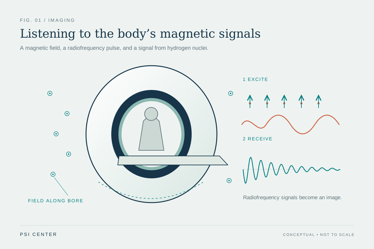
The scanner sends a pulse of radiofrequency energy at the protons. This pulse moves the net magnetization away from alignment with the main magnetic field. When the pulse ends, the protons return toward their prior arrangement and release energy that receiver coils can detect [1]. Radio waves here are non-ionizing; MRI does not use x-ray radiation to make its images [1] [2].
Consider two groups of protons, one in a watery environment and one surrounded by different molecules. After the same pulse, the groups recover at different rates and produce distinguishable signals. The recovery and signal strength depend on the environment and chemical nature of the molecules, providing contrast between tissue types [1]. The scanner can adjust pulse timing to emphasize different aspects of that contrast.
From signal to location
A receiver coil hears the combined signal from a region, but an image requires knowing where it arose. MRI scanners briefly vary magnetic fields across space, called gradients. Because proton resonance depends on magnetic field strength, these gradients make signals from different positions carry distinguishable frequencies or phases. Computer processing then reconstructs a map from the encoded measurements.
This is why staying still matters: motion changes the location of signal while the scanner is assembling its map, smearing boundaries. MRI can image in different directions and provides strong soft-tissue contrast; it often reveals structures such as muscles, ligaments, brain tissue, and nerves more clearly than standard x-rays [1] [2]. It is not universally best for every question: CT is usually better at depicting bone [2].
A powerful field demands care
MRI avoids ionizing radiation, but it is not an environment without physical effects. The static magnetic field can pull on ferromagnetic objects; changing fields can produce loud knocking and sometimes nerve or muscle stimulation; radiofrequency energy can heat tissue [2]. These arise from different parts of the scanner’s operation, so safety involves controlling objects, sound exposure, and scan conditions.
Medical devices and implants need particular attention because magnetic forces, heating, or interference can affect both device function and nearby tissue. FDA guidance says implanted devices should be identified as MR Safe or MR Conditional before scanning; conditional devices are safe only under stated conditions [2]. MRI’s signal-making process is elegant precisely because it is sensitive to magnetic properties, and that sensitivity shapes both the image and the environment in which it is acquired.
Explore the physics: an explanatory diagram
Sources & further reading
Prepared for the independent PSI Center reading library. About edition dates and source checking.
This explainer describes scientific principles. It does not assess individual treatment choices.