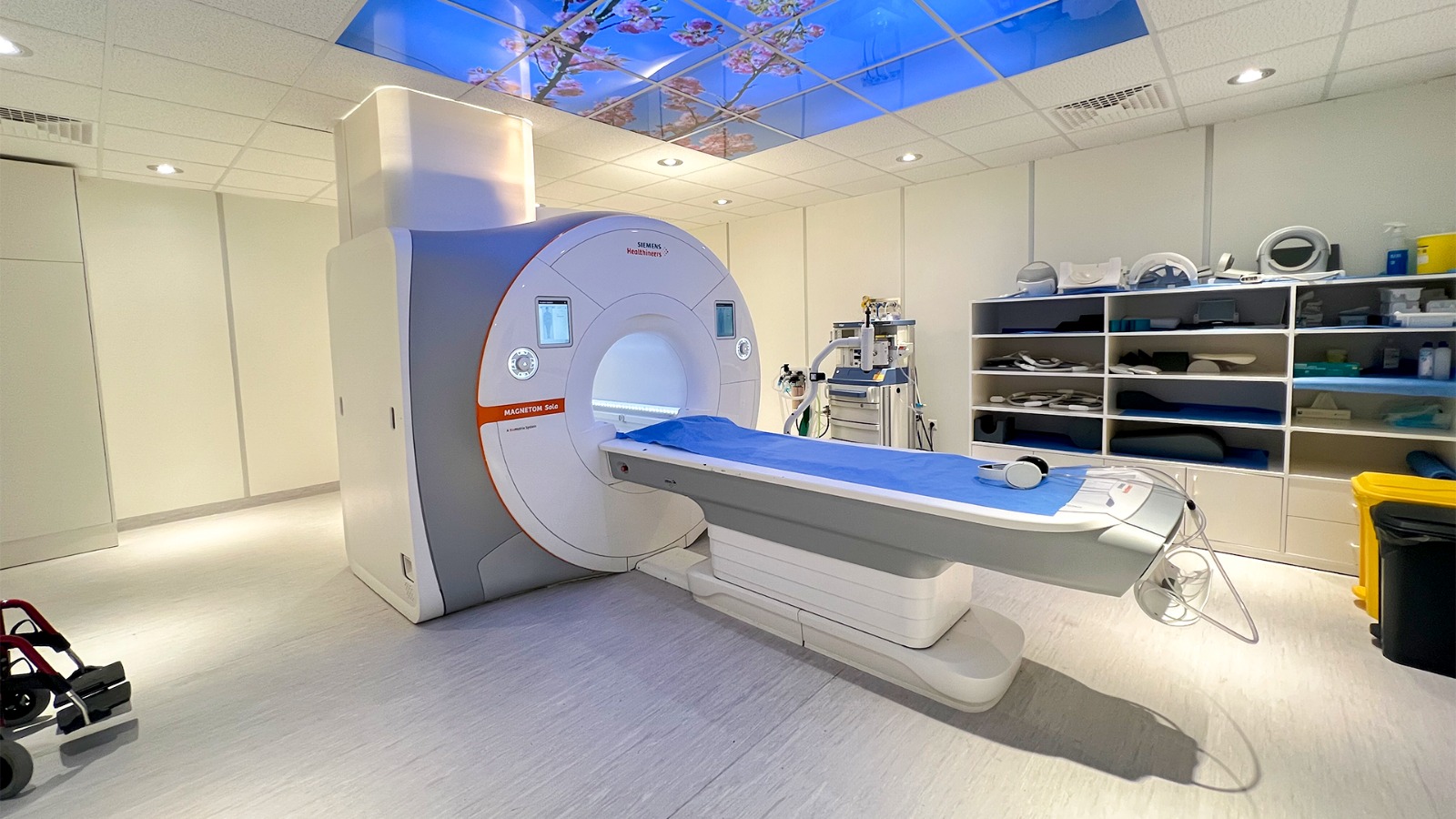
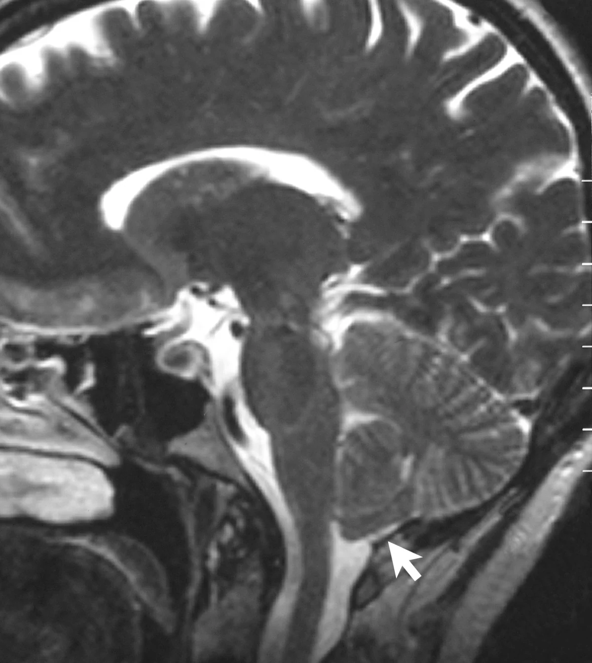
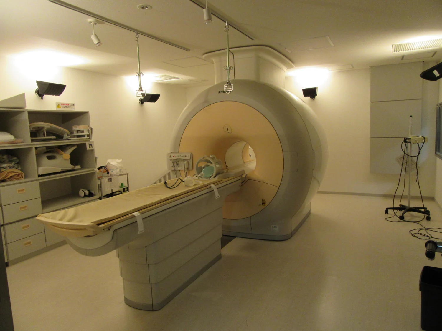
Why can a machine that images the body with magnets sound like a construction site? The noise comes from magnetic fields that switch rapidly while the scanner gathers information. Those switches make parts of the machine vibrate, producing loud knocks and clicks. The sound is a side effect of image-making physics: MRI uses changing fields to encode where signals come from, then listens for radio waves released by hydrogen nuclei in tissue.
A magnetic compass inside the body
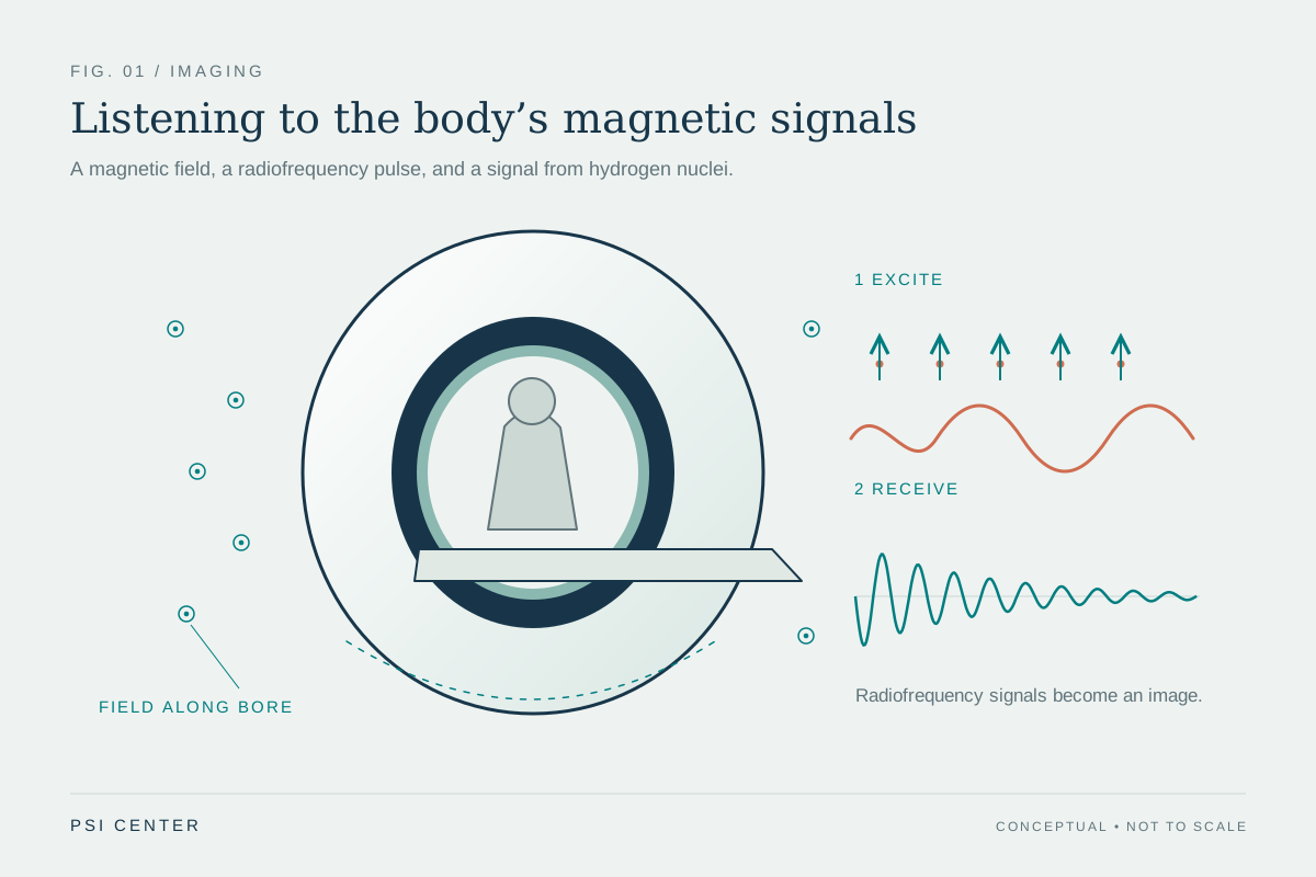
MRI begins with hydrogen, abundant in the water and fat of living tissue. Each hydrogen nucleus has a property called spin, which makes it behave a little like a tiny magnet. In the scanner’s strong static magnetic field, some of these tiny magnets line up more with the field than against it. A radiofrequency pulse nudges them away from that alignment; as they relax back, they release energy that the scanner detects. Differences in this response help distinguish tissues [1].
That signal alone does not say where it came from. The scanner therefore adds controlled changes to the magnetic field across the body. These gradients make hydrogen nuclei at different positions respond at slightly different frequencies or phases, like members of an orchestra playing the same note with staggered timing. The scanner can use those differences to reconstruct a map. MRI uses magnetic fields and radio waves, not plasma or ionizing x-rays [1].
Why a changing field makes a knock
The gradient fields are made by electrical currents in coils. When current changes, it creates a changing magnetic field; that field exerts forces on the coils and nearby structures. The forces arrive in repeated pulses, making components vibrate against the scanner’s frame. The patient hears the resulting knocking, buzzing, or clicking. Different pulse patterns create different rhythms, so a scan can sound like a sequence of mechanical clacks rather than one steady hum.
A useful mental picture is a speaker cone. An electrical signal makes the cone move back and forth, pushing air and producing sound. MRI gradients do not work exactly like a speaker, but repeated electromagnetic forces make scanner parts move and transmit vibrations through the surrounding structure. The noise is thus not the body’s protons audibly spinning; it is machinery responding to the timed fields that help form the image.
The trade between information and sound
Changing gradients quickly can help encode spatial information efficiently, but fast changes can also create stronger or more noticeable vibration. Each MRI sequence uses a planned pattern of radiofrequency pulses and gradients suited to the contrast or detail being sought. That is why the sound may shift during an exam. MRI images can show soft tissues with strong contrast, and the scanner can collect views in different directions; CT, by comparison, is often better at depicting bone [2].
The scanner must also receive faint signals while the patient stays still. Motion can blur the image because the signal is being assigned to locations based on the scan’s timing. In practice, the loud sound and the need for stillness are linked to how the machine gathers data over time. Ear protection is used because changing fields can produce loud knocking that may harm hearing without adequate protection [2].
Sound is one part of the scan environment
The noise is conspicuous, but it is only one feature of the MR environment. A powerful static field attracts some magnetic objects, so people and equipment are screened before entering the scanner area. Time-varying fields can also cause a twitching sensation, and radiofrequency energy can heat the body. Implants and external devices require special attention because fields may move, heat, or disrupt some devices; safety depends on identifying the device and the conditions under which it can be scanned [2].
The loud knocks therefore tell a small story about a complicated measurement: the scanner changes fields in carefully timed ways, those changes help locate tissue signals, and the forces involved make hardware vibrate. The sound is not evidence that the machine is using x-rays or that anything is striking the patient. It is the audible footprint of magnetic-field gradients doing their work.
Explore the physics: an explanatory diagram
Sources & further reading
Prepared for the independent PSI Center reading library. About edition dates and source checking.
This explainer describes scientific principles. It does not assess individual treatment choices.