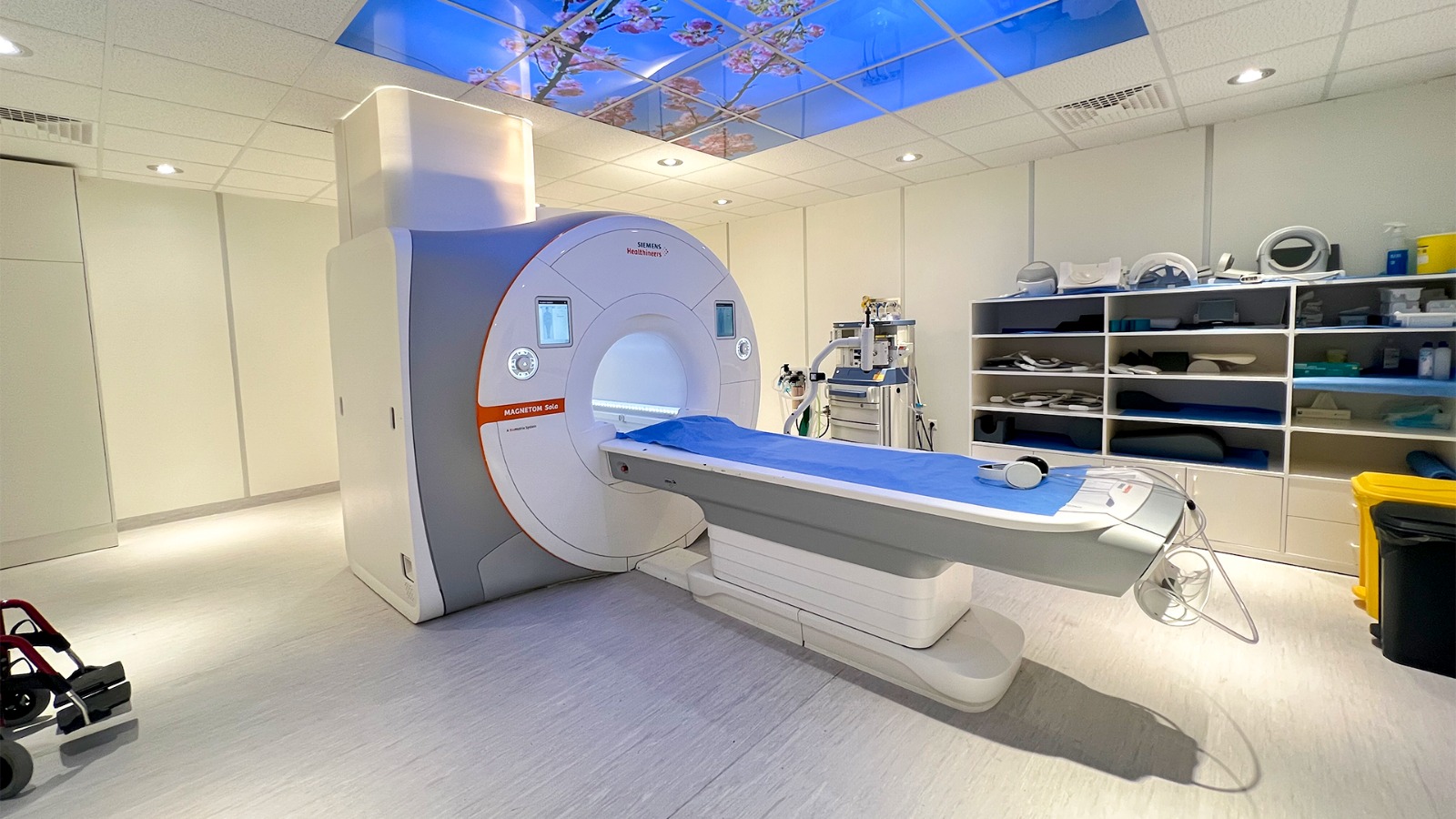
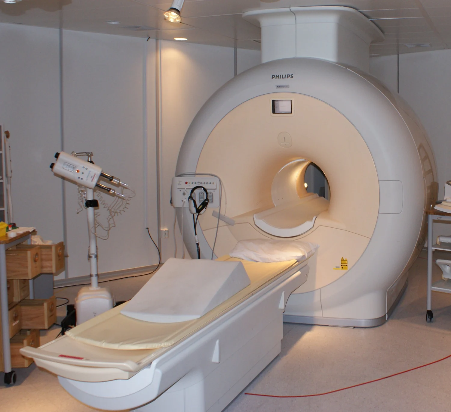
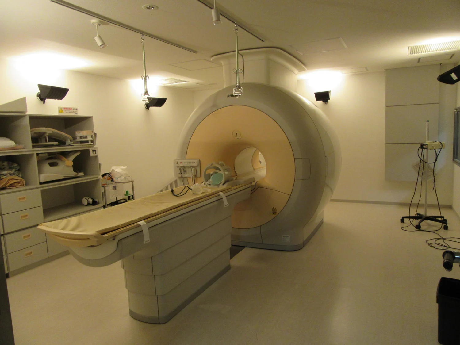
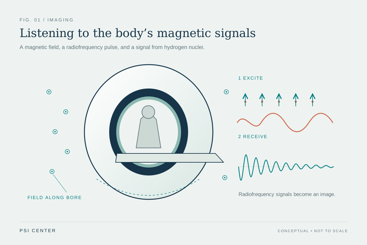
Why does MRI preparation begin before anyone enters the scanner room? An MRI makes images with magnetic fields and radio waves, not ionizing x-rays. That avoids one kind of exposure, but creates a different environment: a powerful magnet is always present, while changing fields and radiofrequency energy operate during imaging. Screening helps staff understand what people and devices may safely encounter those conditions.
A magnet that never switches off
The most vivid MRI hazard is the static magnetic field. It can attract ferromagnetic objects—materials strongly drawn to magnets—and accelerate them toward the scanner. A loose tool or oxygen tank can become a projectile, so screening covers people and objects before they approach the magnet. The key distinction is that the scanner’s magnetic field remains present even between image-taking sequences; pausing the scan does not turn the magnet into an ordinary room.
The magnetic fields that change during imaging create other effects. Their rapid changes produce the loud knocking sounds patients hear, which is why hearing protection matters. They can also stimulate peripheral nerves or muscles, sometimes felt as a twitch. Radiofrequency energy can warm tissue, with potential heating increasing during longer examinations. These are separate physical mechanisms, so one precaution does not address every risk. [1]
Why a device’s exact identity matters
A metal implant is not automatically safe or unsafe simply because it contains metal. Some devices are labeled MR Safe, meaning they pose no known hazard in any MR environment; others are MR Conditional, meaning safe use depends on specified scanner conditions. An unidentified device should be treated as MR Unsafe until its status is established. Magnetic force can move a device, while radiofrequency energy or changing fields may heat it and nearby tissue, or interfere with electrically active devices. [1]
That is why staff need details about implants, external equipment, and accessories rather than a vague description such as “I have a medical device.” A pacemaker, insulin pump, brace, wound dressing, or monitor presents a different question about materials, function, and position. The device itself can also distort the image, potentially making it less useful. Screening connects the physical properties of the equipment to both safety and image quality.
Small magnets can affect implanted electronics
The MRI’s field is not the only magnetic field worth recognizing. Some phones and smart watches contain high-strength magnets. Close to certain implanted heart devices, a magnet may trigger a programmed “magnet mode,” temporarily changing normal operation. A defibrillator, for example, might not detect a dangerous fast rhythm while that mode is active. The FDA advises keeping potentially interfering electronics at least six inches from implanted devices and avoiding carrying them in a pocket over the implant. [2]
This example helps explain why distance and context matter. A magnet can be intentionally used by clinicians to activate a device’s magnet mode for a medical procedure; removal of the field lets the device return to normal operation. A consumer device placed close to the implant can unintentionally create a similar interaction. The FDA describes the risk as low and reports no known adverse events from this issue, while recommending the simple separation precaution.
Preparation is part of image making
MRI also asks patients to remain still so that the scanner can collect clear signals from the intended anatomy. Some people find the enclosed space uncomfortable. An open scanner may be an option for some examinations, though not all systems can perform every scan. Contrast agents, when used, have their own potential side effects, and sedation or anesthesia for people unable to lie still carries risks of its own. These considerations are part of choosing and planning an examination, not consequences of the magnet alone. [1]
The practical sequence is therefore deliberate: identify implants and devices, control what enters the magnet area, plan around the scanner’s conditions, and help the patient remain comfortable and still. MRI’s detailed soft-tissue images can be valuable across many body regions, but those images depend on a carefully controlled environment. Preparation is the way the physics and the clinical task are made to fit together.
Explore the physics: an explanatory diagram
Sources & further reading
Prepared for the independent PSI Center reading library. About edition dates and source checking.
This explainer describes scientific principles. It does not assess individual treatment choices.