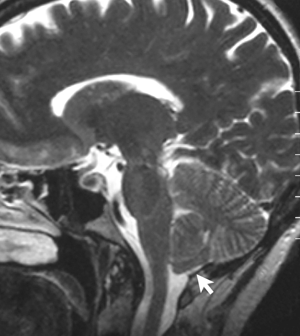
What does a bright patch on a scan mean? By itself, it means that the scanner recorded a particular signal in a particular place. Turning that pattern into a diagnosis takes another kind of reasoning: connecting the image with a person’s symptoms, history, examination, and other evidence. A scan can be crucial, but it is not a self-explanatory picture of disease.
A scan is a measurement translated into a picture
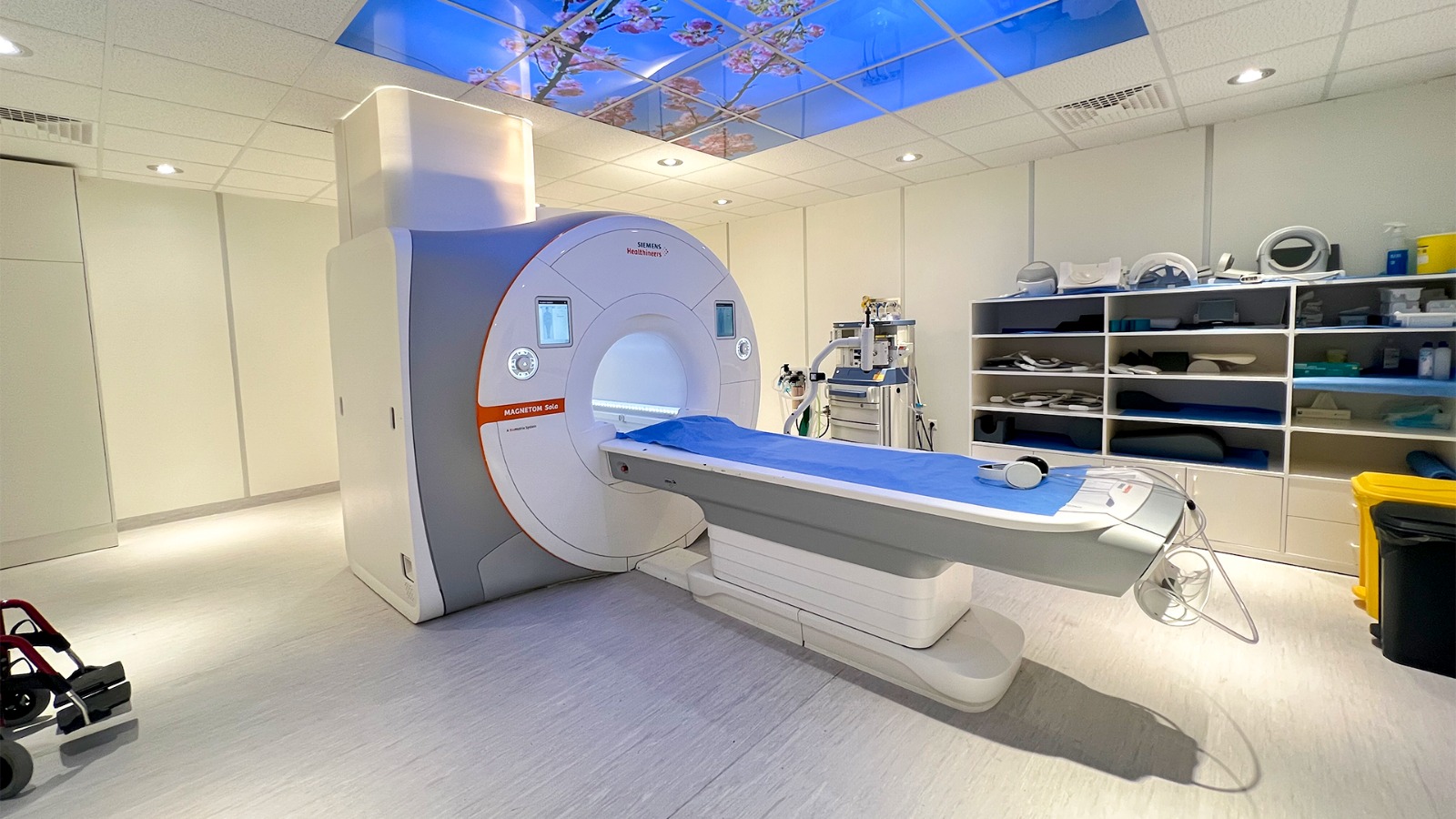
Medical images are constructed from measurements. In magnetic resonance imaging (MRI), magnetic fields and radio waves generate signals from tissues; software processes those signals into image contrast. MRI does not use plasma or ionizing x-rays. The resulting shades and shapes help distinguish structures, but each shade reflects the measurement method and settings as well as the tissue itself.
Consider a small bright area in a brain image. It could reflect several kinds of tissue change, and its appearance may vary with the MRI sequence. The image locates a feature and describes how it appears; it does not automatically tell the reader why it is there or whether it explains a symptom. That second question requires interpretation.
Diagnosis joins several kinds of evidence
A clinician may compare the scan with the patient’s account, examination findings, earlier images, laboratory results, and the pattern of symptoms over time. The same imaging feature can matter differently depending on those details. A finding that fits the location and timing of a symptom may support one explanation; an incidental finding may be unrelated.
This is an example of inference: a conclusion drawn by weighing evidence rather than simply observing one measurement. Computational models can help organize complex information or simulate how systems behave, but model outputs depend on their assumptions and inputs [2]. A model or image can guide a question without settling it on its own.
What image-reading AI actually learns
Machine-learning systems identify patterns in examples and use those patterns to make predictions. For medical imaging, development requires collections of images that have been curated, checked for quality, de-identified, and annotated by clinical experts [1]. An algorithm trained to flag a pattern is learning relationships in those labeled examples; it is not necessarily reasoning about disease in the same way a clinician does.
An AI tool may draw attention to a subtle feature or estimate how closely an image resembles cases in its training data. That can support review, but performance depends on the data and setting. If relevant patient groups or image conditions are poorly represented, predictions may not transfer reliably. Researchers also study explainable AI methods that make features behind some model outputs more inspectable [1].
A useful finding still needs a clinical question
Imagine two people with an identical-looking scan feature. One has a new symptom that matches its location; the other feels well and had the scan for an unrelated reason. The pixels may match, yet the clinical significance can differ. The next step is to ask what explanations fit each person’s full evidence and whether further evaluation is needed.
This distinction also explains why a normal image is not a universal guarantee of health. A scan examines particular structures with particular methods and limits; it cannot answer every clinical question. Images are powerful because they make hidden anatomy measurable. Diagnoses emerge when those measurements are interpreted in context, with uncertainty made explicit.
Sources & further reading
- Artificial Intelligence NIBIB
- Computational Modeling NIBIB
Prepared for the independent PSI Center reading library. About edition dates and source checking.
This explainer describes scientific principles. It does not assess individual treatment choices.