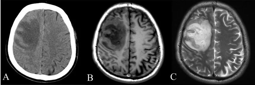
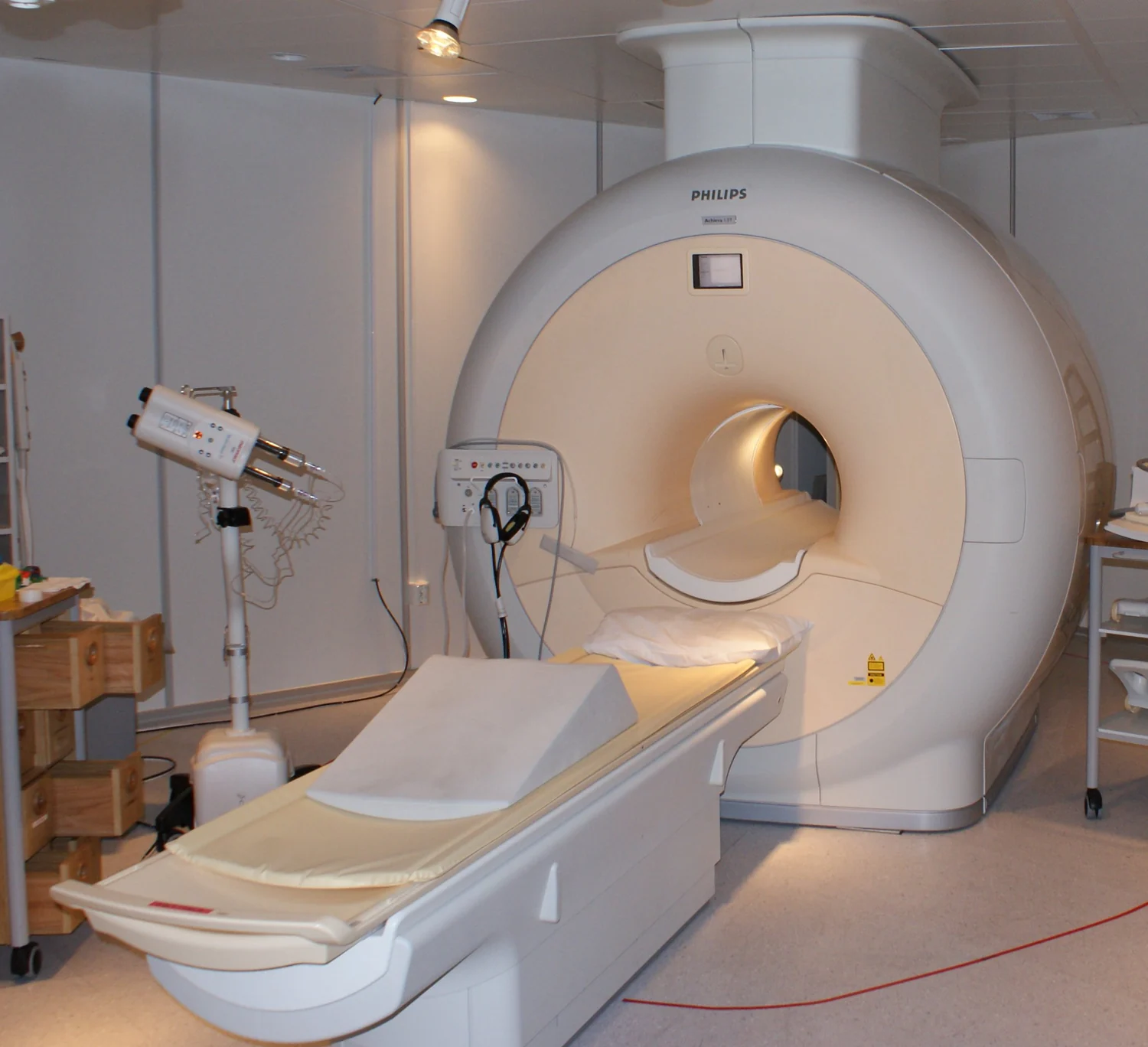
What can light reveal about tissue that a magnetic resonance image does not, and what can MRI show that light cannot? The two methods interact with matter in different ways. MRI uses magnetic fields and radio waves to produce detailed anatomical images; optical imaging follows how light travels through or emerges from tissue. Their strengths overlap in places, but they are not interchangeable.
MRI listens to protons
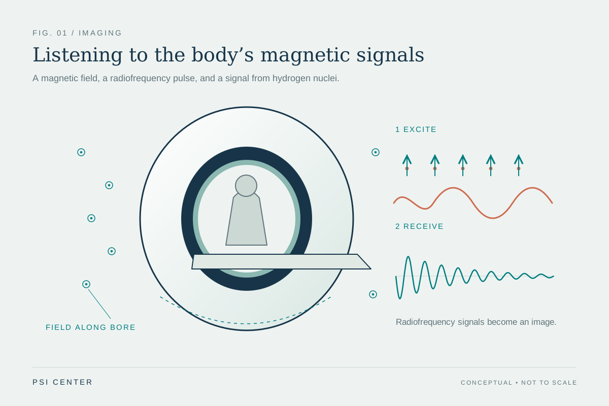
MRI begins with the hydrogen protons abundant in the water of living tissue. A strong magnetic field encourages these protons to align; a radiofrequency pulse then shifts them away from equilibrium. When the pulse ends, sensors detect energy released as the protons realign. The timing and amount of that signal depend on the molecular surroundings, allowing tissues to be distinguished [2].
This is why MRI is useful for detailed views of soft tissues such as the brain, spinal cord, muscles, ligaments, and tendons [2]. The scanner uses magnetic fields and radio waves, not plasma or ionizing x-rays. Its signal reflects magnetic properties of tissue rather than the way light is absorbed or scattered. Stillness matters because motion during data collection can blur the resulting image.
Optical imaging follows light
Optical methods exploit the ways light interacts with biological material: it can be absorbed, scattered, reflected, or emitted [1]. Those interactions carry information about tissue, from small structures seen under a microscope to properties measured across larger regions. Different methods answer different questions; optical coherence tomography uses infrared light for cross-sectional views of structures such as the retina, while diffuse optical methods can estimate tissue hemoglobin concentration and oxygen saturation [1].
A useful mental image is to compare shining a beam through fog with listening for a returning echo. The fog changes how much light gets through and in which directions it scatters. Optical imaging measures those changes; MRI instead detects how protons respond to magnetic fields and radiofrequency excitation. Optical imaging can be repeated to monitor changes, and its many forms can measure multiple soft-tissue properties [1].
Different views, one problem
Suppose researchers want to study a complex tissue. MRI can provide detailed anatomical context, while optical imaging may add information about light-sensitive properties such as blood oxygenation or hemoglobin. Combining methods can therefore add dimensions to an investigation rather than simply producing two versions of the same picture. The NIBIB notes that optical imaging can be combined with MRI to provide enhanced information in complex disease monitoring and research [1].
The complement is practical as well as physical. MRI is suited to soft-tissue anatomy, while optical techniques range across microscopic and larger scales, and some can be used in minimally invasive or non-invasive ways [2] [1]. A modality’s value depends on what a team needs to see, the depth and scale of the target, and how the measurements can be interpreted together. More images do not automatically mean a clearer answer unless each adds relevant information.
Interpretation sets the limits
Neither method reveals every property of a living body. MRI signal depends on tissue magnetic environments and may be enhanced with contrast agents in some examinations [2]. Optical measurements depend on how light travels through the particular tissues and on the chosen technique [1]. Each instrument transforms a physical interaction into data, and each data set needs interpretation in light of the question being asked.
The strongest combined approach is one in which the methods answer distinct, complementary questions. MRI may map anatomy while an optical technique tracks a tissue property or surface structure. Researchers and clinicians can then relate those views, while remaining aware that the combination does not itself prove a diagnosis or treatment effect. Understanding the underlying physics makes the partnership intelligible: one method detects magnetic behavior of protons; the other records how tissue handles light [2] [1].
Explore the physics: an explanatory diagram
Sources & further reading
- Optical Imaging NIBIB
- Magnetic Resonance Imaging NIBIB
Prepared for the independent PSI Center reading library. About edition dates and source checking.
This explainer describes scientific principles. It does not assess individual treatment choices.