If a radioactive atom is placed in the body, what determines where a scanner sees its signal? The atom alone is only part of the answer. A radiotracer pairs it with a carrier molecule whose behavior helps direct the atom through the body or toward a biological target. The scanner detects radiation emitted as the atom decays; the pattern of that signal can then provide information about function. To understand the image, it helps to follow both partners: the molecule that travels and the atom that signals.
The carrier gives the signal a route
A radiotracer consists of a carrier molecule bonded to a radioactive atom. Carriers differ according to the purpose of the scan: some interact with particular biomolecules, and some can be made from a patient’s own cells [1]. The carrier’s properties help determine how the tracer moves, what it binds to, and where it may accumulate. The radioactive atom makes that movement detectable from outside the body.
A useful example is locating intestinal bleeding. Red blood cells taken from a patient can be labeled with radioactive atoms and returned to the bloodstream. A gamma camera can follow the emitted radiation; an accumulation of signal in the intestines can help indicate where blood is collecting [1]. The scanner does not simply photograph the blood. It records emissions and uses their locations to infer where the labeled cells have traveled.
Two cameras, different emissions
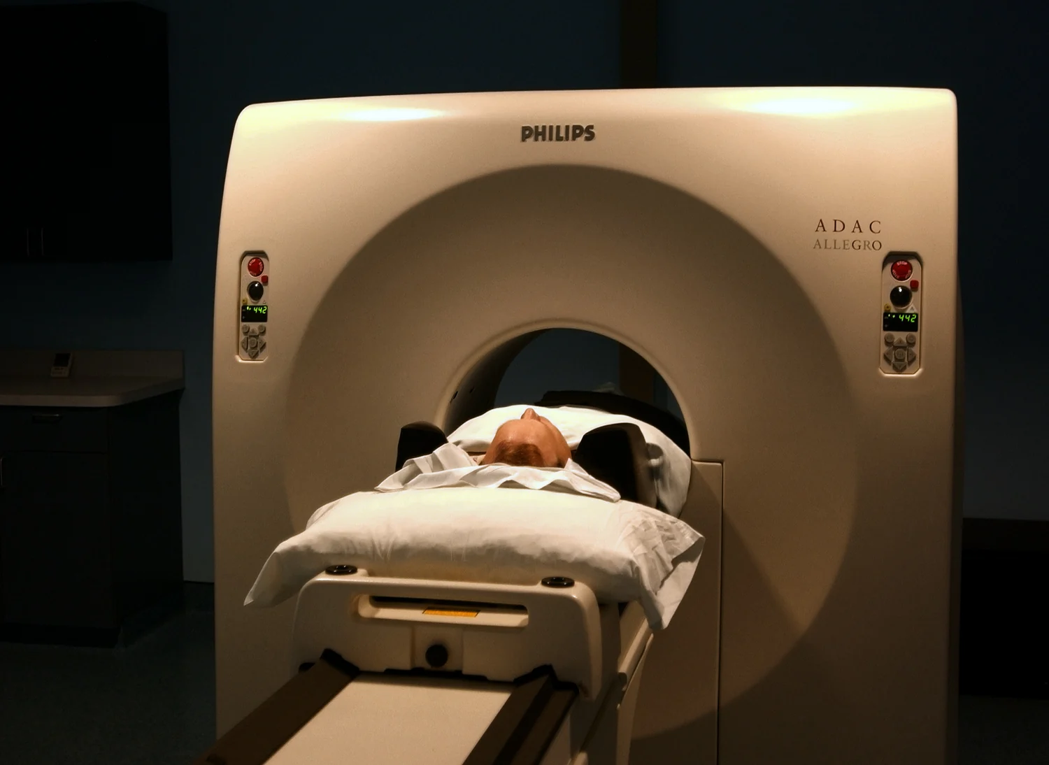
SPECT and PET both create three-dimensional images from radiopharmaceuticals or other radioactive tracers, but they detect different emissions. In SPECT, gamma cameras record gamma rays emitted by tracers inside the body. Detectors rotate around the person, collect two-dimensional views from multiple angles, and use them to construct a three-dimensional distribution [1]. The tracer’s distribution is the central signal; the camera’s measurements provide views from which that distribution is reconstructed.
PET begins with a radionuclide that emits positrons. A positron meets an electron in the body, and the pair annihilates, producing two gamma-ray photons traveling in opposite directions. PET detectors register coincident photons and use their paired detections to locate the event [1]. The distinction between SPECT and PET is therefore rooted in the radioactive decay and detection process, not simply in two different ways to display an otherwise identical signal.
A scan follows biology through physics
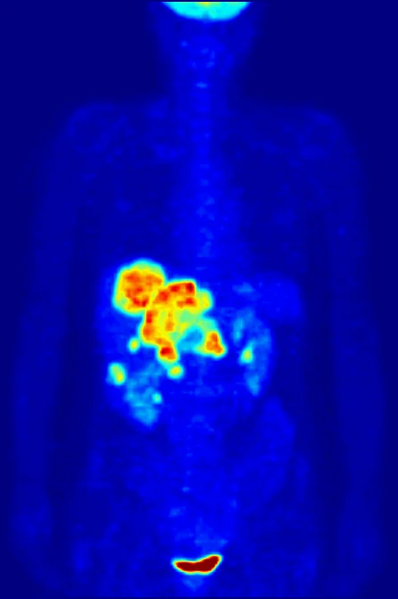
Many PET scans use a radiolabeled form of glucose called fluorodeoxyglucose, or FDG. Because glucose use varies with cellular activity, the tracer can help show regions with greater uptake. PET is used to detect cancer and monitor its progression or response to treatment, among other applications [1]. The image is evidence about tracer distribution and biological processes; interpreting what that pattern means requires clinical context.
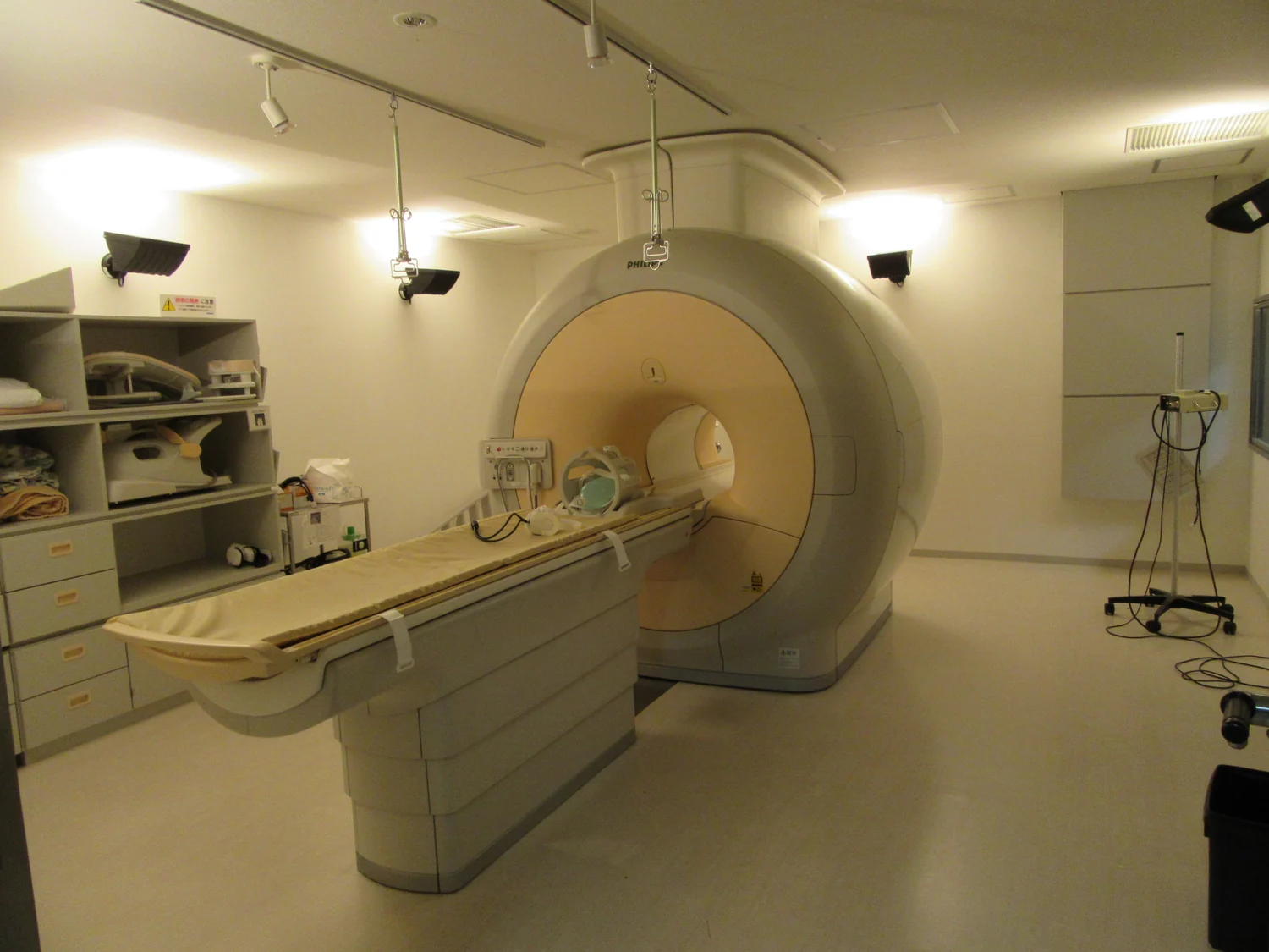
A combined PET/CT examination adds a CT image to the PET view. The two forms of information can complement one another: PET shows where the tracer accumulates, while CT supplies anatomical detail. NIBIB describes PET/CT as widely used for cancer staging [1]. The combination can make it easier to relate a functional signal to a location in the body, but a bright or concentrated signal is not itself a complete diagnosis.
When the tracer becomes treatment
The same basic design—radioactive atom plus carrier—can serve therapy as well as imaging. In radiopharmaceutical treatment, the carrier is intended to bring radioactive material to a target; decay then releases energy that can damage nearby cells. NCI describes engineered compounds with three components: a radioactive molecule, a targeting molecule, and a linker that joins them [2]. Some radiation can affect the targeted cell and neighboring cells, so delivery and the isotope’s emissions both matter.
Imaging can sometimes show whether a targeting molecule reaches a tumor, offering information relevant to a related therapeutic compound [2]. But a diagnostic tracer and a treatment are not interchangeable: they may use different radioactive atoms and radiation doses. The imaging result can inform a treatment strategy without guaranteeing its success. In either role, the radiotracer is more than a radioactive ingredient because its biological carrier determines where the signal—or delivered energy—can go.
Sources & further reading
- Nuclear Medicine NIBIB
- Radiopharmaceuticals: Radiation Therapy Enters the Molecular Age National Cancer Institute
Prepared for the independent PSI Center reading library. About edition dates and source checking.
This explainer describes scientific principles. It does not assess individual treatment choices.