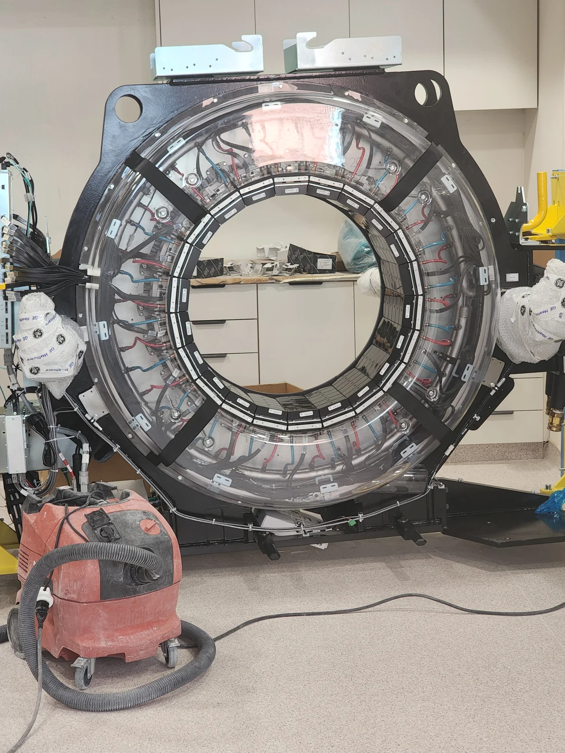
A CT scanner may display CTDIvol and DLP to summarize an examination. These numbers are useful for checking protocols and comparing like with like, but neither is a direct meter reading of energy absorbed throughout a particular person. The distinction begins with the phantom built into the measurement.
A reference phantom gives the index its meaning
CTDIvol is a standardized index of scanner radiation output, derived from measurements in a cylindrical plastic phantom. The phantom is usually 16 or 32 centimeters in diameter; the selected size is part of how the number should be interpreted. It provides a common reference for protocol monitoring and equipment comparisons, rather than a miniature copy of every patient [1].
This is why the word “dose” in the index can mislead. A value in milligray is expressed in dose units, but the reported CTDIvol characterizes the scanner under a specified reference condition. Patient absorbed dose depends on the x-ray output and on how a person’s tissues attenuate and absorb those x-rays. The index does not itself account for that person’s size [1].
DLP adds scan length, not a patient-specific map
Dose-length product, or DLP, is approximately CTDIvol multiplied by the length of the scan in centimeters. Its units are milligray-centimeters. For example, if a protocol has a CTDIvol of 8 mGy across a 30 cm scan range, the corresponding DLP is 240 mGy·cm. This simple multiplication captures that a longer irradiated region generally increases the total amount associated with the reference phantom.
But DLP still does not identify which organs were in the beam, how much energy each absorbed, or how the person’s anatomy altered the exposure. A short scan can have a relatively low DLP even when a local dose question requires closer attention. The FDA specifically distinguishes localized peak-skin-dose assessment from CTDI and DLP indices [3].
Why size changes the interpretation
Imagine the same scanner output used for two body regions, one small and one large. In a smaller body, x-rays generally travel through less material, so the same reference index does not imply the same absorbed dose as in a larger body. The AAPM’s Report 204 describes size-specific dose estimate (SSDE) methods that adjust CTDIvol using patient-size information. That makes SSDE a more tailored estimate of dose to the imaged region than CTDIvol alone [1].
The size measure matters too. A simple width or effective diameter captures geometry; water-equivalent diameter uses CT attenuation information to better represent how much the region attenuates x-rays. Two regions with similar outer dimensions can differ because lungs contain air spaces while abdominal tissues are denser. AAPM Report 220 explains why attenuation-aware size estimates can improve the physical description used for SSDE [2].
An estimate is not a direct measurement
SSDE is useful, but it is still an estimate. It applies a size adjustment to a scanner index; it does not reconstruct the exact absorbed dose in every organ. More detailed organ-dose calculations may use patient anatomy, scanner settings, and computational models such as Monte Carlo transport. Those methods add assumptions and technical work, so the appropriate quantity depends on the question being asked [2].
Effective dose is different again. It combines organ doses with tissue weighting factors to support radiation-protection comparisons across procedures and populations. It is a calculated quantity based on reference models, not a measurement in an individual. Using a generic DLP-to-effective-dose conversion can offer broad context, but it should not be presented as a person’s precise risk estimate [3].
Comparisons need matching conditions
Dose indices are most informative when the comparison is fair: similar examination purpose, body region, scan length, patient-size group, and phantom convention. A CTDIvol value associated with a 16 cm phantom cannot automatically be compared with one referenced to a 32 cm phantom as if both used the same scale. The scanner dose report or metadata may identify the phantom, and AAPM guidance emphasizes checking it when interpreting values [1].
Facilities can also compare protocol distributions with diagnostic reference levels, which are optimization tools for groups of examinations rather than patient dose limits. An unusually high or low index can prompt review of technique, coverage, and image quality. It does not by itself diagnose harm or show that an individual scan was unnecessary; clinical justification and protocol optimization are separate decisions [3].
A practical way to read a dose report
Start by asking what each number represents. CTDIvol is a reference-phantom output index; DLP adds the scan range; SSDE adjusts CTDIvol for a patient-size estimate; organ dose estimates address particular tissues; and effective dose is a population-oriented comparison quantity. Keep the units and the phantom convention alongside the number rather than copying a bare value.
For an individual scan, the report can help a qualified imaging team review the protocol and identify whether a more specific estimate is warranted. It cannot, by itself, tell a patient how much radiation a particular organ absorbed or predict an individual outcome. The useful lesson is not that scanner indices are meaningless; it is that their meaning is precise and bounded.
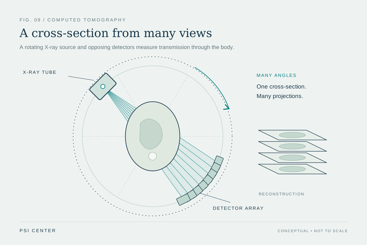
Explore the physics: an explanatory diagram
Sources & further reading
- Size-Specific Dose Estimates (SSDE) in Pediatric and Adult Body CT Examinations (AAPM Report No. 204) American Association of Physicists in Medicine
- Use of Water Equivalent Diameter for Calculating Patient Size and Size-Specific Dose Estimates (SSDE) in CT (AAPM Report No. 220) American Association of Physicists in Medicine
- Radiation Dose Quality Assurance: Questions and Answers U.S. Food and Drug Administration
Sources checked: 1 November 2026. The calculations illustrate physical models; they are not clinical settings.
Prepared for the independent PSI Center reading library. About edition dates and source checking.
This explainer describes scientific principles. It does not assess individual treatment choices.