Can a chest CT use much less radiation and still give similar quantitative measurements? A study published on 15 October 2026 tested that question with photon-counting CT in adults with inflammatory idiopathic myopathy-associated interstitial lung disease. The low-dose scans produced measurements close to those from the study’s standard-dose scans. That is a useful technical result, but the comparison answers a narrower question than whether changing a patient’s imaging protocol improves care.
The comparison began with the measurement task
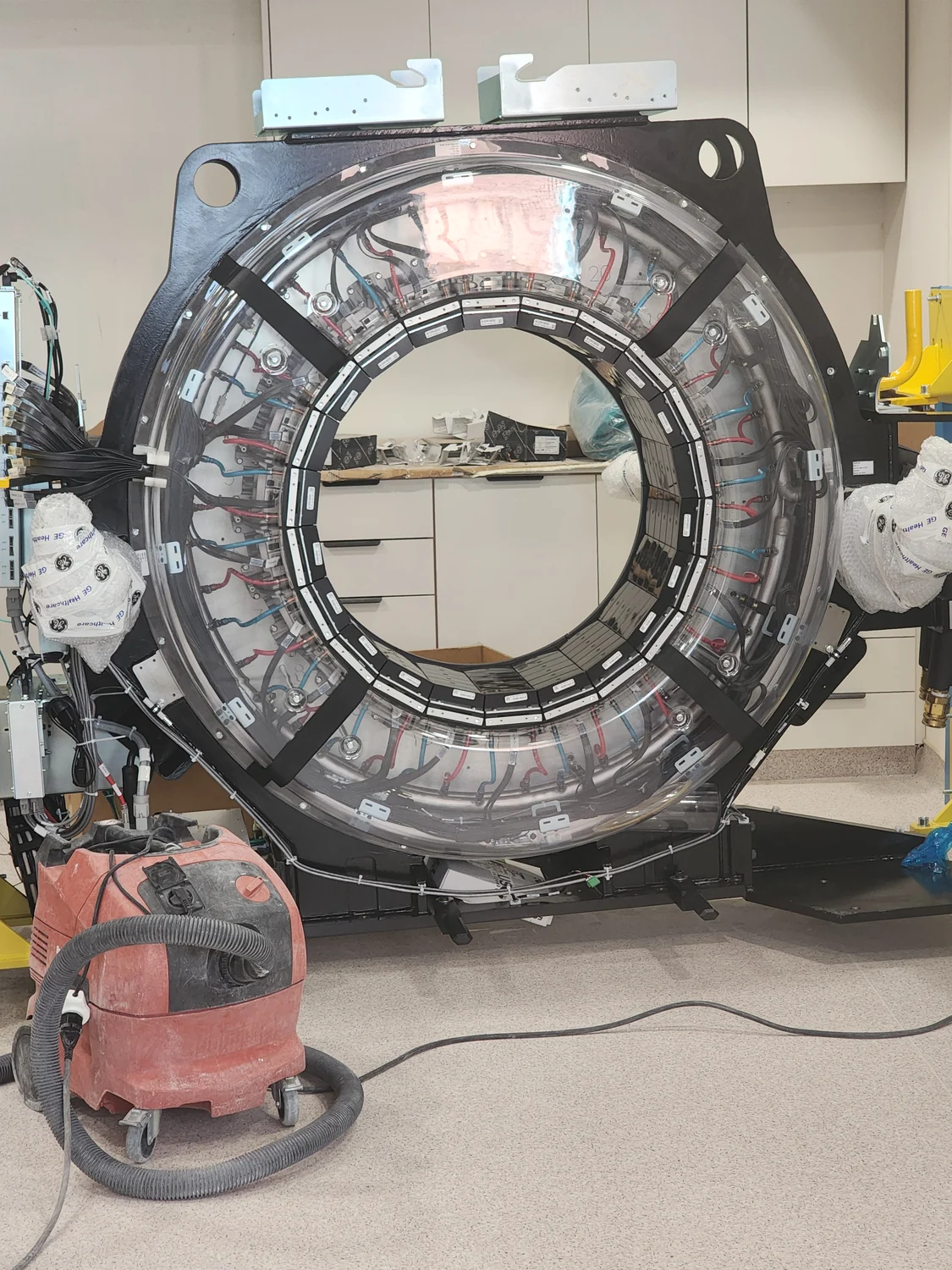
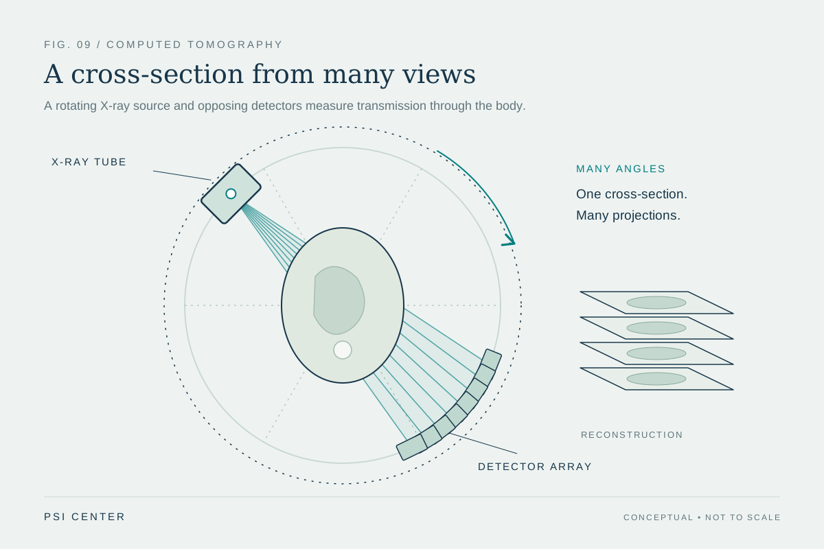
Computed tomography reconstructs cross-sectional images from X-ray measurements taken around the body. Photon-counting CT uses detectors that register individual X-ray photons and their energies. In the new study, investigators asked whether quantitative software could estimate patterns of interstitial lung disease (ILD) from a tin-filtered low-dose scan about as consistently as from a standard-dose scan. ILD is a broad group of conditions involving the tissue around the lung’s air spaces; the study focused on people whose ILD was associated with inflammatory idiopathic myopathy [1].
The researchers enrolled 48 adults at one center. Each participant underwent two consecutive scans during the study period, September 2023 through November 2024. The team compared 1.0-millimeter reconstructions and used software to quantify features such as ground-glass opacity, reticular pattern, and fibrosis extent. This setup makes the standard-dose image a practical comparator for the low-dose protocol; it does not make either scan a direct measurement of microscopic disease.
A large dose-index difference, close agreement
The reported volume CT dose index (CTDIvol) was 0.77 mGy for the low-dose protocol and 5.40 mGy for the standard-dose protocol. CTDIvol is a scanner-output index used to describe and compare CT protocols; it is not an individualized estimate of the absorbed dose to a particular person. The study reported mean net differences below one percentage point for the quantified patterns. The low-dose reconstruction slightly increased measured ground-glass opacity and slightly decreased measured fibrosis extent relative to the standard-dose reconstruction [1].
Those details matter together. A low dose index is not useful by itself if the image cannot answer the intended question. Conversely, close agreement on selected software-derived quantities does not show that every subtle feature is equally visible, that all scanners and protocols behave alike, or that a radiologist would reach the same diagnosis. The American College of Radiology’s CT practice parameter describes protocol design as a balance: obtain diagnostic information of sufficient quality while optimizing dose for the clinical question [2]. The parameter is for head CT, so its general optimization principle should not be mistaken for a lung-specific protocol recommendation.
Agreement is not the same as validation over time
A useful way to interpret the result is to separate three questions. First, can a lower-dose protocol produce measurements similar to a reference scan in this sample? The study provides encouraging evidence for that question. Second, do those measurements accurately track the underlying disease and its changes? The study found that reticular-pattern measurements were associated with pulmonary-function measures such as forced vital capacity and diffusion capacity, with similar correlations for low- and standard-dose acquisitions. Association supports relevance, but it does not establish that a measurement can detect a meaningful change in an individual over time.
Third, would using the low-dose protocol change decisions or outcomes? This study did not test that. It was a single-center feasibility study, not a randomized comparison of care strategies, and the authors explicitly called for future longitudinal validation. A monitoring tool needs repeatability, sensitivity to change, and evidence that observed changes are interpretable in the clinical context—not only agreement between two reconstructions made close together.
Why thinner images can add a trade-off
The paper also explored 0.4-millimeter ultrathin reconstructions. These reconstructions measured somewhat more bronchiectasis, a widening of airways, than the thicker images. Finer slices can make small structures easier to separate, but a more detailed-looking image is not automatically a more reliable measure. Noise, reconstruction settings, segmentation behavior, and the definition of the feature all affect the number produced. The study found limited additional value from ultrathin reconstruction overall, which cautions against equating finer sampling with better clinical information [1].
A promising protocol still needs local and clinical checks
The findings apply to the scanner, software, disease group, and acquisition conditions studied. They do not establish that the same dose or performance will transfer to another scanner, body size, lung condition, or reconstruction pipeline. Software also matters: an automated segmentation can be consistent while still sharing systematic errors with its own training or measurement assumptions.
The practical lesson is not “use the lowest number.” It is to define the imaging question, check whether the images preserve the features needed to answer it, and validate measurements in the setting where they will be used. The 15 October study makes a strong case for further testing of low-dose photon-counting CT in this specific population. Until longitudinal and broader validation is available, its result is best understood as promising technical feasibility—not proof that routine disease monitoring can safely change everywhere, and not evidence of a treatment benefit.
Explore the physics: an explanatory diagram
Sources & further reading
Prepared for the independent PSI Center reading library. About edition dates and source checking.
This explainer describes scientific principles. It does not assess individual treatment choices.