A sharper coronary CT image can make the edge of a calcified artery look easier to inspect. But a scan protocol is a set of linked choices: how the scanner moves, when it records data relative to the heartbeat, how fine its sampling is, and how much radiation is used. A study published on 16 October 2026 compared four acquisition modes on a photon-counting CT system. Its results show why “highest image quality” and “best protocol” are different conclusions. [1]
The scan mode is part of the measurement
Coronary CT angiography reconstructs images of contrast-filled arteries from X-ray measurements collected around a moving heart. Calcium and stents can blur across the apparent vessel boundary, an effect called blooming. Finer detector sampling may reduce that blur, but cardiac motion, reconstruction settings, the patient’s heart rhythm, and the amount of detected signal also influence the final image. A protocol is therefore not one image-quality dial: it is a coordinated way of collecting data for a particular measurement task.
Photon-counting detectors register individual X-ray interactions and sort them by energy, enabling high-resolution and energy-resolved reconstructions. These capabilities do not remove CT’s trade-offs: acquisition path and timing can affect motion robustness and dose as well as sharpness. More detail helps only when it is reliable for the measurement task.
Four acquisition modes, two kinds of evidence
Stein and colleagues tested four modes on a first-generation dual-source photon-counting CT system: high-pitch spiral scanning with prospective ECG gating (Flash), Sequence, Spiral, and retrospective ultra-high-resolution (UHR) spiral acquisition. Their phantom arm used six three-dimensional printed coronary models with stenoses from 21% to 73%. A retrospective clinical arm included 41 coronary CT examinations, with invasive coronary angiography used as the reference for diagnostic-accuracy comparisons. [1]
The arms answer different questions. A phantom with known narrowing isolates how the setup portrays controlled geometry; patient images include anatomy and motion but may reflect local selection and protocol choices. Together they make the engineering comparison more informative without turning it into a prospective trial.
Sharper images came with a dose cost
In the reported comparison, UHR had the highest clinical image-quality score and the lowest quantitative blooming. Its mean score was 4.12 on the study’s scale, compared with 3.00 for Flash. Yet UHR also had the highest reported radiation dose: 26.44 mSv on average, versus 13.65 mSv for Sequence and 1.40 mSv for Flash. These are study-specific values from the tested scanner and cohort, not dose predictions for another person or a benchmark for every institution. [1]
A score cannot stand alone. If the task is to decide whether a narrowing crosses a clinically meaningful threshold, the question is whether added detail improves classification enough to justify the other costs. A low dose also fails the task if motion or noise makes the measurement unreliable.
Accuracy depends on the threshold and the reference
The authors reported that diagnostic accuracy varied by mode when detecting stenoses at or above 50% and 70%. For example, Flash accuracy was 76.5% at both thresholds; Sequence was 100% at the 50% threshold and 57.1% at 70%; UHR was 64.7% and 82.4%, respectively. The pattern cautions against treating “diagnostic accuracy” as a single property of a scanner. It depends on which threshold is tested, how cases are distributed around it, and how the CT result is compared with the reference method. [1]
A separate retrospective study in Taiwan compared photon-counting CT with energy-integrating CT in 205 asymptomatic health-screening participants. It found lower dose-length-product values for the photon-counting group, but the scans were acquired in helical mode and the study compared detector generations rather than the four modes in Stein’s work. The different questions matter: it supplies context about detector comparisons, not an independent replication of the UHR-versus-Flash result. [2]
What the dose numbers do—and do not—mean
The reported mSv values summarize radiation dose using the study’s stated dose calculation. Effective dose is a population-level quantity used for broad comparisons; it is not a direct measurement of an individual patient’s organ dose or personal risk. A scanner’s displayed output, the anatomy scanned, patient size, scan length, and conversion assumptions all matter. Comparing two modes within one study can reveal a relative trade-off under those conditions, but the numbers should not be carried over as universal protocol settings.
Likewise, using invasive angiography as a reference gives the comparison a clinically relevant anchor, but the reference answers a particular question about lumen narrowing. The study did not test whether selecting one CT mode improves treatment decisions, symptoms, or health outcomes. Nor does an accuracy estimate from 41 retrospective examinations establish how well a mode performs across different scanners, heart rates, calcium burdens, or hospitals.
A useful reading rule for protocol comparisons
When reading an imaging comparison, track the setup, image-quality measure, diagnostic task, and dose metric. Ask whether results came from phantoms or patients, whether comparators used similar conditions, and whether the tested threshold matches the intended task. A protocol that wins one measure and loses another maps a trade-off; it does not choose a universal winner.
The October study is best read as a task-specific evaluation of acquisition modes on one system, with a helpful phantom component and a limited retrospective clinical component. It suggests that very high spatial resolution can reduce visible blooming and improve image ratings while increasing radiation exposure. The reported diagnostic results do not collapse into a simple “sharpest is most accurate” rule. Prospective comparisons with clearly defined patient groups and protocol-selection criteria are needed before translating these findings into a general clinical preference.
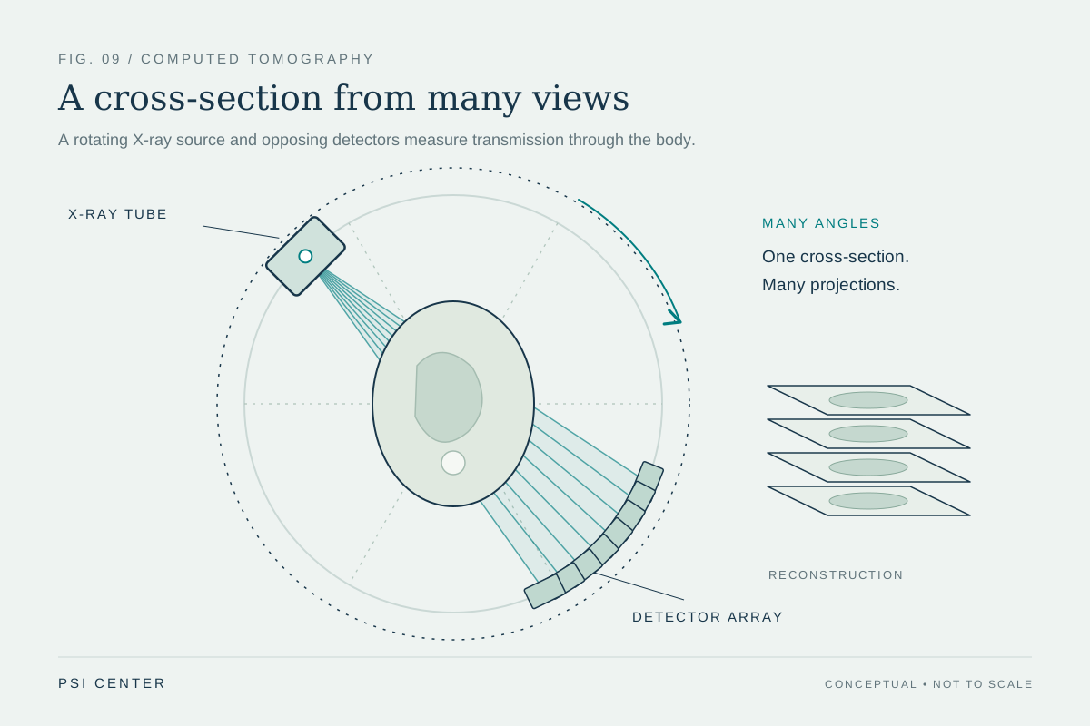
Explore the physics: an explanatory diagram
Sources & further reading
- Impact of Photon-Counting CT Acquisition Modes on Coronary Stenosis Assessment and Radiation Dose: A Phantom and Clinical Evaluation PubMed / Zeitschrift für Medizinische Physik
- Initial clinical evaluation of photon-counting detector computed tomography for coronary artery disease in Taiwan PubMed / Coronary Artery Disease
Sources checked: 11 November 2026. The calculations illustrate physical models; they are not clinical settings.
Prepared for the independent PSI Center reading library. About edition dates and source checking.
This explainer describes scientific principles. It does not assess individual treatment choices.