After pancreatic surgery, a soft-tissue finding near the operation site can be difficult to interpret: recurrent tumor and healing-related fibrosis may occupy similar-looking space. A study published on 12 October 2026 asked whether photon-counting CT could add quantitative clues by measuring iodine-related signal and extracellular volume. Its results are an early measurement study, not a validated rule for deciding whether an individual has recurrent cancer. [1]
Why energy-resolved CT can add another measurement
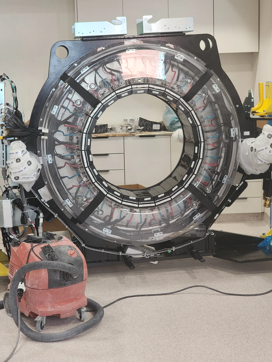
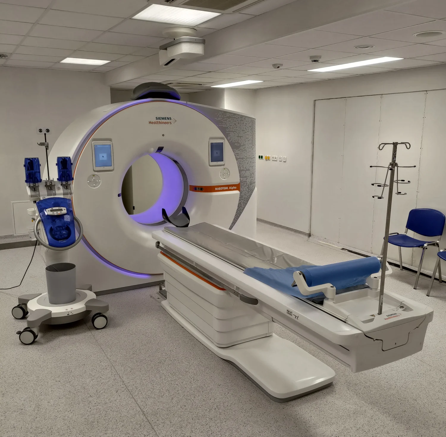
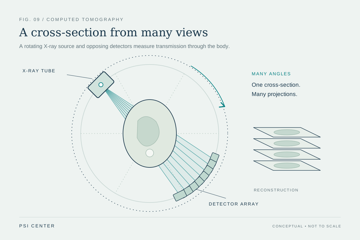
A conventional CT image represents how strongly each voxel attenuates X-rays. With photon-counting CT, the detector records individual X-ray interactions and sorts them into energy ranges. Those measurements can support reconstructions that emphasize a selected energy or estimate the contribution of materials such as iodine. The output is still calculated from detector data through a reconstruction model; it is not a direct chemical assay of each tiny piece of tissue. [2]
Iodine is useful because contrast material changes X-ray attenuation in an energy-dependent way. A virtual monoenergetic image displays an estimate of what the scan would look like at a chosen photon energy. An iodine-density map instead estimates iodine concentration under the decomposition model. The two products answer related but different questions: one changes the displayed energy weighting, while the other attempts a material-specific measurement.
What the 2026 pilot compared
Platt and colleagues retrospectively identified 22 people who had undergone pancreas-protocol photon-counting CT after surgery for pancreatic ductal adenocarcinoma. Two blinded readers measured soft tissue in the surgical bed. They assessed iodine density, iodine normalized to aortic enhancement, fat fraction, extracellular volume and ordinary post-contrast attenuation. Recurrence classification relied on tissue pathology or enlarging FDG-avid tissue; nonrecurrence required shrinking soft tissue together with stable or falling CA 19-9. [1]
The reported comparison included 11 recurrence cases and 10 nonrecurrence cases. Median iodine density was higher in the recurrence group, as were several other enhancement-related measurements. This is physically plausible: tissue with a different blood supply, vascular permeability and extracellular structure can retain or distribute contrast differently. But iodine signal is not a tumor-specific molecule. It summarizes contrast behavior within a region and can vary with acquisition timing, injection, circulation, tissue composition and reconstruction.
A cutoff that fits a cohort is not yet a clinical boundary
The study also calculated thresholds that separated its groups well, including an iodine-density threshold of 1.27 mg/mL and an extracellular-volume threshold of 14.0 mL. Such numbers can look definitive when reported beside high sensitivity or specificity. Yet the thresholds were selected and evaluated in this same small retrospective sample. A handful of different cases, measurement choices or patient characteristics could materially change their performance. The reported separation is therefore hypothesis-generating; it does not tell us how reliably the cutoffs would work at another hospital or in a broader population. [1]
There is also a distinction between reader agreement and diagnostic validity. The study reported high agreement between its two readers, which suggests they measured the selected regions similarly. Agreement does not establish that the regions capture all relevant disease, that the classification reference is error-free, or that applying a threshold improves decisions or outcomes.
Maps depend on calibration and assumptions
An iodine map is produced through material decomposition: an algorithm uses energy-dependent attenuation data to estimate the proportions of selected basis materials. The answer depends on which materials the model includes and on calibration. A 2025 pancreatic CT study comparing iodine-density images with virtual monoenergetic images describes this spectral workflow and its image-quality trade-offs. In photon-counting CT, as in other quantitative imaging, noise, partial-volume effects and the chosen reconstruction can influence a concentration estimate. [2]
A useful analogy is separating colors of light with a limited set of filters. Measurements across energy bands help distinguish likely contributors, but the calculation still assumes a set of ingredients and a model of how each changes the signal. If real tissue contains mixtures or artifacts outside those assumptions, the displayed material value may be biased. Phantom work and repeatability checks help characterize such errors, but patient-level performance requires its own validation.
What would make the result clinically useful?
The next evidential step is independent testing with a prespecified protocol and threshold in a larger, preferably multicenter group. Investigators would need to define how recurrence is confirmed, include the full range of postoperative appearances, and report uncertainty around sensitivity, specificity and predictive values. Since the consequences of a false alarm and a missed recurrence differ, performance also needs to be judged in the population where the test would actually be considered—not only by how far two study-group averages are apart.
Finally, a better measurement is not automatically a better care pathway. A prospective evaluation would need to show whether adding spectral measurements changes interpretation, follow-up testing or treatment decisions in a beneficial way. For now, the study demonstrates a promising way to quantify iodine-related contrast in a difficult imaging setting. It does not establish that photon-counting CT can independently diagnose recurrence, replace pathology or guide an individual patient’s treatment.
Explore the physics: an explanatory diagram
Sources & further reading
- A Pilot Study of Photon Counting CT Spectral Analysis Following Pancreatic Adenocarcinoma Resection to Differentiate Local Recurrence From Postsurgical Fibrosis Journal of Computer Assisted Tomography (2026), indexed by PubMed
- Photon-counting detector computed tomography: iodine density versus virtual monoenergetic imaging of pancreatic ductal adenocarcinoma Abdominal Radiology (2025), PubMed Central
Sources checked: 7 November 2026. The calculations illustrate physical models; they are not clinical settings.
Prepared for the independent PSI Center reading library. About edition dates and source checking.
This explainer describes scientific principles. It does not assess individual treatment choices.