Why not simply turn up an imaging machine until every detail is crisp? Because an image is produced through physical measurements, and collecting more information can carry a cost. In computed tomography, X-rays pass through the body from many angles. The goal is not maximum sharpness in isolation; it is enough useful detail to answer the medical question with an appropriate exposure.
A CT slice is reconstructed from many views
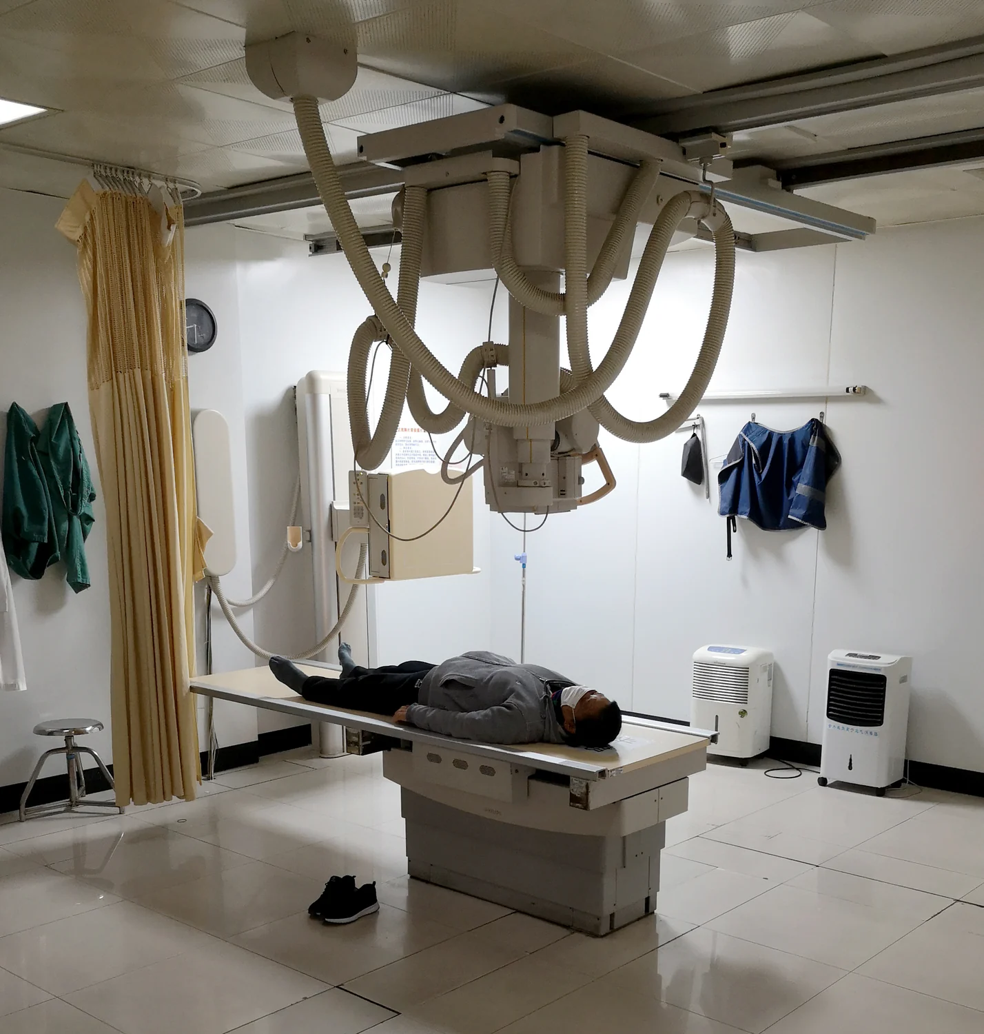
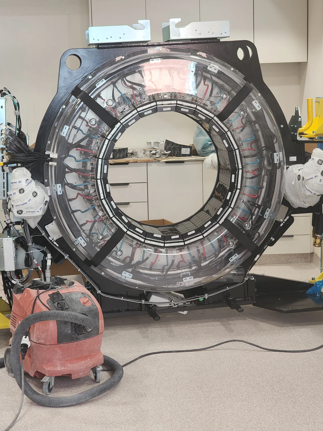
A conventional radiograph records a projection: structures along the X-ray path overlap in one image. CT takes a different approach. As the source and detectors rotate around the body, they collect many snapshots from different angles. A computer combines those measurements to reconstruct cross-sectional slices, revealing internal structures with more detail than a conventional X-ray image [1].
Imagine trying to locate a small object hidden inside a layered loaf. One view may show its shadow but not its depth. Looking from multiple directions gives more clues about where it sits. CT uses the same broad logic, with X-ray attenuation—the reduction of beam intensity as it passes through tissue—measured at many angles. The reconstructed slices can help diagnose conditions, plan procedures, or evaluate treatment [1].
More measurements involve more radiation
The tradeoff is that CT uses ionizing radiation, which has enough energy to potentially damage DNA. Because CT builds images from many X-ray projections, it generally involves a higher radiation dose than conventional radiography [2]. A medically justified scan can provide important information, but repeated or unnecessary exposure should be avoided, and younger patients are more sensitive to radiation effects than adults [1].
Image quality and dose are connected through the measurements available to the reconstruction. Too little information can make a scan less useful; increasing exposure can improve the signal in some conditions, but it increases radiation dose. The practical target is an image adequate for its diagnostic or procedural purpose, not a technically perfect picture regardless of cost. FDA guidance describes optimization as using the lowest dose that yields image quality adequate for diagnosis or intervention [2].
A useful image depends on the question
Consider two different tasks: checking a bone injury and guiding a procedure near blood vessels. The relevant anatomy and needed detail may differ. The scan protocol should be selected for the clinical question and the person’s size and anatomy. A broad scan that is not needed can add exposure without contributing useful information; a carefully chosen examination aims to collect the measurements that matter.
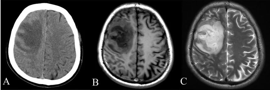
The choice of imaging method also matters. FDA guidance says clinicians should consider examinations with less or no radiation, such as ultrasound or MRI, when appropriate [1]. MRI uses magnetic fields and radio waves rather than ionizing X-rays. It is not a substitute in every situation, because different methods reveal different properties and have different practical limits. The best image is the one suited to the question, not necessarily the one with the most pixels or slices.
Sharper is not the same as more informative
A detailed scan may also reveal findings unrelated to the original reason for imaging. These incidental findings can require further assessment, so additional detail is not automatically a benefit. The decision to image weighs the likelihood that the result will help diagnosis or treatment against exposure and other burdens. For medically necessary exams, the FDA emphasizes that clinical benefit generally outweighs the small radiation risk, while encouraging efforts to avoid unnecessary exposure [2].
This balance is easiest to see as a measurement problem. A detector records a pattern, a computer reconstructs anatomy, and a clinician interprets that image in context. More measurements can sharpen the reconstruction, but the scan must still be justified and optimized. Precision comes from matching the imaging method and acquisition to the question, while keeping dose no higher than needed to obtain a useful answer.
Sources & further reading
Prepared for the independent PSI Center reading library. About edition dates and source checking.
This explainer describes scientific principles. It does not assess individual treatment choices.