Could an MRI measure several aspects of brain blood flow without collecting every possible data point? A paper published on 27 October 2026 tested a reconstruction method for multi-parameter arterial spin labeling (ASL), an MRI technique that uses magnetically labeled blood water as its own tracer. In eight healthy volunteers, the method reconstructed data sampled across several dimensions and preserved quantitative maps more closely than a matched compressed-sensing approach. The result is a technical advance, not evidence that a clinical scan is already 28 times shorter or that the maps improve diagnosis. [1]
A blood-flow image made by subtraction
Arterial spin labeling (ASL) turns the water in arterial blood into a temporary MRI tracer. Radiofrequency pulses change the magnetic state of incoming blood; the scanner compares a labeled acquisition with a control acquisition made without that change. Their small signal difference carries information about blood reaching brain tissue. No injected tracer or ionizing radiation is needed for this measurement. [2]
The difference is faint and time-sensitive. Labeled blood takes time to travel from the neck to tissue, and its magnetic signal fades as it relaxes. A post-labeling delay that is too short can leave labeled blood still in arteries; a very long delay loses signal. Multi-delay measurements can help estimate both cerebral blood flow (CBF) and arterial transit time (ATT), the travel time from labeling plane to tissue. Each additional setting means more data to collect and reconstruct. [2]
Why measure several dimensions at once?
The October study extended ASL beyond a single flow map. It combined four post-labeling delays with 14 echo times and repeated control–label measurements to estimate CBF, ATT, blood–brain-barrier (BBB) water-exchange time, and intravascular transit time (ITT). Echo time changes how long the MRI signal is allowed to evolve before measurement; the different decay patterns help separate signal components. The authors describe their acquisition as echo-time-resolved ASL, or TASL. [1]
More dimensions can make a measurement richer, but each combination can add acquisition steps. In the study’s example, each of eight healthy volunteers had two independent sessions. The fully sampled data contained 36 slices, 14 echo times, eight control/label measurements (four pairs), and four delays. This is a deliberately information-dense research protocol, not a routine scan time for ordinary ASL. [1]
Reconstruction looks for shared structure
The proposed method treats neighboring measurements as related rather than rebuilding each image in isolation. Echoes from the same tissue share a broad spatial structure, while control and label images contain much of the same anatomy plus a smaller perfusion difference. A low-rank subspace model represents these repeated patterns with a smaller set of basis signals. The reconstruction then estimates the coefficients of those bases from fewer sampled points, with sparsity regularization to constrain the solution. [1]
The authors coordinated undersampling across in-plane and through-plane spatial data, echo time, and control–label measurements. Their combined reduction factor was about 28: the design combined a factor of roughly four in one set of dimensions with about seven in another. This is a sampling and reconstruction factor in the tested framework. It does not mean every clinical ASL scan will become exactly 28 times shorter, because scan duration also depends on sequence timing, calibration, hardware, and how many parameter maps are required. [1]
What the comparison showed
At the strongest undersampling, the subspace reconstruction produced higher structural-similarity and peak-signal-to-noise scores than a matched compressed-sensing reconstruction, particularly at later echo times. When the researchers fitted CBF, ATT, BBB exchange, and ITT maps, the subspace-derived maps were generally closer to their fully sampled references. For example, across subjects and sessions, mean map similarity was 0.875 for CBF with the subspace method and 0.857 with compressed sensing; the advantage was larger for ITT (0.753 versus 0.698). These metrics compare reconstructions with a reference dataset; they are not diagnostic accuracy scores. [1]
The study also compared repeatability across two sessions. Intraclass correlation values for the accelerated parameter estimates were reported as high and broadly comparable to those from fully sampled data. That is useful evidence that the reconstruction did not obviously destroy repeatability in this small experiment. With eight healthy volunteers, it cannot establish how well the method performs in patients, across scanners, or in regions with disease-related abnormalities. [1]
The remaining gap is clinical validation
ASL measurements already have established clinical protocols, but this study asks whether a much more information-rich research acquisition can be reconstructed from substantially fewer samples. Its participants were healthy, and the authors tested retrospectively undersampled datasets against fully sampled references. They did not show that a prospective clinical examination was shortened by a measured amount, that BBB water-exchange estimates diagnose a condition, or that using these maps changes care. The distinction matters: a reconstruction that resembles its reference is a technical result, not proof of a patient benefit. [1]
The next useful checks are broader and practical: test the sequence prospectively, include people with varied anatomy and pathology, compare scanners and reconstruction settings, and evaluate whether the maps remain stable for the clinical question being asked. The 2014 ISMRM consensus explains why ASL timing and signal-to-noise trade-offs matter; this new work explores one way to make richer acquisitions more manageable. It is a promising reconstruction study whose clinical role remains to be established. [2]
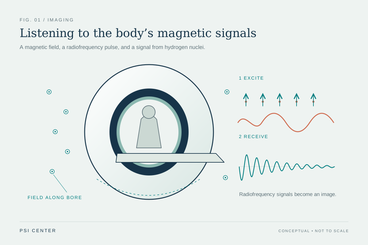
Explore the physics: an explanatory diagram
Sources & further reading
- Fast Multiparametric ASL MRI via Multi-Dimensional Acceleration and Subspace Reconstruction Magnetic Resonance in Medicine (2026), original research
- Recommended Implementation of Arterial Spin Labeled Perfusion MRI for Clinical Applications Magnetic Resonance in Medicine (2014); ISMRM Perfusion Study Group and European Consortium for ASL in Dementia consensus
Sources checked: 13 November 2026. The calculations illustrate physical models; they are not clinical settings.
Prepared for the independent PSI Center reading library. About edition dates and source checking.
This explainer describes scientific principles. It does not assess individual treatment choices.