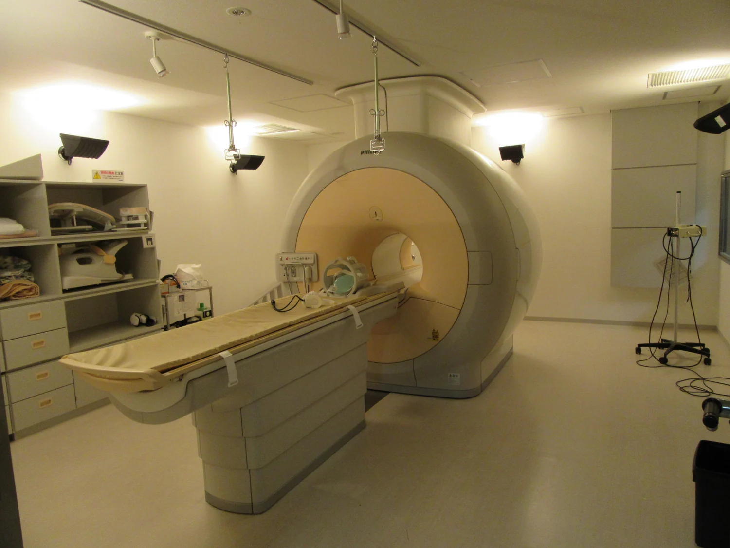
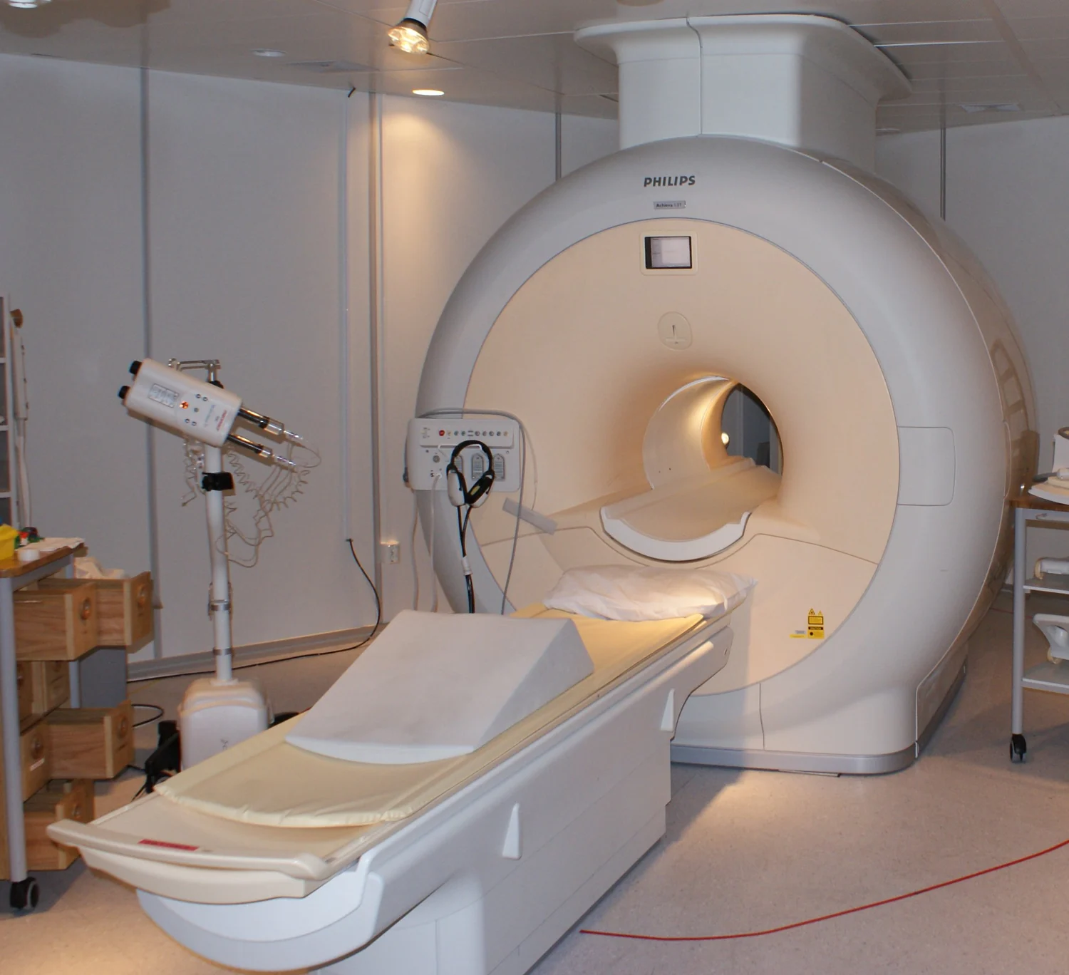
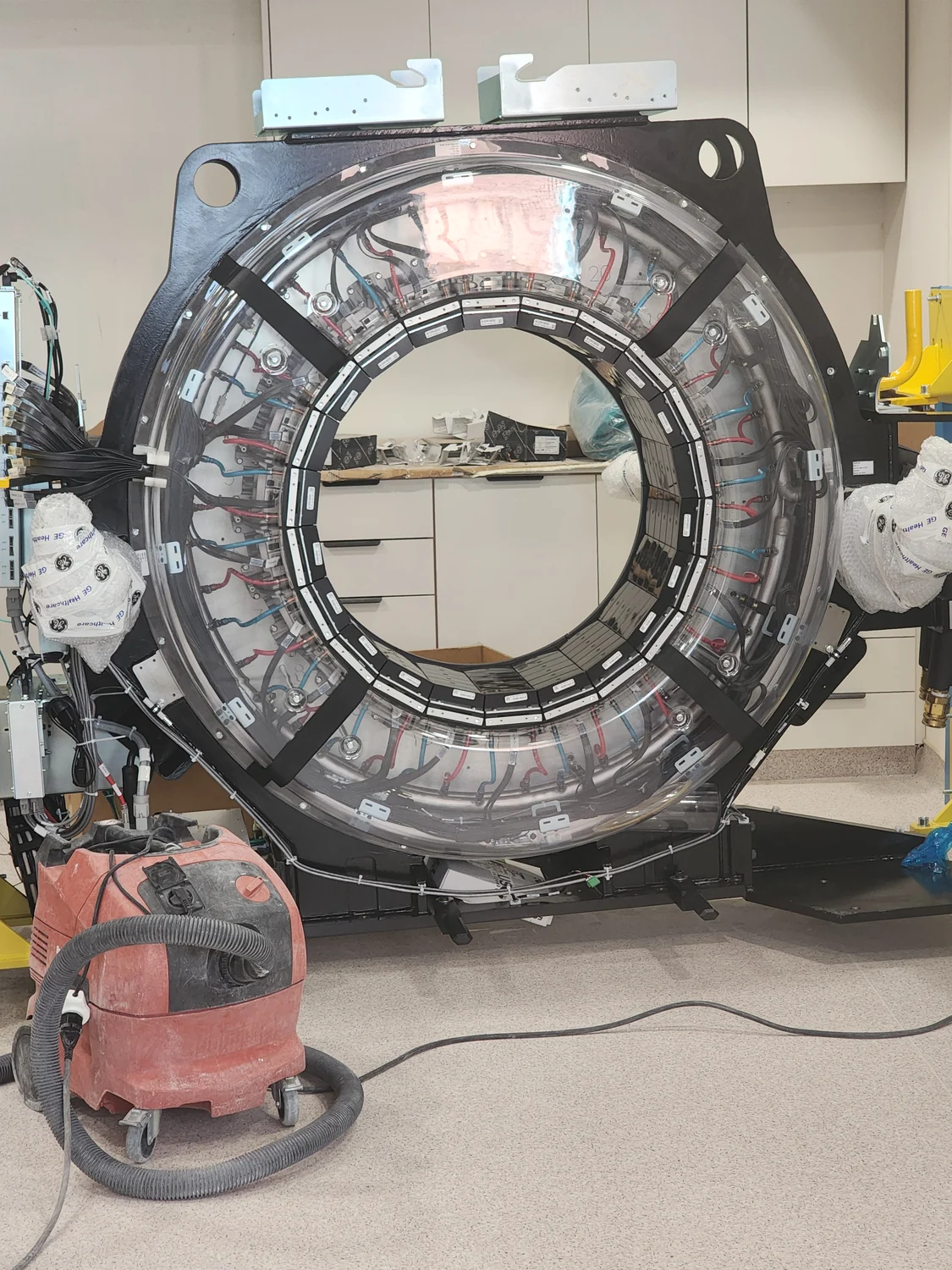
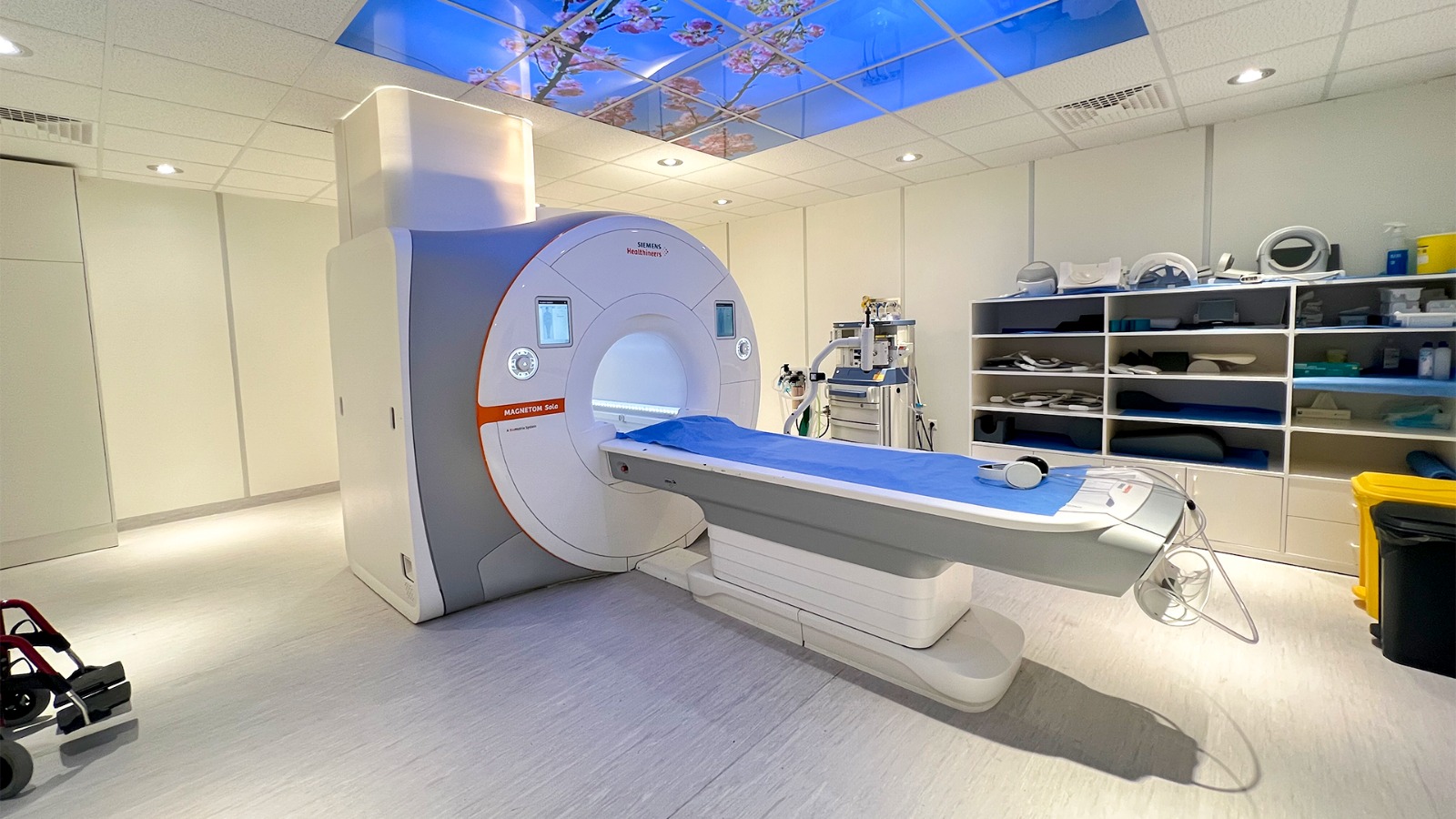
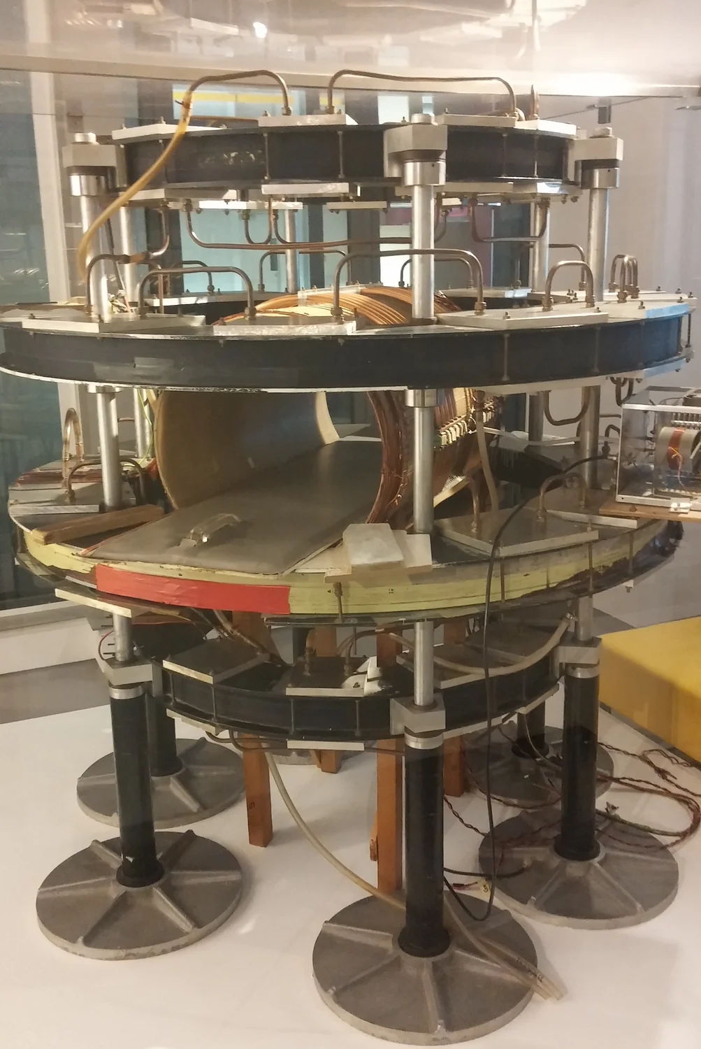
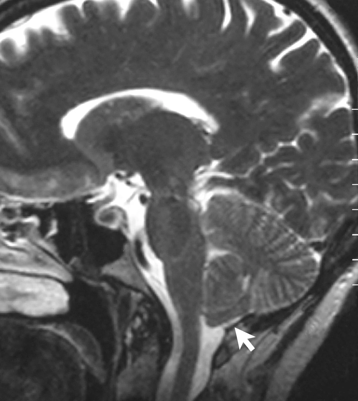
MRI uses a powerful magnetic field to orient hydrogen protons, radiofrequency pulses to disturb that alignment, and receiver coils to detect returning signals. Tissue-dependent signal timing helps form images.
Computational models let researchers explore how an imaging device may behave before every question can be answered in a clinic. Their predictions are useful only when grounded in data and checked against physical measurements.
A nuclear medicine scan follows a tagged molecule through the body, revealing where biological activity occurs. Its signal can be paired with structural imaging to place that activity in anatomical context.
A CT scanner reconstructs cross-sections from many X-ray measurements taken around the body. The changing angles help distinguish overlapping structures that a single projection would collapse into one shadow.
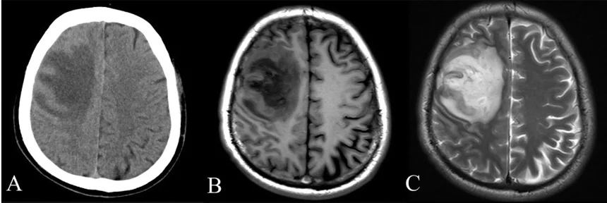
MRI uses a strong magnetic field to organize proton spins and radiofrequency pulses to disturb them. Sensors detect returning signals that vary with tissue chemistry, allowing soft-tissue images without ionizing X-rays.
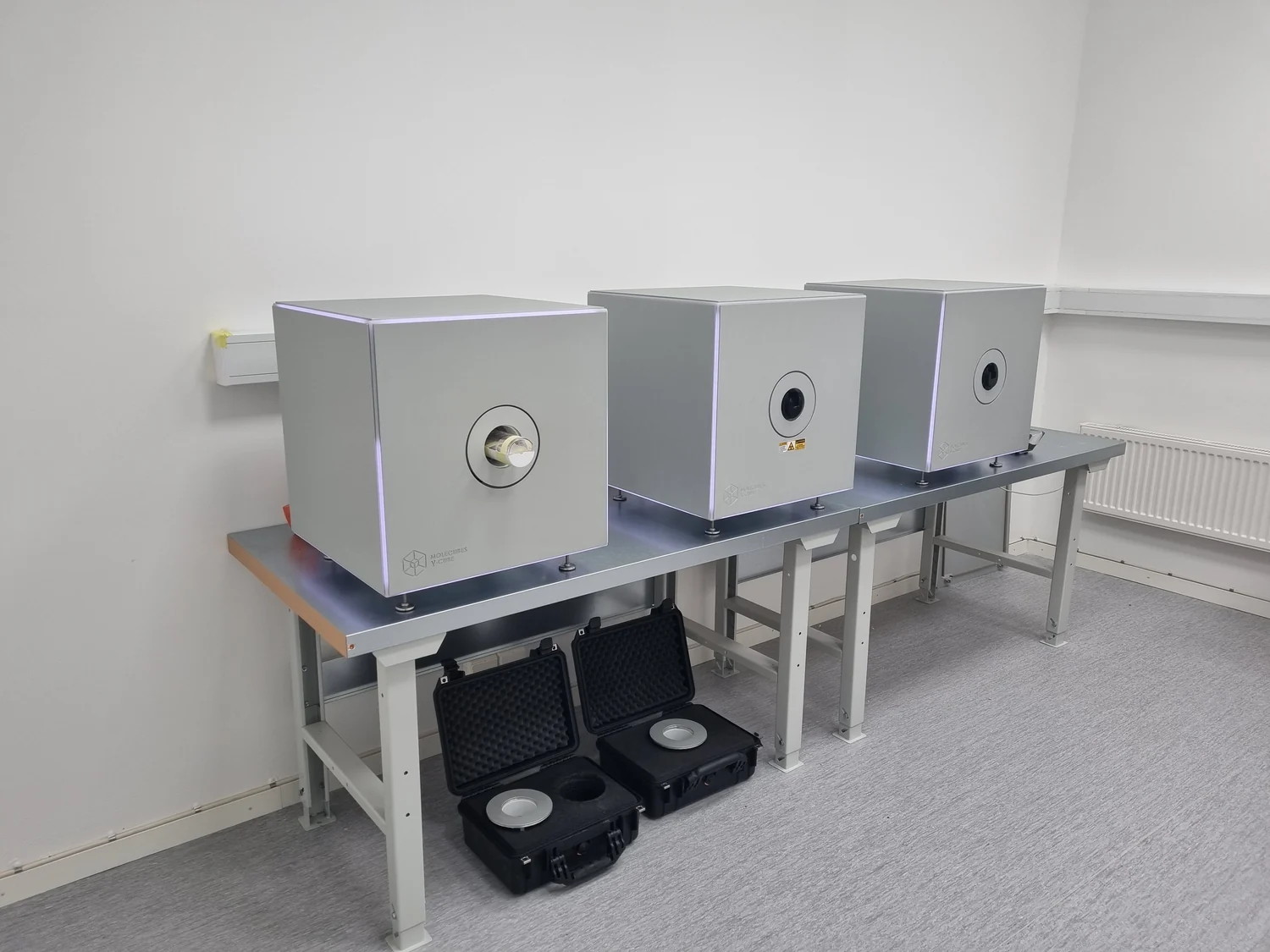
Photoacoustic imaging uses absorbed laser energy to create sound inside tissue, then detects those waves with ultrasound equipment. The hybrid method may reveal blood vessels, oxygenation, and other features below the surface.
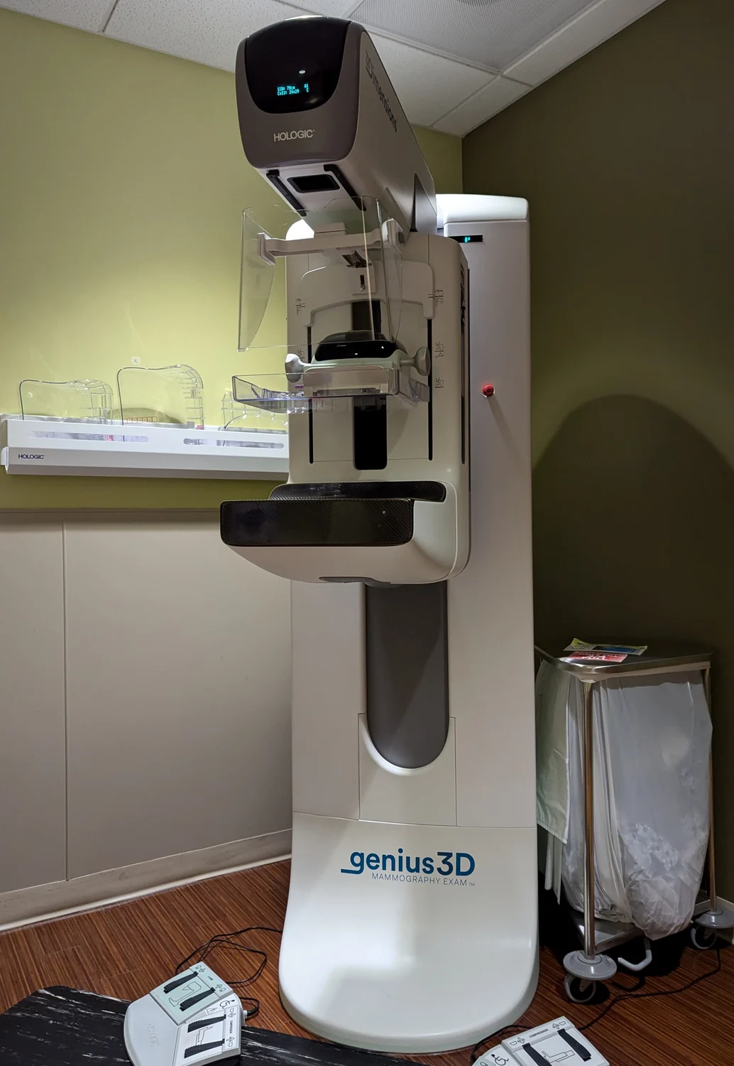
Mammography is radiography tailored to breast structures, with dedicated image receptors and quality requirements. Its aim and image geometry distinguish it from CT and live fluoroscopy, though all use ionizing X-rays.
A CT image is reconstructed from many x-ray measurements taken around the body. Computation turns those projections into slices, and a stack of slices can reveal how anatomy is arranged in three dimensions.
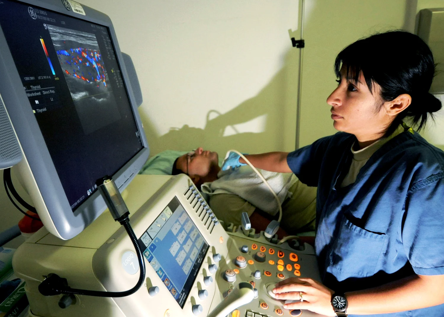
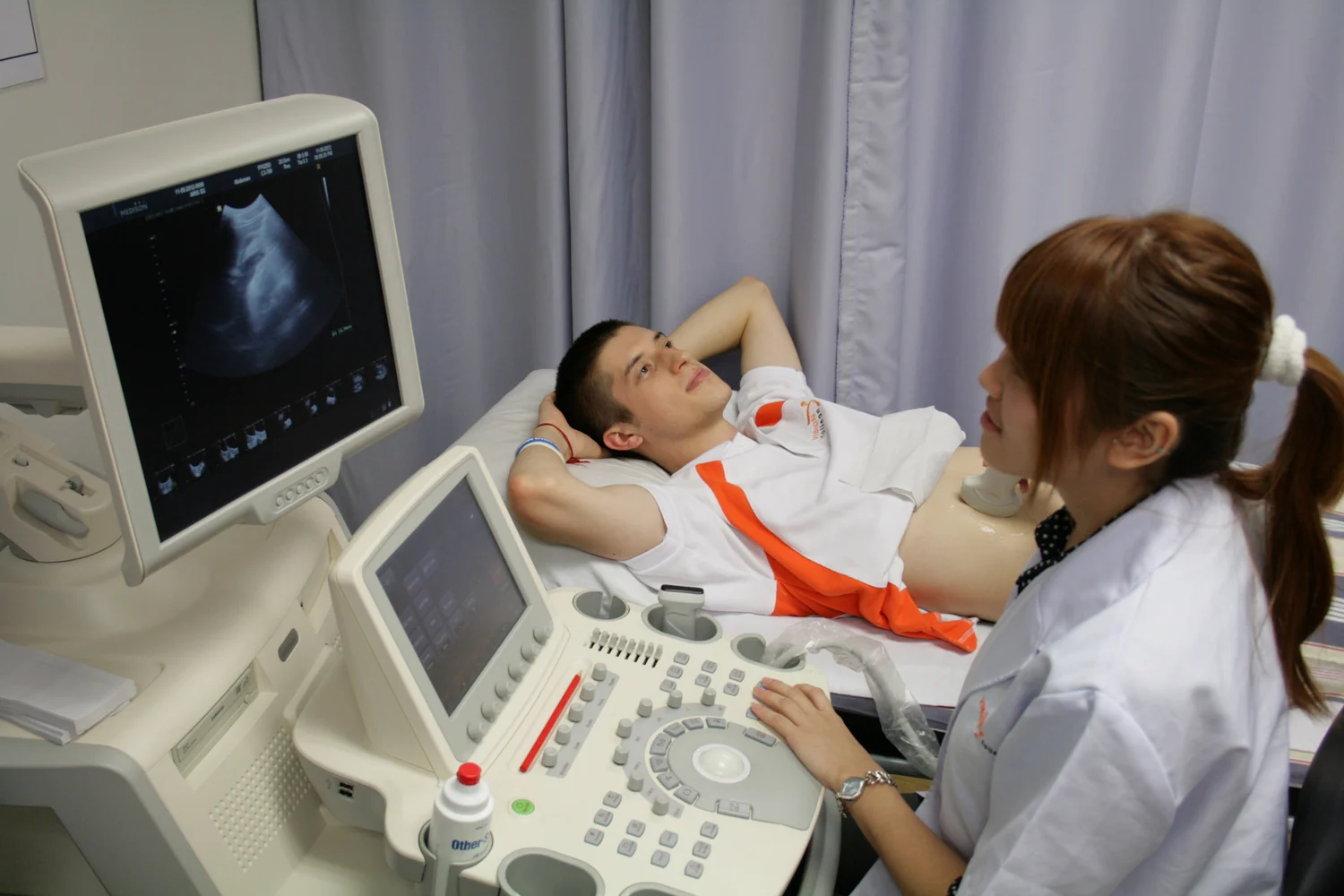
Ultrasound turns returning echoes into a live view of internal structures and motion. Its portability and real-time images can support assessment and guidance, while physics limits what the beam can see.
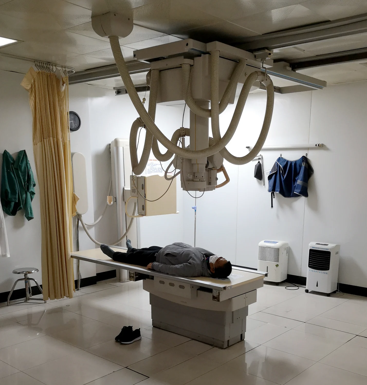
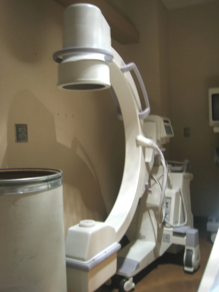
Fluoroscopy creates live images with X-rays, so exposure accumulates during a procedure. Time is one part of dose management, alongside clinical need, image quality, patient factors, and optimized technique.
Both techniques use x-rays, but they collect different views. A plain radiograph compresses anatomy into a projection; CT gathers measurements around the body and reconstructs cross-sections that reveal where structures sit.
MRI maps tissue through magnetic behavior, while optical imaging measures how light is absorbed, scattered, reflected, or emitted. Together, these methods can reveal different aspects of the same biological problem.
Fluoroscopy creates a sequence of X-ray images that lets clinicians watch anatomy, contrast material, or instruments move in real time. Its usefulness comes with a need to balance visibility against radiation exposure.
An implant can respond to MRI’s static and changing magnetic fields and radiofrequency energy, while also affecting the image. Identifying its exact status and conditions is part of safe, useful scanning.
MRI and ultrasound turn different physical interactions into pictures: magnetic behavior of tissue water in one case, returning sound echoes in the other. Their strengths depend on what lies between probe and target.
Medical images must answer a specific question while managing exposure and image quality. CT gathers many X-ray views to build slices, so detail, radiation dose, patient factors, and clinical purpose all matter.
An MRI scanner combines powerful static and changing magnetic fields with radiofrequency energy. Understanding how those forces interact with metal, implants, and the body explains why preparation is part of the imaging procedure.
Images are reconstructed from measurements gathered over time. When a body part moves while those measurements are being assembled, the resulting picture can contain blur, ghosting, or mismatched edges.