How can a probe resting on the skin show a clinician what is happening inside the body right now? It sends sound into tissue and listens for echoes. Because the image updates as those echoes return, ultrasound can add a moving view at the bedside: a changing heart chamber, blood moving through a vessel, or a needle approaching a target.
Echoes become locations
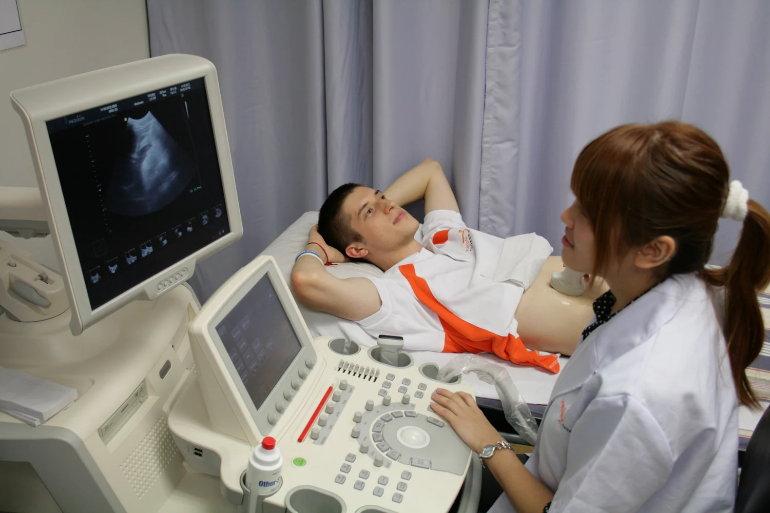
A transducer is the probe that both emits sound waves and detects their echoes. Diagnostic ultrasound uses frequencies above human hearing, commonly in the megahertz range. When a sound wave meets a boundary between materials—such as fluid and soft tissue—some energy reflects back toward the probe. The scanner measures how long the echo takes to return and estimates how far away the boundary lies.
The basic mental experiment is to clap in a large room. A delayed echo suggests a distant wall; a quick echo suggests a nearby one. Ultrasound uses the travel time of sound inside the body, along with the assumed speed of sound, to place returning signals into an image. Their strength also helps determine how bright a feature appears. Gel helps sound pass between skin and probe by preventing air pockets that would block transmission [1].
Why a live image helps
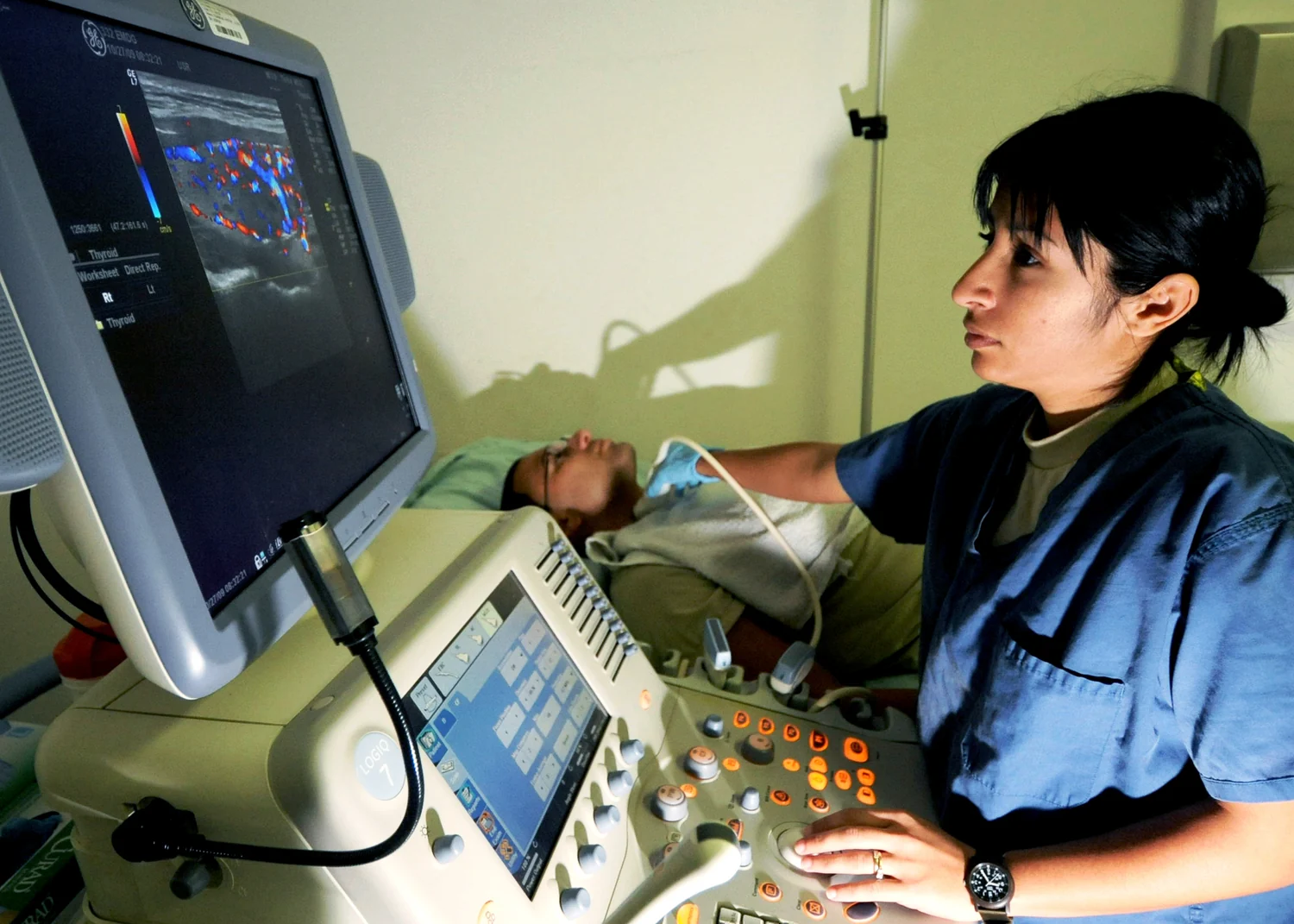
Unlike a still photograph, ultrasound imaging can show motion as it happens. A clinician can watch a heart move, see blood flow, or track the position of a needle during an image-guided procedure. Doppler ultrasound uses changes in returning sound to visualize blood-flow direction and speed; color maps can display this information alongside anatomical structure [2].
That combination can be useful when a question depends on movement or location. For example, while a needle is guided toward a target, repeated images show its changing position relative to nearby tissues. Ultrasound can also be brought into different care settings because its probe is relatively compact. Its value at the bedside comes from pairing a physical examination with an image that can change as the patient or probe moves.
The view has blind spots
Sound does not travel equally well through every material. Bone and air can block or distort the beam, so ultrasound is generally poor for imaging structures behind them. This is why a clear view of some organs is possible while the lungs or adult bones can be difficult to assess. The limitation is physical: echoes from an obstructing boundary may prevent useful information from reaching deeper tissue.
The scan is also an interpretation, not an automatic diagnosis. Probe position, image settings, anatomy, and the clinical question affect what can be seen. Functional images such as elastography estimate relative tissue stiffness, while standard anatomical imaging maps structure; neither picture alone answers every medical question. The method’s strengths and limitations both shape whether it is a suitable tool for a particular examination.
Sound energy, used with purpose
Diagnostic ultrasound uses non-ionizing sound rather than x-rays, and it is generally regarded as safe when used prudently by trained providers. But “non-ionizing” does not mean the beam has no interaction with tissue. Ultrasound energy can produce slight heating and, under some conditions, cavitation—small gas pockets in fluids or tissues. The FDA recommends minimizing exposure while maintaining diagnostic quality [2].
Ultrasound also has therapeutic uses, which should not be confused with imaging. High-intensity focused ultrasound concentrates acoustic energy to heat or alter targeted tissue; diagnostic scans instead use returning echoes to create pictures. In routine imaging, the point is to gather information in real time, not to treat tissue. What ultrasound adds at the bedside is a portable, dynamic window, bounded by the path sound can take through the body.
Explore the physics: an explanatory diagram

Sources & further reading
- Ultrasound NIBIB
- Ultrasound Imaging FDA
Prepared for the independent PSI Center reading library. About edition dates and source checking.
This explainer describes scientific principles. It does not assess individual treatment choices.