How can a flash of light help make an image several centimeters inside the body? In photoacoustic imaging, a brief laser pulse enters tissue and is absorbed by molecules such as hemoglobin in blood. That tiny energy deposit makes the tissue expand momentarily and produce a pressure wave. A sensor listens for the resulting ultrasound, allowing researchers to map where light was absorbed. The method joins two familiar kinds of imaging in an unusual sequence: light provides contrast, while sound carries the signal back out.
A pulse becomes a pressure wave
Light does not travel through tissue in a perfectly straight, transparent path. It can be absorbed, scattered, or reflected, and different tissue components interact with different wavelengths in distinct ways. In photoacoustic imaging, a short laser pulse is sent into the region of interest. Molecules that absorb some of that light warm by a very small amount and expand briefly. Their expansion launches ultrasound waves, which are pressure disturbances moving through tissue.
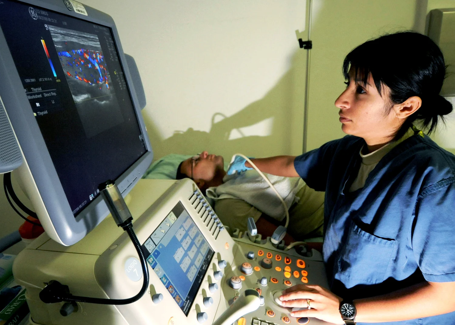
A transducer detects the returning waves. Ultrasound systems ordinarily send sound into the body and interpret echoes from tissue boundaries; here, the tissue itself generates the sound after absorbing light. The transducer records when the waves arrive and where they are detected. By combining those measurements, a computer can reconstruct a map of the sources. The approach is called photoacoustic because photons—particles of light—initiate the event, and acoustic waves carry the measurable signal [1] [2].
Why use sound to carry light’s information?
Light can distinguish substances through their absorption patterns, but scattering makes it difficult to form a sharp conventional optical picture deep in tissue. Ultrasound scatters less strongly in many soft tissues and can travel farther. Photoacoustic imaging exploits this division of labor: light helps mark the location and type of absorbing material, while sound can transport that information toward a detector. It is a useful mental experiment to imagine shining a brief flashlight into fog, then listening for tiny clicks from the spots that absorb the flash.
The signal is not a direct photograph of every cell. It is a reconstruction shaped by the pulse, tissue properties, sensor geometry, and image processing. Different wavelengths can emphasize different absorbers, including hemoglobin, and changes in signal can help estimate blood oxygenation. Those estimates are physical measurements that may illuminate biology; whether a particular measurement can reliably answer a clinical question requires separate validation.
A view of vessels and oxygen
Blood vessels are promising targets because hemoglobin absorbs light. The resulting sound signals can help reveal vessel location and, under suitable methods, characteristics of blood flow. Researchers supported by NIBIB have developed a noninvasive three-dimensional technique called PACTER that visualized vessels and calculated blood-flow speeds in human hands and feet. The work suggests a possible way to study circulation in regions commonly assessed in vascular disease and diabetes, but does not by itself establish a routine diagnostic test or better health outcomes [1].
The distinction between anatomy and function matters. A map can show where vessels lie, while repeated or wavelength-sensitive measurements may provide information about flow or oxygenation. Researchers hope photoacoustic methods could eventually assess oxygen in arteries of the neck or track brain metabolism. These are prospective applications. Demonstrating that an instrument can capture a signal is an early step; clinicians also need evidence that the signal is dependable, interpretable, and useful for decisions.
How it differs from ultrasound alone
Conventional diagnostic ultrasound sends sound waves into the body and uses returning echoes to draw structures. Photoacoustic imaging instead uses a light pulse to generate sound within the tissue. The detector may resemble an ultrasound transducer, but the origin of the signal differs. It is also distinct from optical coherence tomography, which uses reflected infrared light to produce subsurface images rather than converting absorbed light into ultrasound [2] [1].
Because light is scattered and absorbed, depth and image quality depend on tissue and wavelength; the method cannot simply illuminate any deep structure with equal clarity. Its potential strength is contrast linked to light-absorbing molecules, paired with acoustic detection. The research frontier is to refine the physics, sensors, and reconstruction so that meaningful patterns can be measured consistently. Photoacoustic imaging thus offers a bridge between optical contrast and ultrasound’s ability to listen inside soft tissue, with its clinical role still dependent on evidence for each use.
Sources & further reading
- Optical Imaging NIBIB
- Ultrasound NIBIB
Prepared for the independent PSI Center reading library. About edition dates and source checking.
This explainer describes scientific principles. It does not assess individual treatment choices.