Follow the signal from a small imbalance in nuclear spins to spatial encoding and reconstruction, with worked examples showing why frequency, resolution, field of view, noise, and scan time are different quantities.
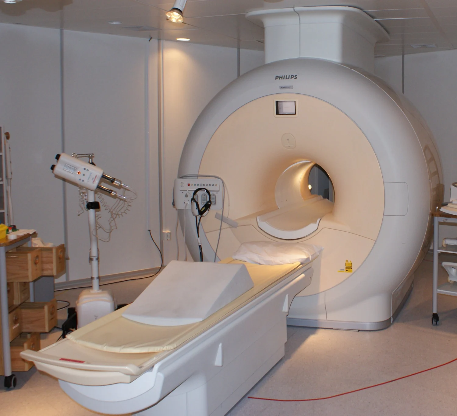
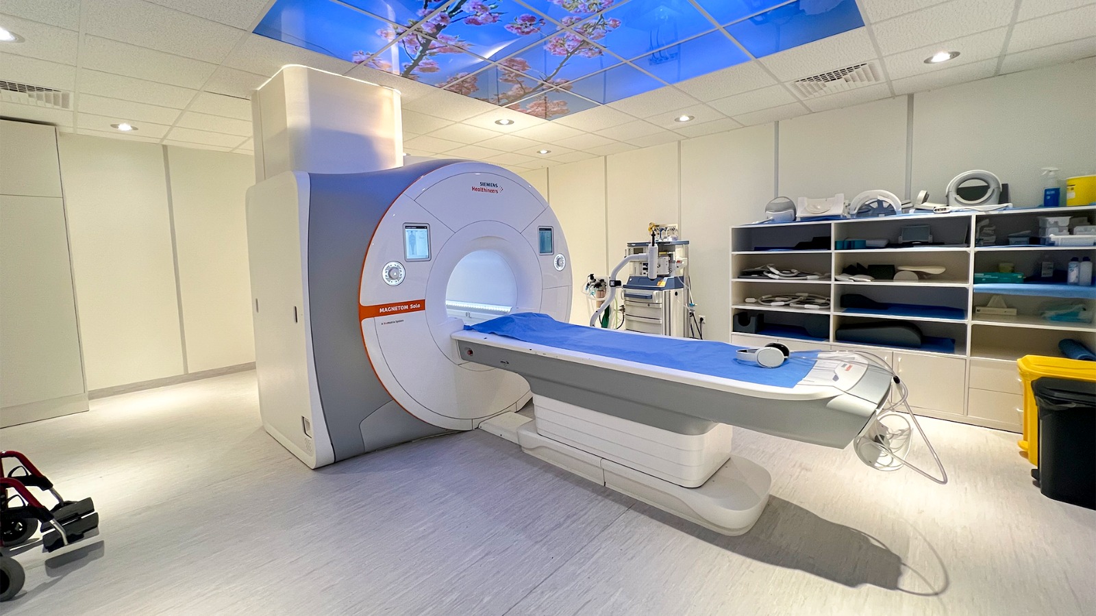
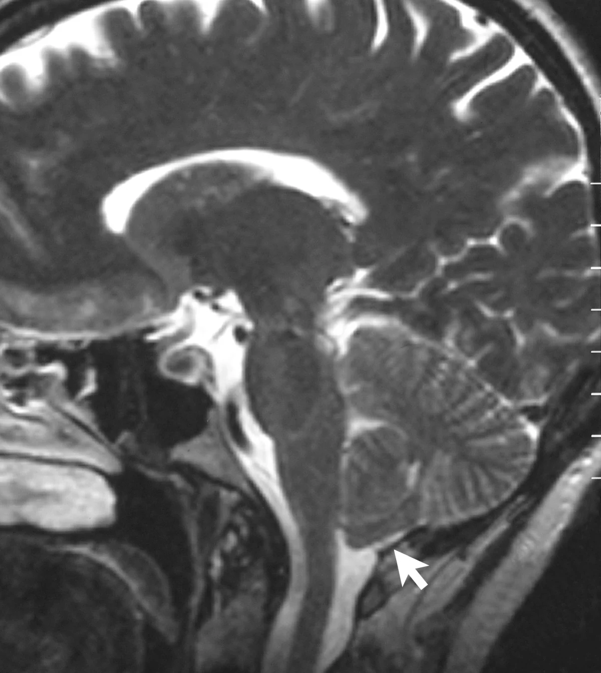
MRI converts tiny changes in hydrogen-proton behavior into spatial detail. Magnetic fields align the signal, radiofrequency pulses disturb it, and carefully timed measurements distinguish tissue environments without x-rays.
MRI contrast comes from how protons respond and recover in different tissue environments, as well as from scan settings and image processing. Optical imaging measures light interactions and can add complementary information at other scales.
Children’s imaging asks a practical physics question: how can a scanner gather enough information to answer a clinical question while accounting for a smaller body and a longer lifetime ahead?
A radiotracer combines a radioactive atom with a carrier that helps determine where it travels. The resulting signal can reveal biological activity, while the carrier and isotope shape what the scan can show.
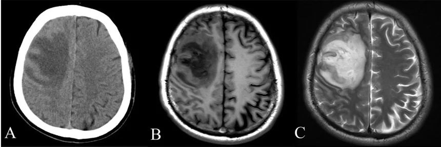
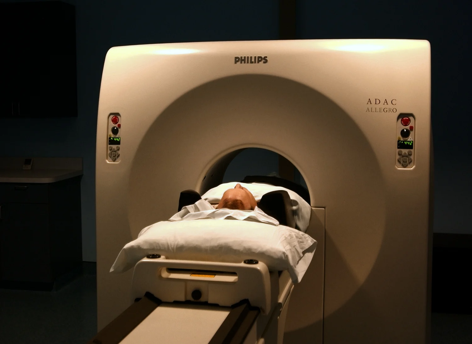
Medical images turn measurements into a grid of picture elements, but visible detail depends on more than screen sharpness. CT and MRI collect different signals and build images through distinct physical processes.
MRI and CT contrast agents make images clearer through different physical interactions. One changes how nearby hydrogen protons return to alignment; the other changes how strongly a material attenuates X-rays.
A sequence of medical images can reveal how anatomy or biological activity changes over time. MRI and nuclear medicine create those images through different physical signals, so their comparisons answer different questions.
SPECT turns emissions from a radiotracer inside the body into a three-dimensional map. Its rotating detectors gather projections from many directions, allowing clinicians to see patterns of function as well as anatomy.
Medical scanners collect indirect measurements, then mathematics turns those signals into a view of the body. Reconstruction combines physical assumptions, computation, and choices about what the resulting image can reliably show.
MRI builds images from signals produced as hydrogen protons respond to magnetic fields and radio pulses. Even small movements can misalign the measurements that a computer combines into a picture.
An imaging algorithm learns statistical patterns from examples. If those examples fail to reflect the people, scanners, or conditions encountered later, performance can become uneven, even when the training set is large.
PET maps where a radioactive tracer participates in bodily processes; CT supplies detailed structure. Their paired images help locate functional signals in anatomy, while each method contributes different information and limitations.
Contrast agents help imaging systems distinguish structures that would otherwise look similar. In CT and X-ray exams, their visibility comes from how they alter the passage of X-rays through selected parts of the body.
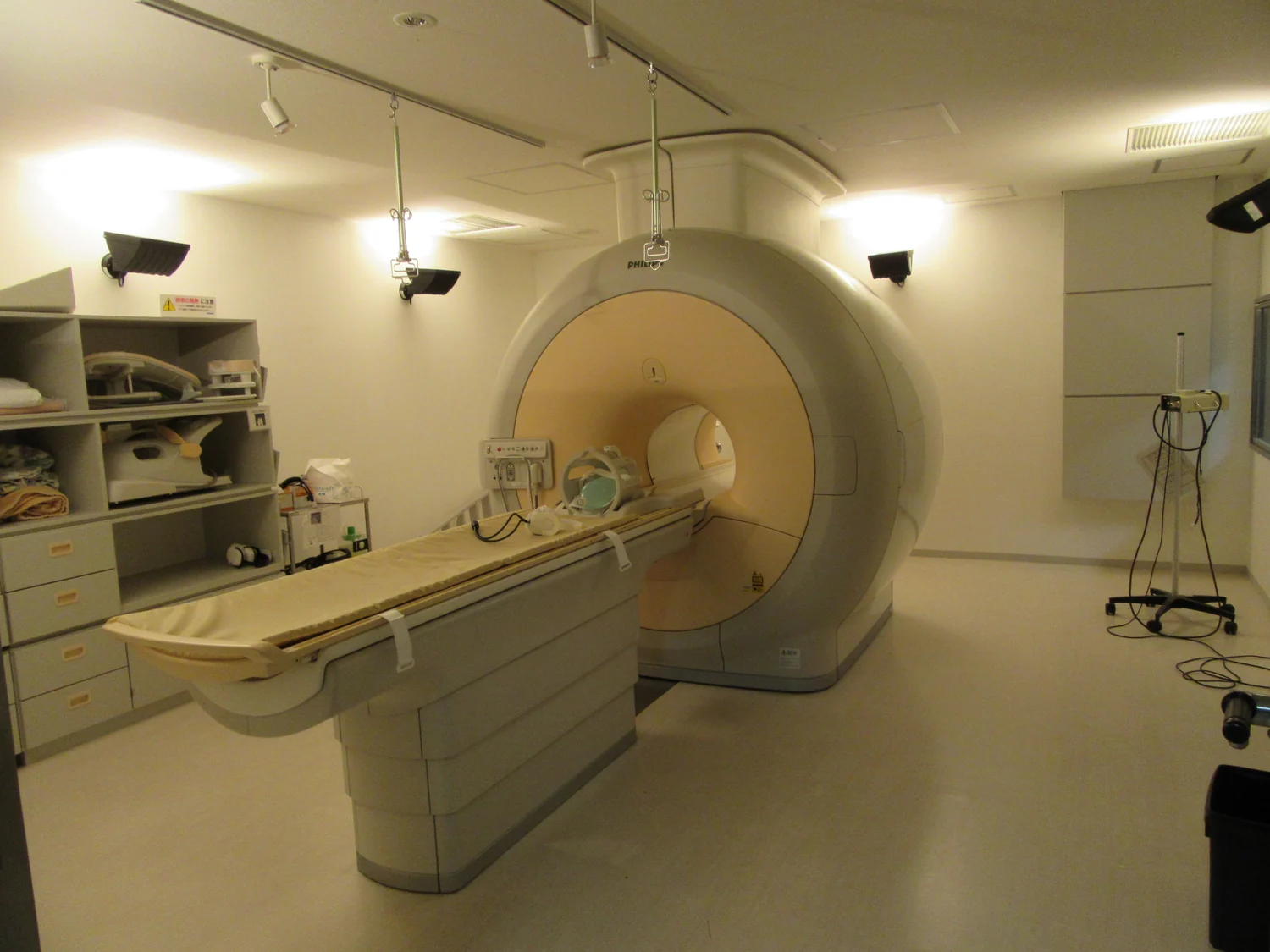
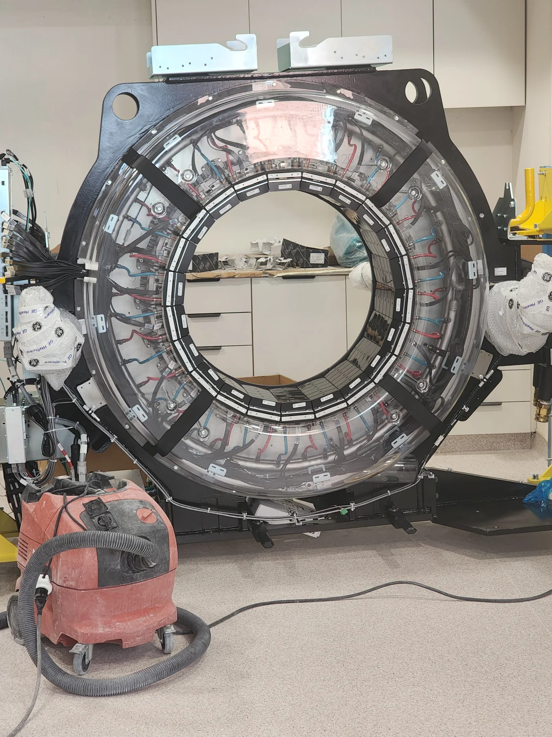
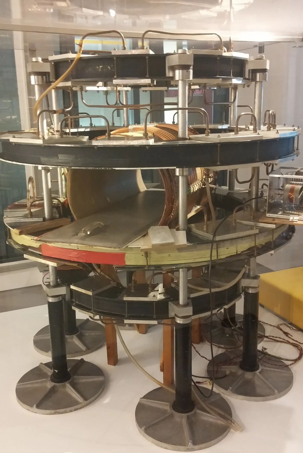
The knocks of an MRI scanner are a by-product of rapidly changing magnetic fields. Their force makes the machine vibrate, while the same fields help locate signals used to build detailed images.
AI can help turn incomplete measurements into usable medical images, but reconstruction depends on the data and assumptions supplied. Better-looking output does not automatically mean more faithful anatomy or a better diagnosis.
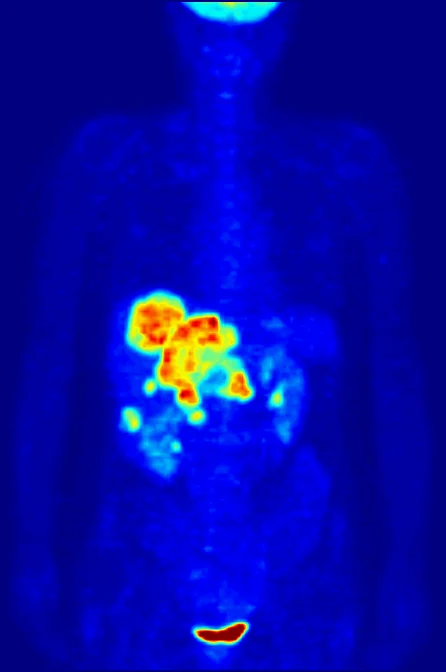
PET converts a tiny event inside the body into a line of likely origin: a positron meets an electron, producing two gamma photons detected in coincidence. Many such events build a functional image.
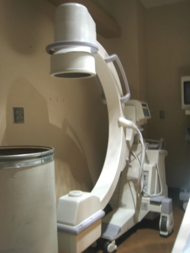
An X-ray image is a map of how much radiation reaches a detector. Bone stands out because its composition and thickness remove more X-rays from the beam than many surrounding tissues do.