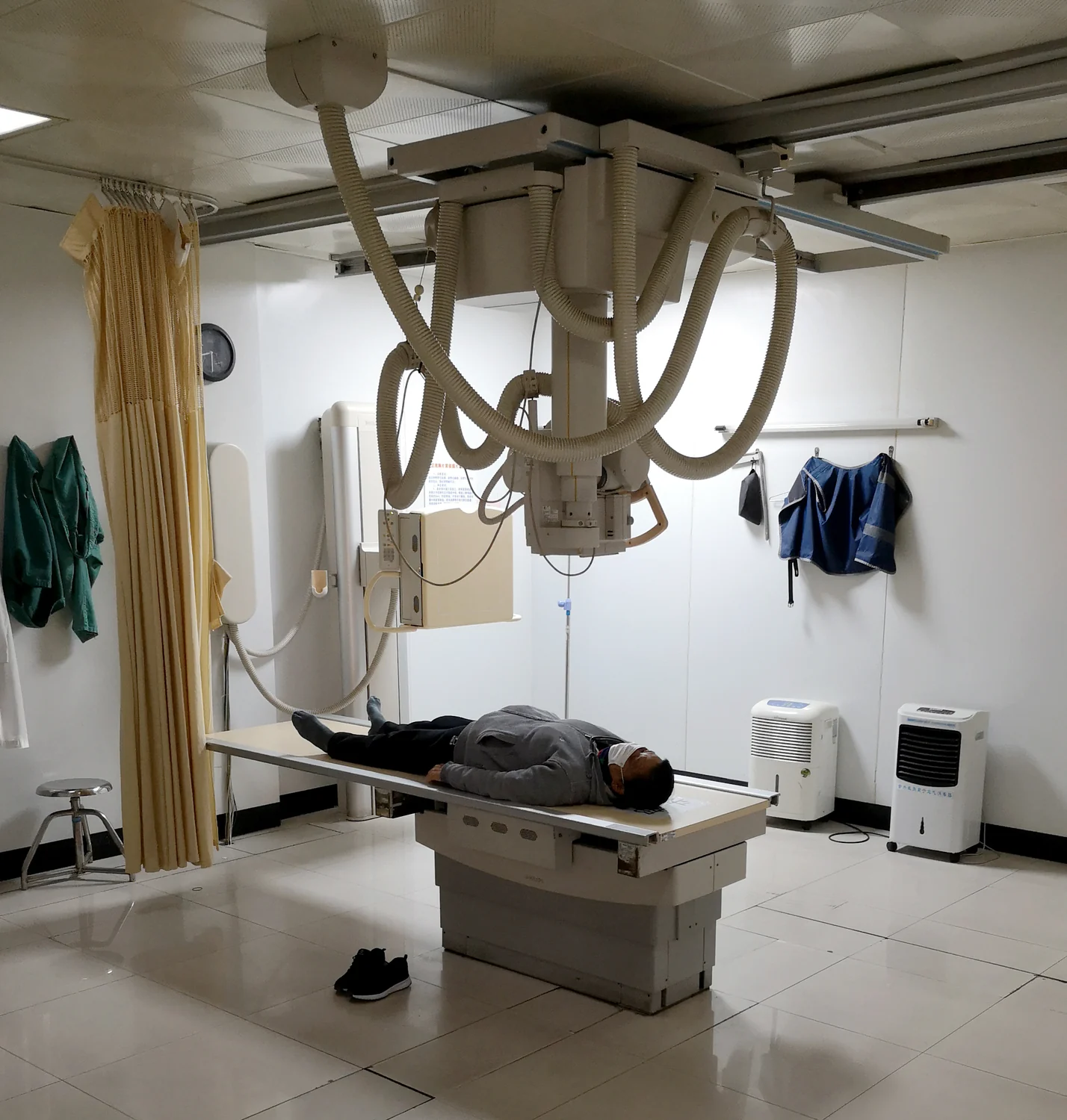
Why does a hand’s skeleton appear pale against the darker outline of its soft tissues? The image is not a photograph of bone reflecting light. It is a record of how an X-ray beam changes as it passes through the body. Bone absorbs and scatters more of that beam than many tissues around it, so fewer X-rays reach the detector behind it. The resulting contrast turns differences in hidden material into visible shapes.
A shadow made by a beam
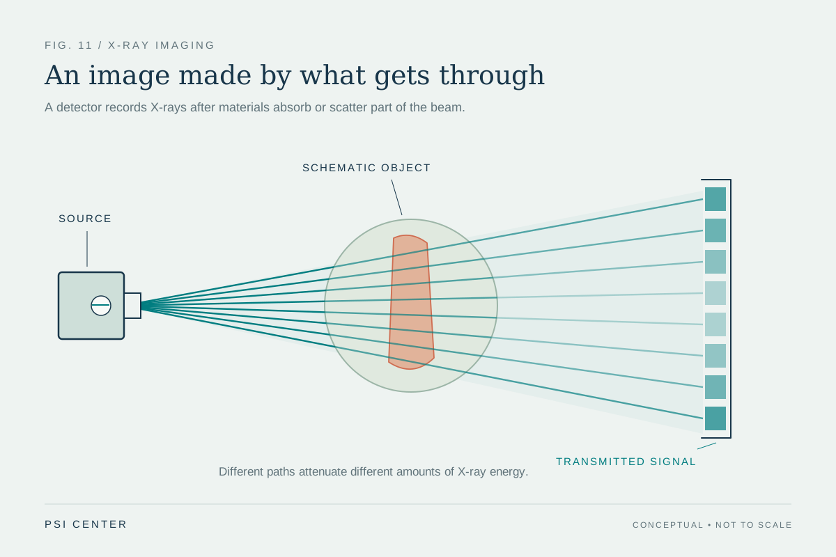
X-rays are electromagnetic radiation, like visible light, but carry more energy and can pass through the body. In a conventional radiograph, an X-ray source sends a burst through the patient toward a detector on the other side. The detector records the pattern that emerges, creating an image of internal “shadows.” Where more radiation arrives, the image is generally darker; where less arrives, it is lighter. [1]
Imagine placing a thin sheet of paper and a thick book in front of a lamp. The paper dims the beam a little; the book blocks more. An X-ray uses a different kind of radiation and detector, but the basic comparison helps: what the detector sees depends partly on what the beam crossed. The image is therefore a projection, with structures at different depths layered over one another.
Why bone removes more X-rays
Bone is mineral-rich and often thicker than nearby soft tissue. Its mineral content, including calcium-containing compounds, interacts with X-rays strongly. Some photons are absorbed, transferring energy to matter; others are scattered away from their original direction. Both effects reduce the number that continue toward the detector. The surrounding muscle and fat generally attenuate the beam less, so more radiation passes through those regions.
This difference is called attenuation: the reduction in a beam’s intensity as it travels through material. It depends not only on what a material is made of, but also on how much material the beam crosses and on the beam’s energy. A dense, broad bone may therefore appear especially bright, while a thin bone or one viewed edge-on can produce a subtler line. Brightness is a comparative signal, not a direct color or a simple measure of bone health. [2]
A two-dimensional overlap
A plain X-ray compresses a three-dimensional body into a two-dimensional image. If a wrist is turned, bones that were separated in one view can overlap in another. That overlap can hide a small fracture or make normal anatomy look crowded. Clinicians often use more than one projection, shifting the angle so the same structures cast different shadows. Each view adds information about position, though it remains a projection rather than a full depth-resolved map.
Computed tomography addresses this limitation by collecting many X-ray projections as the source and detectors rotate around the body. A computer reconstructs cross-sectional slices, which can be viewed individually or assembled into a three-dimensional representation. CT is still X-ray imaging, but its multiple angles help separate structures that a single radiograph piles together. [1]
Contrast, usefulness, and exposure
Soft tissues can be harder to distinguish on X-rays because their attenuation may be similar. In some examinations, contrast agents that stop X-rays more strongly help outline structures such as blood vessels or parts of the digestive tract. The material changes the beam’s passage, and the detector records that difference. The result depends on the question being asked; a contrast study and a plain bone image are not interchangeable techniques. [2]
X-rays are ionizing radiation, meaning they carry enough energy to potentially damage DNA. The FDA describes the cancer risk from medical imaging as generally small, with risk depending on factors such as exposure, age, and body region. Imaging teams weigh whether an exam is needed to answer a clinical question and use settings intended to keep exposure as low as reasonably achievable while retaining useful image quality. Bone’s brightness comes from physics; deciding when an image is worthwhile is a matter of clinical judgment. [2]
Explore the physics: an explanatory diagram
Sources & further reading
- Medical X-rays NIBIB
- Medical X-ray Imaging FDA
Prepared for the independent PSI Center reading library. About edition dates and source checking.
This explainer describes scientific principles. It does not assess individual treatment choices.