How can a camera outside the body locate a molecule moving through tissue? In SPECT, a radioactive tracer emits gamma rays that pass out of the body and reach detectors arranged around the patient. The scanner records many views as its cameras rotate, then a computer reconstructs a three-dimensional picture of where the tracer accumulated. The image is therefore a map of a biological process, not simply a photograph of anatomy.
A tracer follows a biological route
A radiotracer combines a carrier molecule with a radioactive atom. The carrier is selected for how it behaves in the body, while the radioactive component provides a detectable signal. Depending on the question, tracers may be administered in different ways, including injection, inhalation, or oral intake [1]. As the molecule moves or binds in tissue, the pattern of its emissions offers clues about the process being studied.
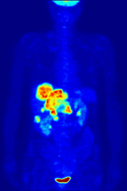
For example, NIBIB describes radiolabeled red blood cells used to trace the source of intestinal bleeding: cameras can follow the cells’ gamma emissions, and an accumulation in the intestine can help indicate where blood is escaping [1]. The physical logic is indirect but powerful. The detector does not see blood itself; it detects emissions from labeled cells whose movement carries information about the underlying event.
Why the camera rotates
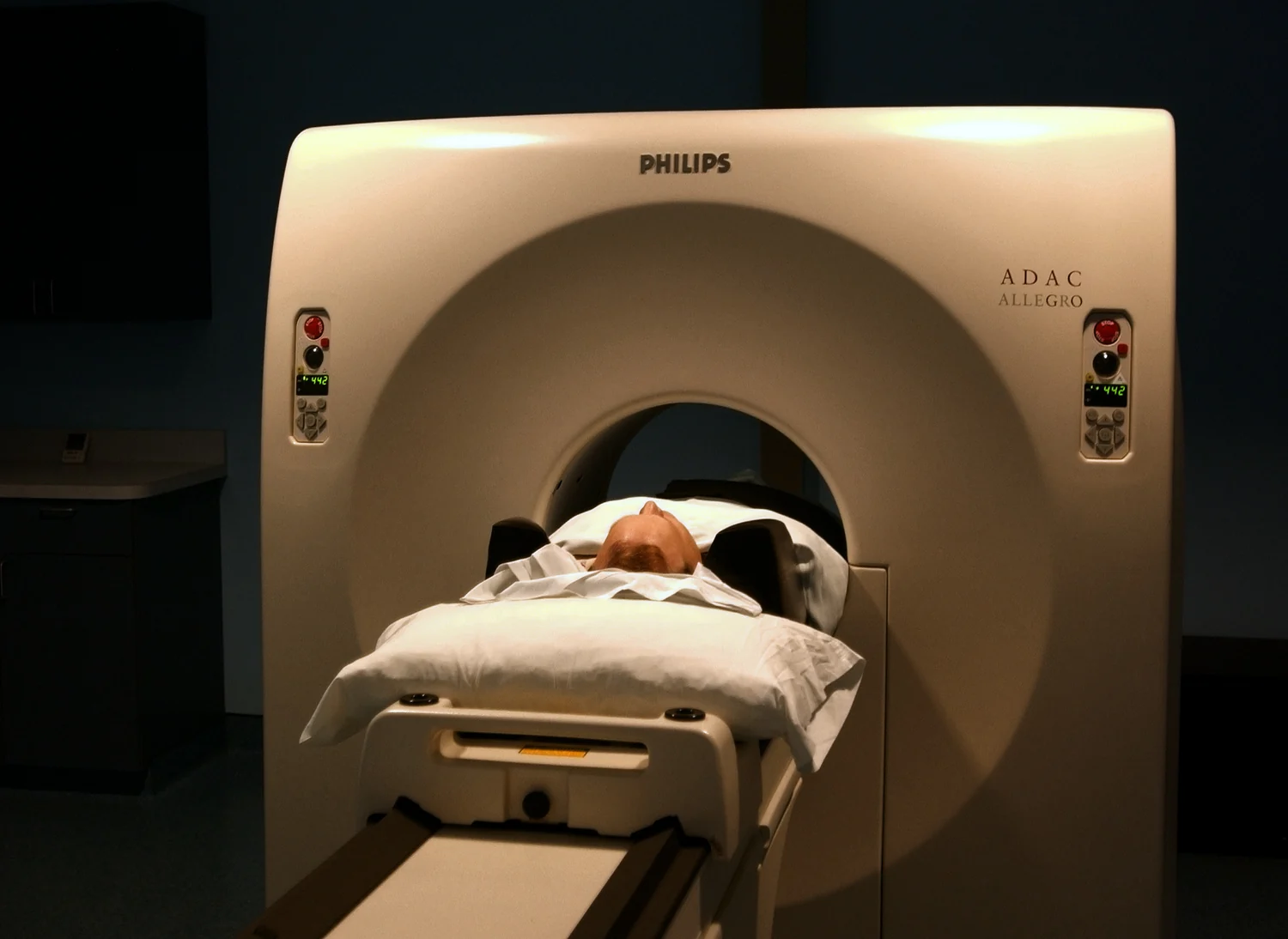
A single detector view compresses depth. A bright region in a flat projection could come from a shallow structure or from one deeper in the body, because both lie along the same line of sight. SPECT addresses that ambiguity by collecting many two-dimensional projections from different angles as gamma camera detectors rotate around the patient [1].
A computer combines those views to estimate the three-dimensional distribution of the tracer. A mental model is to imagine taking silhouettes of a vase from many positions around a room: each view hides some details, but the set constrains the shape. SPECT reconstruction uses measured emission patterns rather than visible silhouettes, and the result depends on how those signals are collected and processed.
Function comes into focus
Because the tracer’s distribution reflects its biological route, SPECT can reveal functional patterns. NIBIB describes its use with particular radiopharmaceuticals to assess heart disease, bone disorders, gallbladder disease, intestinal bleeding, and certain movement disorders [1]. The choice of tracer determines what biological process the scan can meaningfully represent; one tracer’s image cannot answer every question.
This is different from imaging that primarily maps anatomy. A functional image can show where a molecule travels or accumulates, while a structural image shows physical form. Combining modalities can add context, but the interpretations remain distinct. MRI, for example, uses magnetic fields and radio waves rather than radioactive tracers or ionizing x-rays; SPECT depends on emissions from a radiotracer.
Signal and radiation are linked
The same radioactive decay that makes a tracer detectable also means the patient receives ionizing radiation. Ionizing radiation has enough energy to damage DNA; x-rays and gamma rays are examples, and some medical imaging procedures involve exposure [2]. In nuclear medicine, the amount and type of radiopharmaceutical are selected for the clinical question, balancing a useful signal against exposure.
NIBIB notes that radiation doses from most diagnostic radiopharmaceutical studies are generally comparable to common imaging exposures, while also emphasizing minimizing the amount needed for a diagnostically useful examination [1]. The reconstructed image should therefore be understood as the outcome of a designed measurement: a tracer supplies biological specificity, gamma detectors collect projections, and computation reconstructs the spatial pattern. SPECT’s strength is following function through that chain, with interpretation grounded in what the chosen tracer actually tracks.
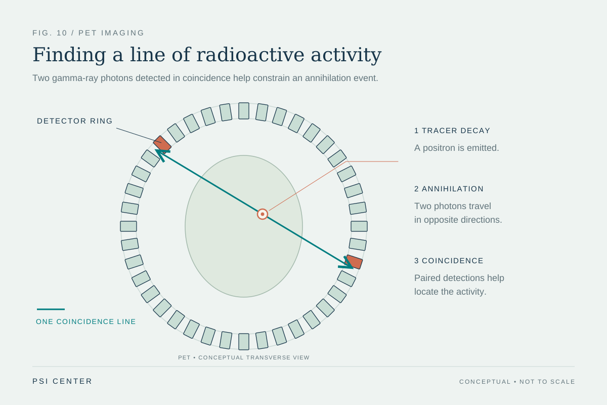
Explore the physics: an explanatory diagram
Sources & further reading
- Nuclear Medicine NIBIB
- Radiation National Cancer Institute
Prepared for the independent PSI Center reading library. About edition dates and source checking.
This explainer describes scientific principles. It does not assess individual treatment choices.