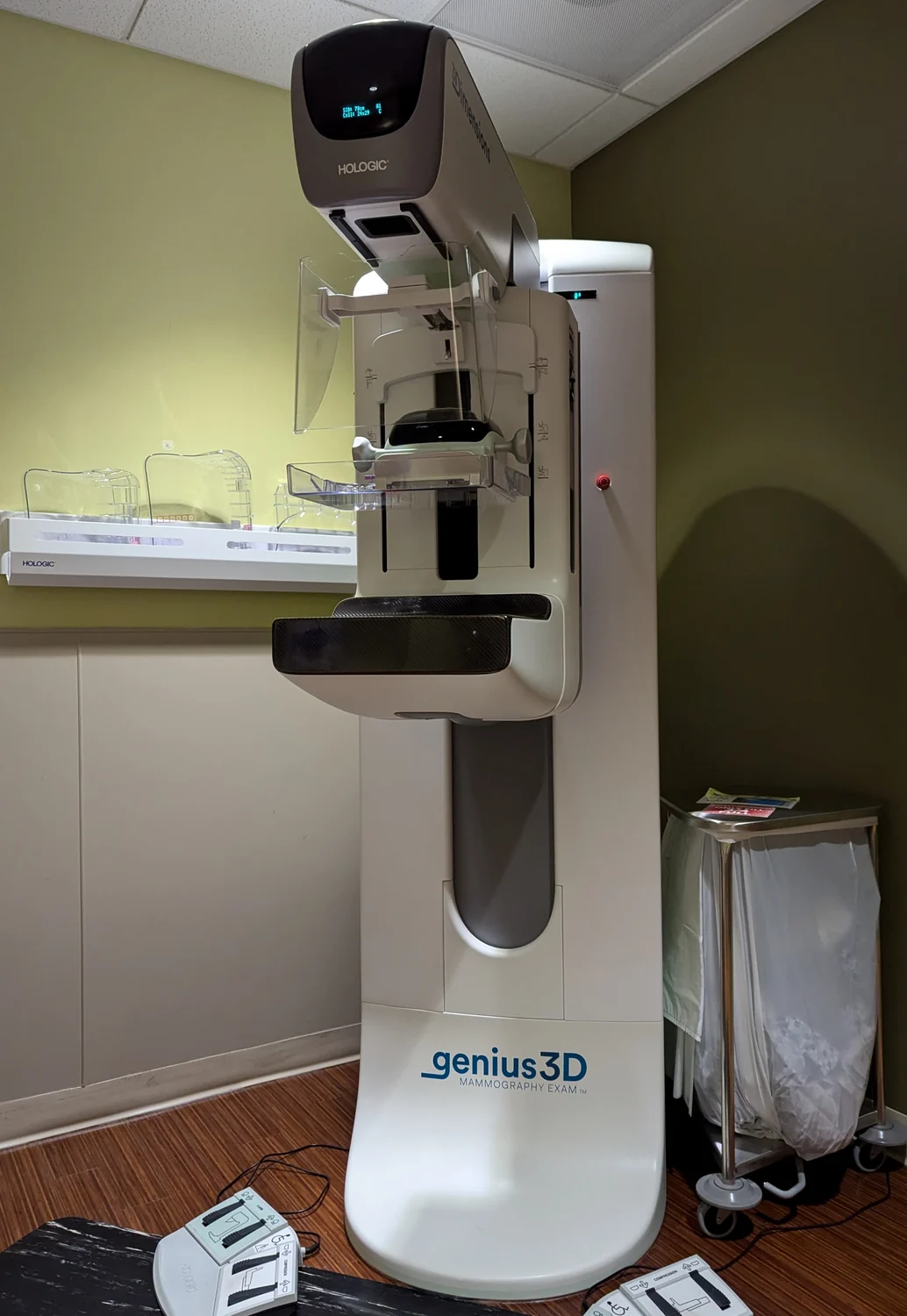
Would an image-reading algorithm recognize the same finding if the scan came from a different hospital or patient group? It can only learn from the examples it receives. Medical images vary with anatomy, equipment, acquisition choices, and disease, while training labels reflect human judgments and criteria. A large collection may still leave out important variation. Representative data help test whether a model’s apparent skill travels beyond the examples on which it learned.
Examples become learned patterns
Machine learning is an approach in which an algorithm analyzes data and makes predictions by identifying patterns. Deep learning uses many layers of computation to learn more complex patterns from large, often unstructured datasets [1]. In medical imaging, the input might be an X-ray, CT scan, or MRI, and the output could be a classification, a highlighted region, or a prediction intended to support a clinician.
The model does not understand an image in the same way a radiologist does. It adjusts internal parameters so that its outputs align with examples and labels in its training data. If a feature appears consistently alongside a label, the model may rely on that feature—even if it is incidental, such as a scanner artifact or a marking associated with a hospital workflow. The algorithm can therefore appear accurate while learning a shortcut that fails in a new setting.
Data quality includes the labels
A useful imaging dataset takes substantial preparation. NIBIB describes curation, de-identification, quality assessment, annotation, and labeling by clinical experts as steps in creating high-quality data for AI development [1]. Curation means selecting and organizing the material; annotation adds information about what is visible or clinically relevant. Labels provide the target the algorithm is trained to predict.
These steps shape the model’s learning. If experts disagree about a subtle finding, a dataset may contain inconsistent labels. If low-quality scans are excluded, the resulting model may perform less reliably on lower-quality images encountered in practice. If cases are labeled using a definition that differs from later clinical practice, the model may learn the wrong target. A very large dataset can reduce some forms of random uncertainty, but volume alone does not correct biased sampling or mistaken labels.
Representation is a performance question
Representative data should reflect the range of people and circumstances where a tool is meant to be used. That includes variation across populations and potentially across scanners, clinical sites, and image quality. If one group is sparsely represented, researchers may have too little evidence to know whether performance differs for that group. NIBIB specifically notes the need for AI tools to be fair and to perform equally across groups and populations [1].
Imagine training a model mostly on images from one hospital, then using it in another hospital with different equipment and patient populations. A model might mistake image-processing differences for meaningful anatomy, or encounter patterns it rarely saw during training. Evaluation on data separated from training—and on relevant subgroups—helps expose such gaps. The goal is not merely a high average score, but evidence that the tool behaves reliably across the situations included in its intended use.
Support requires inspection
A model can assist with imaging work by drawing attention to subtle changes, but its output still needs evaluation in context. Some AI systems are difficult to interpret because they do not show how they arrived at a prediction. NIBIB describes explainable-AI research that links concepts to corresponding clinical images, helping people inspect how a model reached a conclusion [1]. Such methods can support review, though explanation alone does not prove that the prediction is correct.
Medical devices range from tools that monitor or support bodily functions to wearable sensors and implanted devices [2]. Imaging algorithms likewise occupy a defined role: they may aid assessment, but their performance depends on the data and task for which they were developed. Representative datasets, careful labels, external evaluation, and human inspection address different failure points in the chain. None guarantees perfect results; together they make it more possible to see where a model works, where it may falter, and what claims the evidence can support.
Sources & further reading
Prepared for the independent PSI Center reading library. About edition dates and source checking.
This explainer describes scientific principles. It does not assess individual treatment choices.