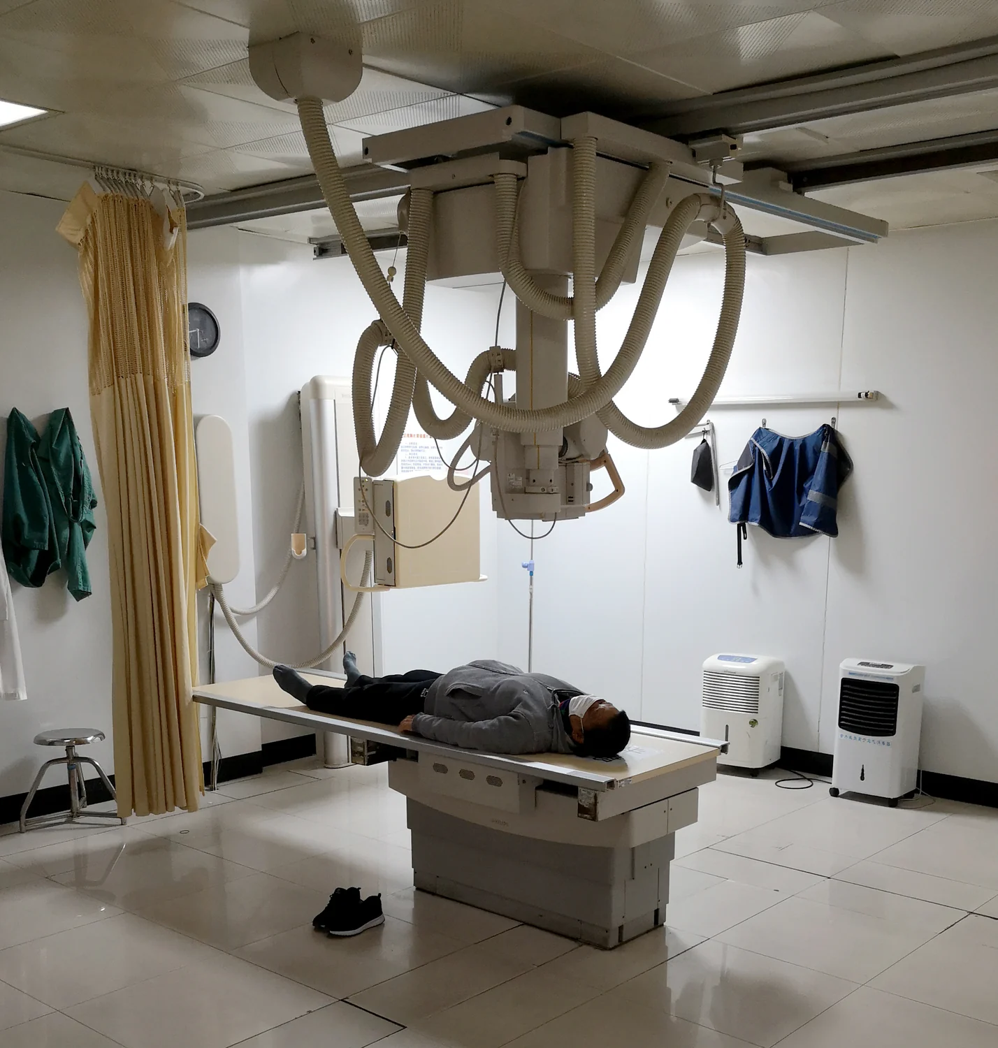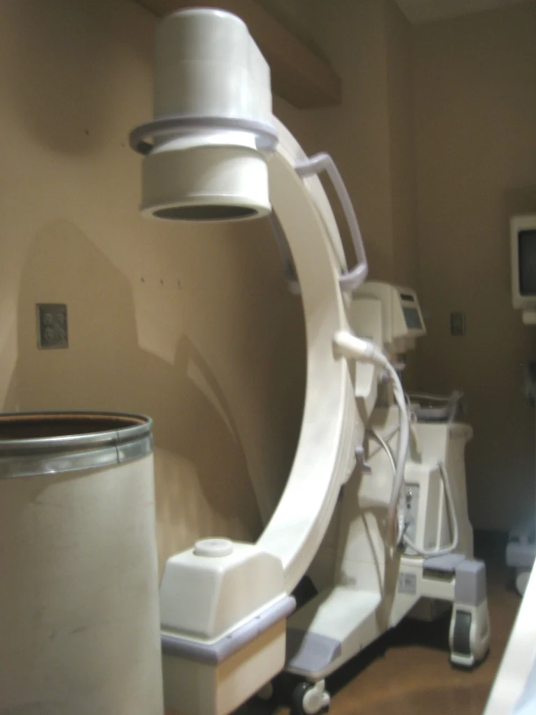
Why does the time spent using fluoroscopy matter if the monitor shows only an image? Fluoroscopy displays a continuing X-ray picture that lets a clinical team watch movement inside the body or guide an instrument. Because the image is produced while X-rays pass through the patient, longer use can mean more exposure. Understanding that link helps explain why teams balance useful visibility against radiation dose.
A live image requires repeated exposure
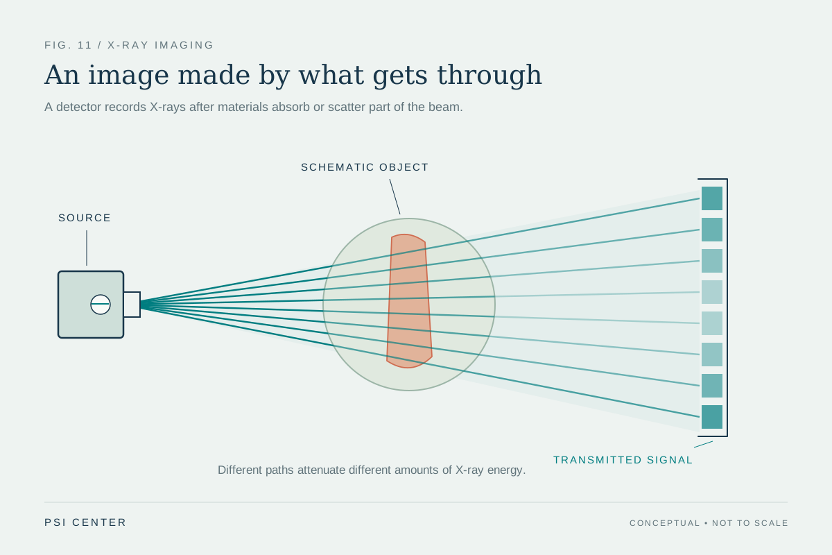
In fluoroscopy, an X-ray beam passes through the body, and the transmitted pattern appears on a monitor. The continuous image can show the movement of a body part, contrast agent, or instrument [1]. This is useful for tasks such as guiding a catheter or placing a device, where the operator needs to see changes as they happen.
A still radiograph records a single image, while fluoroscopy presents an ongoing view during a procedure [2]. The practical difference is that fluoroscopy can keep supplying images while the team works. In complex interventions, longer periods of use can lead to relatively high radiation doses [1]. Time matters because the imaging beam is part of the process that produces each view.
Dose and risk are connected
X-rays are ionizing radiation: they carry enough energy to potentially damage DNA. Exposure can slightly increase the chance of cancer later in life, while tissue effects such as skin injury are associated with higher exposures and are uncommon in many imaging exams [2]. The FDA notes that long, complex interventional fluoroscopy procedures may, in some circumstances, deliver skin doses high enough for tissue effects [2].
The probability of harm from a fluoroscopic procedure is statistically very small, and when the procedure is medically needed, its benefits outweigh the radiation risks [1]. That balance is not a reason to ignore exposure. It is a reason to use the imaging that answers the clinical question and to manage the beam carefully while preserving the visibility needed for the procedure.
Time is one part of optimization
The FDA describes optimization as using the lowest radiation dose that still gives image quality adequate for the task [2]. In a live procedure, that means the team must consider how much imaging is needed to guide the next action. Fluoroscopy should be performed for the shortest time necessary with the lowest acceptable exposure [1]. The aim is not simply the fewest seconds; it is sufficient information with minimized exposure.
Other factors also shape dose. The FDA notes that dose varies by procedure and that risk depends on exposure as well as patient age, sex, and the body region involved [2]. Beam location and direction affect which tissues are exposed. This is why exposure time cannot be treated as a complete measure on its own: procedure complexity, equipment settings, and patient factors also matter.
Visibility has a clinical purpose
Fluoroscopy can help clinicians direct catheters through vessels, place stents, visualize blood vessels, and guide orthopedic procedures [1]. In each case the image is useful because it can guide an action in real time. The relevant question is whether the imaging provides information needed to diagnose, treat, or guide the procedure, and whether the dose is optimized for that purpose [2].
A useful mental picture is a moving map used while navigating a difficult route. Looking at the map can prevent mistakes, but watching it continuously when no decision depends on it adds little information. Fluoroscopy’s live view is valuable when it guides a step; exposure management asks how to keep the view useful while avoiding unnecessary beam use.
Explore the physics: an explanatory diagram
Sources & further reading
- Fluoroscopy FDA
- Medical X-ray Imaging FDA
Prepared for the independent PSI Center reading library. About edition dates and source checking.
This explainer describes scientific principles. It does not assess individual treatment choices.