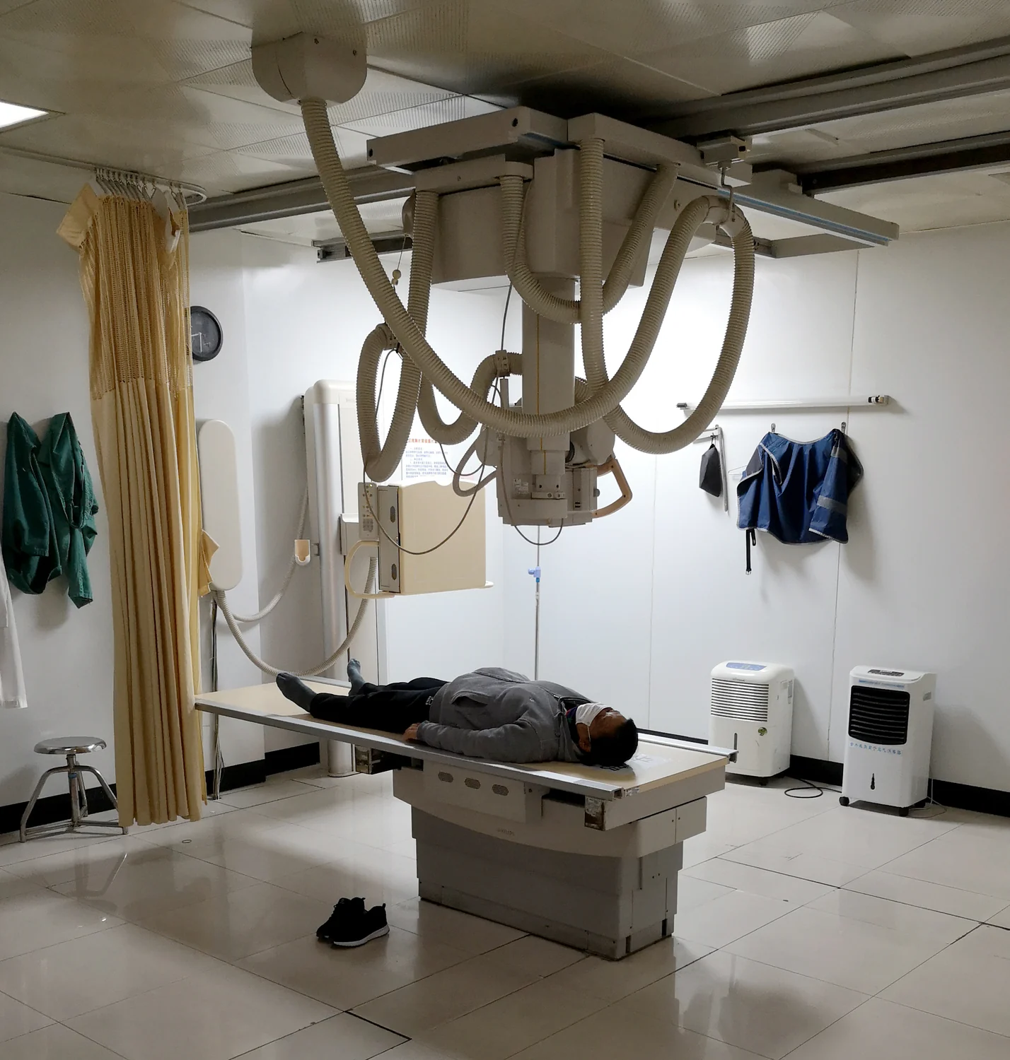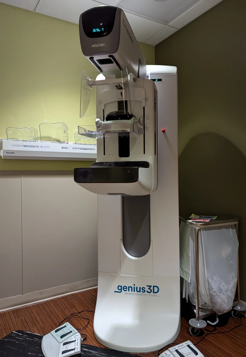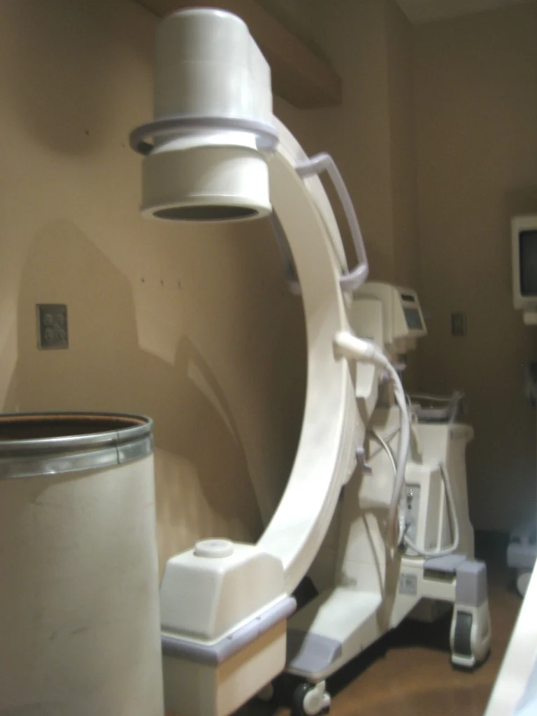
How can a clinician watch a catheter move through a blood vessel while it is still inside the body? Fluoroscopy answers with a moving sequence of X-ray images. Each frame is a snapshot made as X-rays pass through the patient, and together the snapshots reveal motion. The technique makes hidden movement visible, but the image is produced with ionizing radiation, so both image quality and exposure matter.
A movie made from X-rays
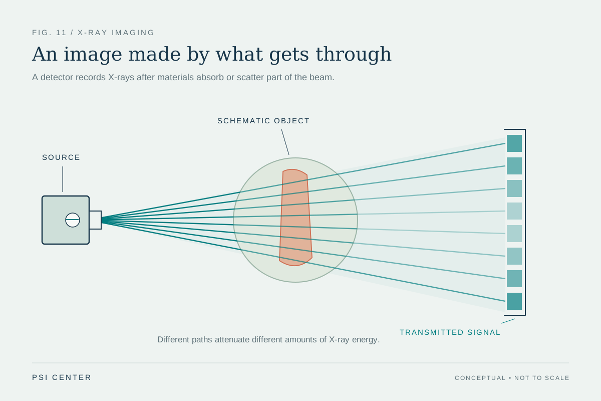
An ordinary radiograph records how much X-ray beam reaches a detector after passing through the body. Different tissues absorb or scatter different amounts, creating contrasts that appear as shadows. Fluoroscopy applies the same basic principle repeatedly: the system displays a continuing series of images on a monitor, much like an X-ray movie [1].
The moving picture is especially useful when position changes from moment to moment. A clinician can see a contrast agent travel through a passage, follow a catheter along a vessel, or check the placement of a device. The image does not show every structure equally well; it shows differences in how strongly they affect the beam. Contrast material can make certain spaces easier to follow by changing how much X-ray reaches the detector.
Following a tool through the body
Picture a flexible catheter entering a blood vessel. Without a live image, the operator would know where the instrument began but have limited information about its route through branching anatomy. Fluoroscopy supplies updated views as the catheter advances. If it turns toward an unintended branch, the changing image can reveal that shift while adjustment is still possible. The image guides an action rather than merely documenting anatomy after the fact.
Fluoroscopy is used in varied procedures, including catheter manipulation, angiography, stent placement, and orthopedic work. Its role depends on what needs to be seen: a moving instrument, a changing flow of contrast, or the alignment of bone fragments. The imaging team selects views and exposure settings suited to the task. Repeated images provide temporal information, but the number and duration of exposures also shape the radiation dose.
Why dose and image quality are linked
X-rays are electromagnetic radiation with enough energy to ionize atoms, meaning they can remove electrons. That property makes them useful for imaging but also means exposure can affect living tissue. In fluoroscopy, dose varies with the procedure; long, complex interventions can involve relatively high exposures. Potential effects include skin and underlying-tissue injury and a small increase in the chance of cancer later in life [1].
Reducing exposure is not simply a matter of making every image faint. If the moving anatomy or instrument becomes impossible to distinguish, the image may no longer serve its purpose. The goal is an acceptable image for the clinical question with exposure kept as low as reasonably possible and time kept to what is necessary. This is why teams use trained operators, optimized protocols, and dose monitoring.
Motion is information, not decoration
A fluoroscopic sequence can show how a structure behaves, not just where it sits. In a swallowing examination, for example, a moving contrast pattern can reveal passage through the throat and esophagus. In a procedure, the sequence can show whether a device follows the intended route. Still, a moving image is not automatically a complete diagnosis: interpretation depends on the clinical question and the anatomy visible in that projection.
Fluoroscopy belongs to the family of X-ray imaging. A continuous or rapidly repeated detector record can show internal movement in real time, while other X-ray methods emphasize still images or cross-sectional slices [2]. The central tradeoff is therefore concrete: enough successive information to guide a decision, while avoiding unnecessary exposure. The live image is powerful precisely because it connects what the clinician does with what the body does next.
Explore the physics: an explanatory diagram
Sources & further reading
- Fluoroscopy FDA
- Medical X-rays NIBIB
Prepared for the independent PSI Center reading library. About edition dates and source checking.
This explainer describes scientific principles. It does not assess individual treatment choices.