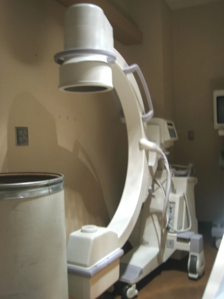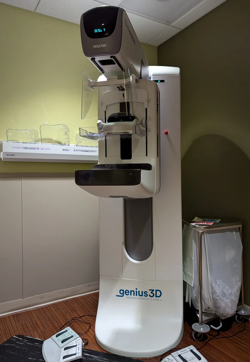
Why does a mammogram require the breast to be positioned in a particular way when an ordinary chest X-ray does not? The answer begins with the imaging question: mammography seeks detail in breast structures, including small abnormalities, across tissue that can overlap in a two-dimensional picture. It uses the same basic physics as radiography, but its target shapes how the image is made and checked.
The shared physics of an X-ray
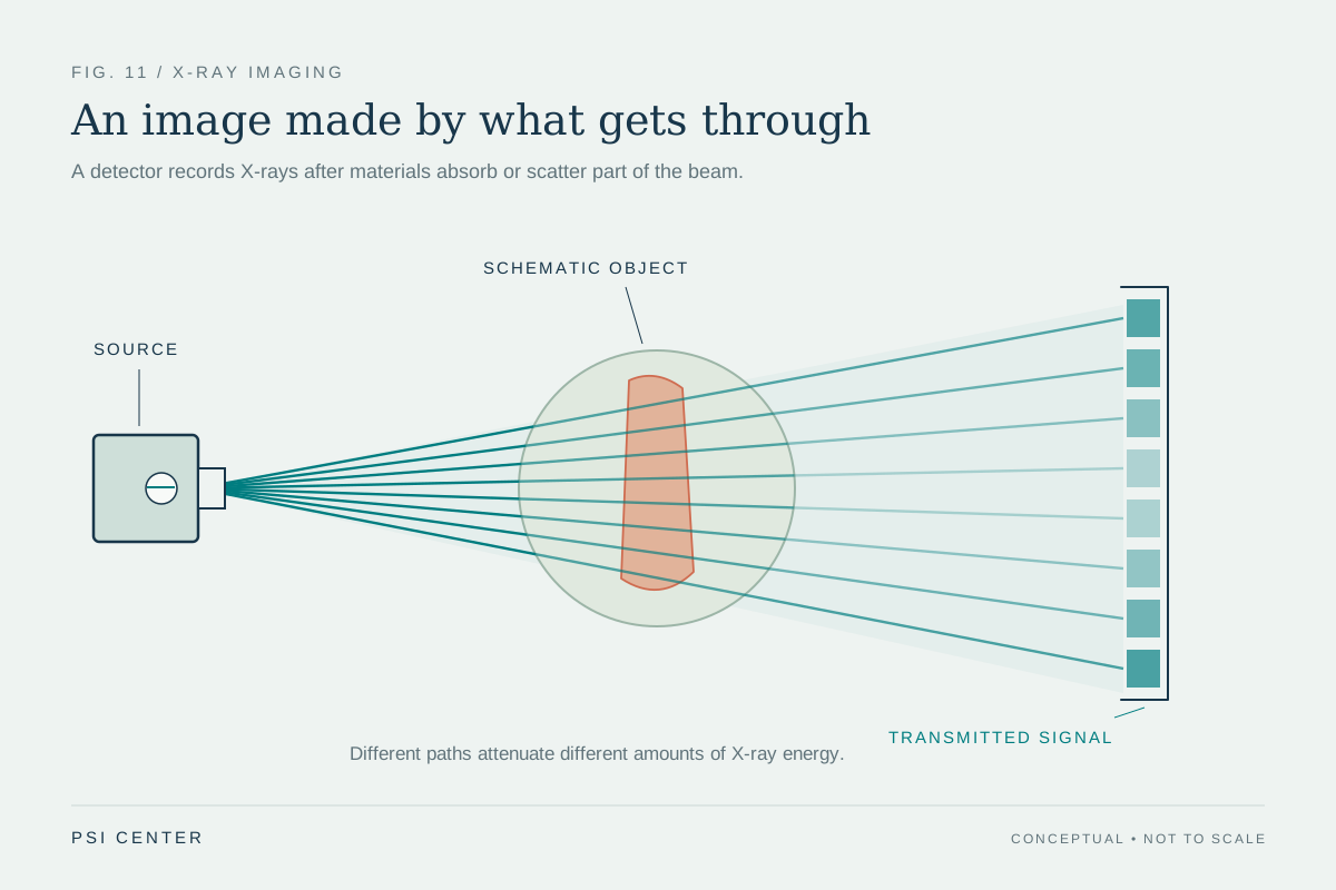
An X-ray beam passes through the body. Some photons are absorbed or scattered by tissue; the pattern that emerges reaches a detector and is recorded. Differences in how much radiation gets through create contrast in the image. Mammography is a special type of radiography aimed at imaging the internal structures of the breast [2].
X-rays are ionizing radiation, meaning their energy can potentially damage DNA. That does not make every exposure equally risky: the concern depends on factors including dose and the person’s age and body region. The FDA describes the radiation dose from mammography as small and says the benefit of early detection outweighs the exposure risk [1].
Why the breast needs a tailored view
Breast tissue contains structures that can be subtle and can overlap when viewed from one direction. Positioning and compressing the breast helps spread tissue for imaging and reduce motion, making internal detail easier to record. The objective is not simply to make a generic picture, but to capture images suitable for examining breast structures.
This is a useful distinction from a chest radiograph, which creates a broad projection of the chest. A mammogram concentrates on the breast and uses equipment and quality processes suited to that task. The FDA describes both screen-film mammography, where an image is recorded on film, and full-field digital mammography, where a receptor’s signal is converted into a digital picture [1].
One projection, many projections, or a live view
A conventional radiograph records an image for later evaluation. Mammography belongs to this category. Computed tomography (CT) records many X-ray projections as a detector moves around the body; a computer reconstructs cross-sectional slices. Fluoroscopy displays a continuing X-ray image on a monitor, allowing clinicians to watch motion or guide a procedure [2].
A mental model is to compare looking at a loaf from the side with slicing it and inspecting each cross-section. A projection combines structures along the beam’s path; CT uses multiple angles to estimate where structures lie in depth. Fluoroscopy adds time, showing movement as it happens. These different tasks explain why the modalities use X-rays differently and why CT generally involves more exposure than conventional radiography.
Quality and dose are linked to the question
An image must be good enough to answer its clinical question, while unnecessary radiation exposure should be avoided. The FDA describes two radiation-protection principles: justification, meaning the exam should be clinically warranted, and optimization, meaning settings should use the lowest dose that yields adequate image quality [2].
Mammography also has dedicated quality standards. The Mammography Quality Standards Act established baseline requirements for radiation dose, personnel, equipment, and image quality [1]. Good image quality matters because early detection depends on seeing structures clearly; radiation control matters because X-rays carry a small potential harm. The balance is specific to the exam and its purpose, not a claim that every imaging method is interchangeable.
Explore the physics: an explanatory diagram
Sources & further reading
- Mammography FDA
- Medical X-ray Imaging FDA
Prepared for the independent PSI Center reading library. About edition dates and source checking.
This explainer describes scientific principles. It does not assess individual treatment choices.