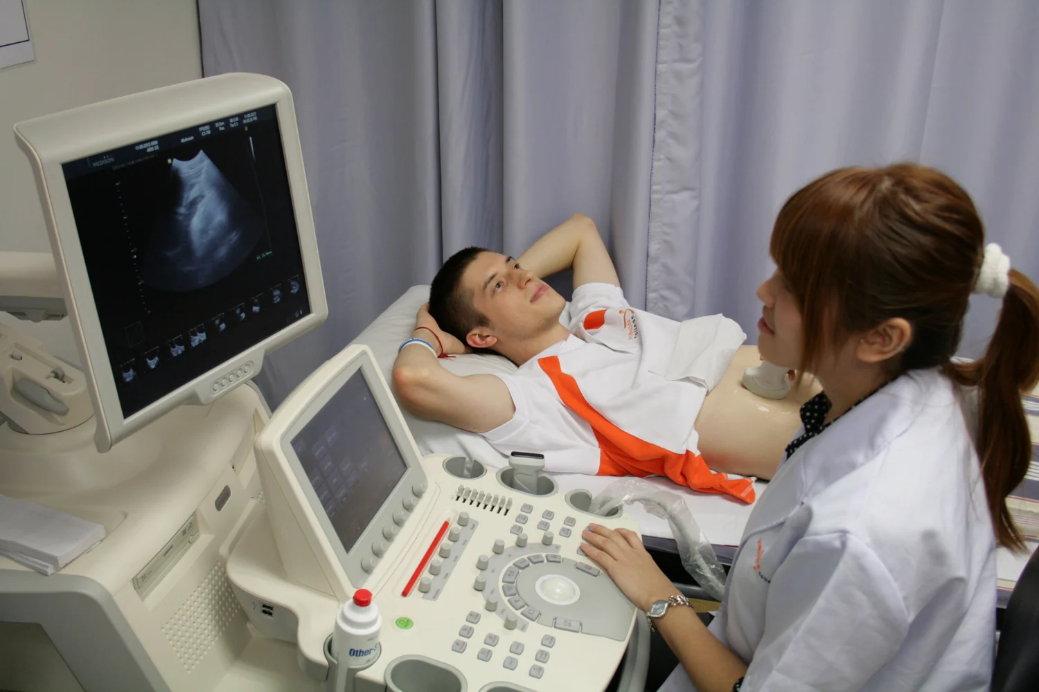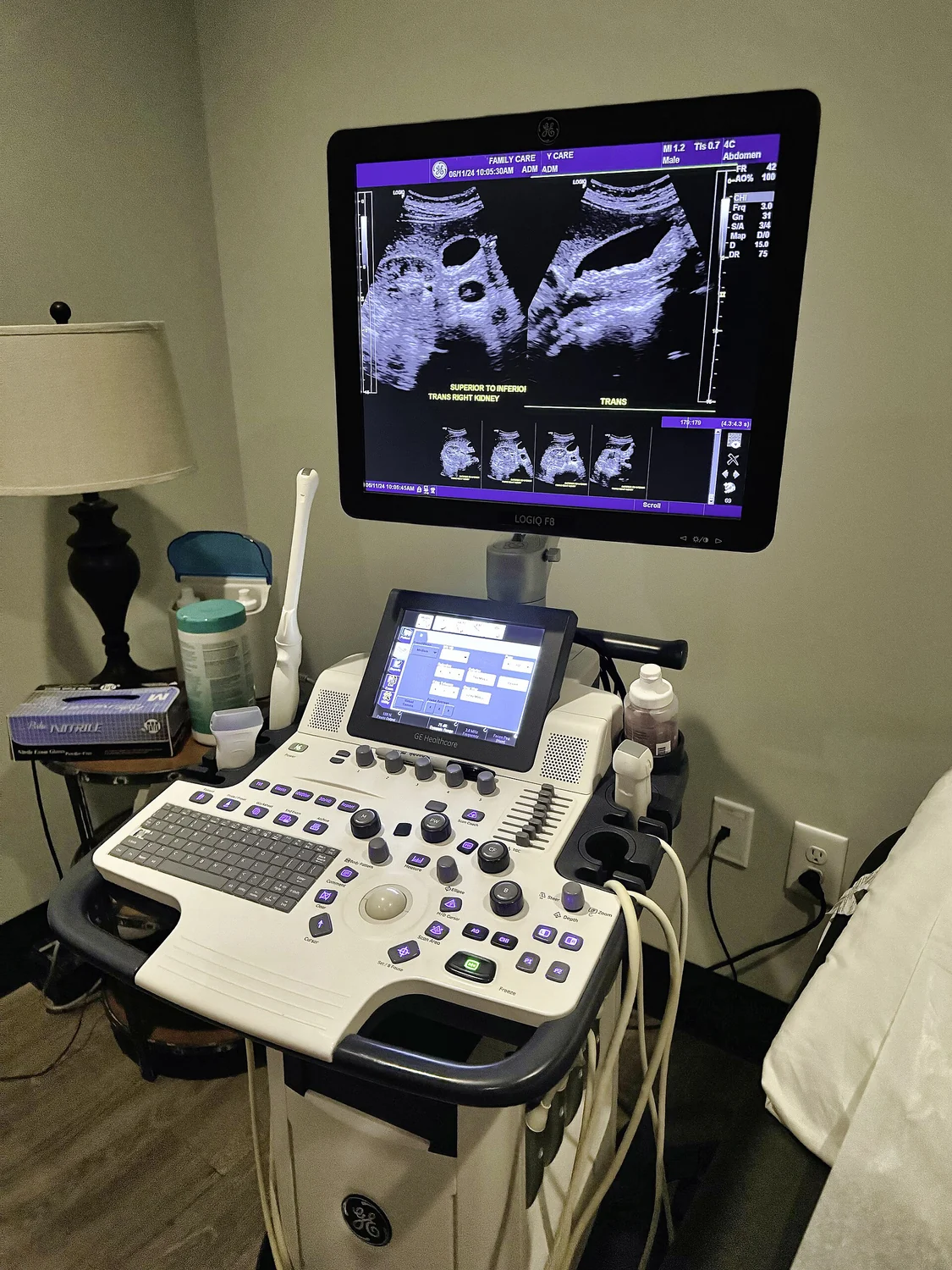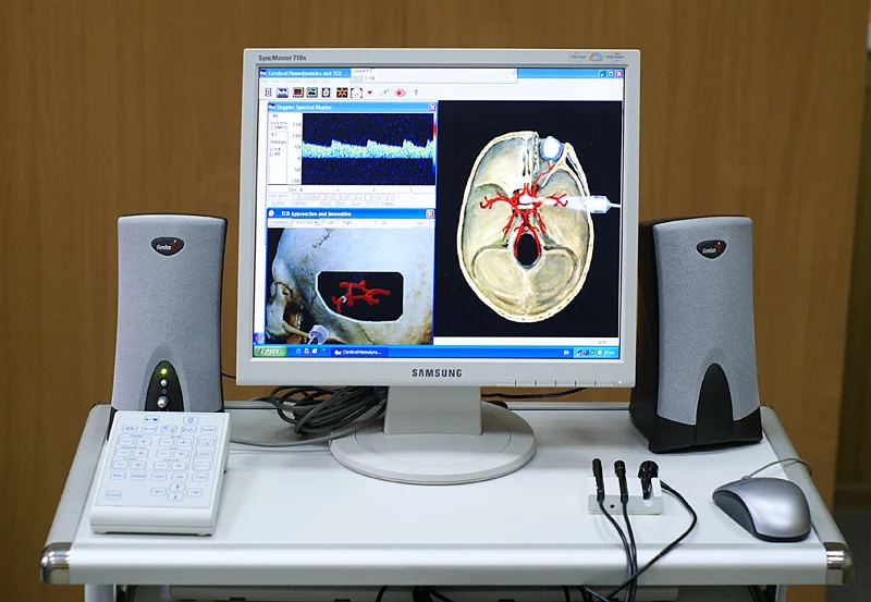
A medical ultrasound probe can listen for echoes, concentrate heat, or generate a cloud of microscopic bubbles that mechanically breaks tissue apart. These effects present different control problems inside a moving, heterogeneous body. [1] Focused ultrasound already has established clinical applications for selected indications. Evidence for one device, organ, or endpoint does not establish every other proposed use. Here we follow the physics from wave propagation to thermal ablation and cavitation-based histotripsy, then examine what the pivotal human studies actually measured. FDA records and the cited literature were checked on October 30, 2026.
1. A pressure wave carries energy through tissue
Ultrasound consists of alternating compression and rarefaction: tiny local changes in pressure that travel through a material. Tissue particles move around their resting positions as the disturbance passes; a plug of tissue does not travel from the skin to the target. Frequency describes how rapidly the pressure oscillates. It says little by itself about the eventual biological effect, which also depends on amplitude, timing, geometry, and the material receiving the wave. [3]
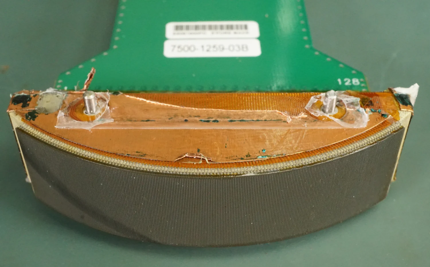
A transducer converts electrical energy into sound, commonly through a piezoelectric material that changes shape under an electric field. The reverse effect can convert returning echoes into electrical signals. An imaging system interprets those echoes to estimate where boundaries and scatterers are located. A therapeutic system instead delivers sound for a chosen interaction with tissue, often alongside a separate imaging function. Sharing the word ultrasound does not make diagnostic imaging and tissue ablation interchangeable exposures. [1]
As a wave travels, some energy is absorbed and some is scattered or reflected. Absorption converts organized acoustic motion into heat. Reflection depends on how acoustic properties change across an interface. The familiar coupling gel has a physical purpose: it removes an intervening air gap that would hinder transmission. Ultrasound avoids ionizing radiation, but that does not make its energy biologically inactive. The FDA distinguishes the favorable safety record of prudent diagnostic use from the capacity of sound to cause heating and cavitation. [2]
2. Focusing is a problem of arrival times
Picture ripples produced around a pond. Their starting times must compensate for different travel distances if the crests are to meet at one location. A focused transducer applies the same principle to pressure waves. A curved emitting surface can supply the geometry, while an array of independently driven elements can adjust the timing electronically. At the intended focus, contributions reinforce one another. [4]
This concentration does not produce an isolated point surrounded by complete silence. A real beam has a central region and weaker energy elsewhere. Array layout, aperture, and electronic steering affect those secondary regions. In their original simulations of high-intensity arrays, Stephens and colleagues examined both useful focal energy and unwanted off-target lobes, including their possible heating. The engineering question includes where unwanted energy goes. [4]
The body complicates arrival-time matching. Fat, muscle, fluid, and bone do not offer identical acoustic travel times. The skull is an especially demanding barrier because it can distort both phase and amplitude. Aubry and colleagues experimentally demonstrated that information from prior CT scans could support adaptive focusing through skull bone. That work established a physical correction strategy, rather than proving that any intracranial target can be reached safely. Patient-specific propagation remains part of the problem. [5]
Focusing is thus a prediction followed by a measurement. The predicted overlap tells a system where energy should concentrate. Feedback has to establish whether the intended biological effect is occurring there. A visually neat beam sketch represents only the first half of that task.
3. A wavelength calculation explains the scale, not a treatment plan
For a deliberately simplified example, assume sound travels through a uniform, water-like material at 1,500 meters per second. Choose a hypothetical frequency of 1 million cycles per second. Wavelength equals speed divided by frequency: λ = c/f = 1,500/1,000,000 meters = 0.0015 meters, or 1.5 millimeters. At twice that frequency in the same hypothetical material, the wavelength would be 0.75 millimeters. These are educational assumptions, not device settings or a measurement of a particular patient's tissue.
The calculation explains why wavelength enters discussions of spatial control. A finite aperture cannot concentrate a wave into an infinitely narrow spot; interference produces a pattern with a finite extent. Changing frequency or aperture changes that pattern. But wavelength alone does not give the dimensions of a clinical lesion. The emitting geometry, propagation distortions, the definition used for beam width, and the mechanism of tissue damage also matter. [3] [4]
There is another tradeoff: higher-frequency ultrasound is generally attenuated more strongly in soft tissue. A shorter wavelength can therefore come with less penetration. Precision cannot be reduced to choosing the largest frequency. The useful design balances transmission to the target with spatial concentration and protection of everything along the path. [3]
4. Thermal ablation depends on the history of heat
In thermal focused ultrasound, absorption deposits heat preferentially in the focused region, damaging tissue. The original FDA assessment of the ExAblate fibroid system describes this combination of acoustic delivery, thermal destruction, and MR guidance. The emitter can remain outside the skin. [18]
The temperature at a location is a balance. Acoustic absorption supplies heat; conduction spreads it; flowing blood can carry energy away. Pennes's classic measurements and heat-transfer analysis of the human forearm helped establish a framework for representing the interaction of tissue temperature and perfusion. That framework is a useful conceptual starting point, although a simplified distributed-perfusion model cannot capture every nearby large vessel or every patient-specific flow pattern. [6]
Imagine heating two otherwise similar regions, with one next to substantial blood flow. An equal amount of deposited acoustic energy need not produce equal temperature histories. Conversely, heat can spread beyond the region of strongest sound. The acoustic focus and the eventual boundary of injury are related objects, rather than identical shapes. This is a reason to measure a changing temperature field instead of treating the planned focus as the complete answer. [6] [18]
A thermal map also has a time dimension. A brief maximum temperature and a sustained lower elevation are not equivalent biological histories. Monitoring systems therefore track temperature evolution and may estimate accumulated thermal injury using models. Such estimates still depend on measurement quality and tissue assumptions. A colorful overlay is a model-informed measurement, not a microscopic census of surviving cells. The feedback must be interpreted together with anatomy and the device's validated clinical purpose. [16]
5. Histotripsy makes mechanical disruption the intended effect
Cavitation begins when an acoustic rarefaction allows a small bubble nucleus to grow. Depending on the acoustic field and surrounding material, bubbles can oscillate, expand substantially, and collapse. The resulting local motions and stresses can damage tissue. Maxwell and colleagues measured how the probability of cavitation changed with pressure in liquids, tissue-mimicking materials, and excised tissue. Their experiment shows why cavitation is a probabilistic material interaction, rather than a simple guarantee attached to the word ultrasound. [11]
Cavitation-based histotripsy deliberately generates a confined bubble cloud and uses its mechanical activity to fractionate tissue. The Edison FDA record identifies the device mechanism as nonthermal mechanical ablation. Its ultrasound delivery is structured to make mechanical breakdown, rather than accumulated bulk heating, the intended route to tissue destruction. Calling this nonthermal describes the dominant treatment mechanism; it does not imply that physical energy transfer or every local temperature change vanishes. [19] The physical target is a volume of tissue; the mechanism does not inherently recognize malignant cells and leave every normal cell intact.
The distinction matters for feedback. A thermal technique asks whether the target experienced the required heating history. Cavitation-based histotripsy asks where a bubble cloud formed and whether the planned tissue volume was mechanically disrupted. Both require targeting and confirmation, but the relevant measurements differ. The primary liver trial used ultrasound visualization during treatment and subsequent contrast-enhanced imaging to assess coverage. [21]
Tissue mechanics can also influence susceptibility. In laboratory and animal work, Vlaisavljevich and colleagues found that stronger structures could resist fractionation better than softer tissue, with some larger vessels remaining within treated liver regions. This helps explain interest in selective effects near vessels. It does not establish that histotripsy universally spares every vessel, duct, or nerve. The same study found destruction of smaller vessels, and preclinical selectivity is not a substitute for clinical complication data. [12]
6. Two ablation mechanisms, different failure modes
Thermal ablation and cavitation-based histotripsy begin with a focused acoustic field, yet they transform tissue differently. One builds a damaging thermal history; the other uses bubble-mediated mechanical forces. The comparison is useful because it identifies which uncertainties a measurement must resolve. A temperature map cannot directly certify mechanical fractionation, and the appearance of a bubble cloud cannot certify that a thermal ablation boundary is adequate. [16] [19]
Blood flow is a good example of a mechanism-dependent constraint. It can remove heat from a thermal target, but cooling alone does not prevent mechanically driven bubble damage. That gives nonthermal approaches a different relationship to the thermal heat-sink problem. It does not eliminate the importance of vessels: blood supply, bleeding, vascular injury, and liver reserve remain clinical concerns. A mechanism can bypass one limitation while introducing or retaining others. [6] [12]
Nor does the absence of an incision mean that an intervention is reversible. Ablation intentionally destroys tissue. Appropriate targeting can make that destruction useful, but a placement error can injure a functional structure. A rigorous comparison therefore follows the whole chain from energy delivery through tissue effect to patient outcome, rather than ranking treatments by how dramatic their equipment looks.
| Question | Thermal focused ultrasound | Cavitation-based histotripsy |
|---|---|---|
| Intended dominant effect | Heat-mediated tissue injury | Bubble-mediated mechanical fractionation |
| Important feedback | Anatomy and temperature evolution | Anatomy and bubble-cloud location, with follow-up assessment |
| Mechanism-specific concern | Uneven heating, conduction, and perfusion losses | Inadequate or misplaced mechanical disruption |
| Shared limit | Target access and protection of adjacent structures | Target access and protection of adjacent structures |
7. The route to a target is part of the target
An organ visible on a scan is not automatically accessible to a therapeutic beam. Sound must reach it through the intervening anatomy. Gas-filled bowel can interrupt a useful acoustic path. Ribs, scars, and interfaces may redirect or absorb energy. The original fibroid FDA assessment explicitly treated the inability to avoid important intervening structures as a contraindication for that device. This is a concrete example of physics limiting eligibility, rather than merely reducing image quality. [18]
The distinction between imaging visibility and therapeutic access is easy to miss. An image may be collected from a convenient angle or with a different modality. A therapy system still needs a usable path from its own emitter to the planned volume. A clear MRI depiction does not make an obstructing gas layer acoustically transparent. Conversely, a favorable beam path is insufficient if the target cannot be reliably distinguished and tracked. [16] [20]
The skull illustrates why there is no universal list of sound-transparent and sound-opaque structures. Bone presents attenuation and distortion, yet specialized transcranial arrays can compensate for aspects of it. Correction redistributes phases to restore a focus; it does not undo every transmission loss or make skull heating irrelevant. A correction validated for one anatomical problem is not a general solution to all body barriers. [5]
For an editorial description, the important consequence is that noninvasive refers to how the energy enters the body. It does not describe universal reach, absence of pain, freedom from anesthesia-related burdens, or the probability of a successful clinical outcome. Those questions belong to a specific procedure and population.
8. Images must show anatomy, effect, and uncertainty
MRI can supply detailed anatomy and, in appropriate tissues and sequences, information about temperature change. A major method uses the temperature dependence of the water proton resonance frequency. Ishihara and colleagues demonstrated rapid phase-based temperature mapping in phantoms and an animal model. The basic idea is that heating alters a measurable aspect of the MR signal; it is not that the scanner sees heat as a separate visible substance. [7]
That distinction exposes possible errors. A change in magnetic field, sample position, or other signal conditions can resemble part of the change attributed to temperature. A temperature estimate often refers to a baseline or a modeled background phase, so the validity of that reference matters. Water-based phase thermometry also does not behave identically in every tissue component. A single uniform color scale can hide considerable variation in how well different regions were measured. [7] [8]
Holbrook and colleagues developed and tested a real-time liver thermometry approach using moving heated phantoms and nonheated volunteers. Those experiments addressed measurement performance under motion; they were not a clinical trial proving liver-cancer control. That distinction is instructive: a system must establish that its thermometer works before a treatment trial can meaningfully connect the displayed heating to outcomes. [8]
Different observations answer different questions: intraprocedural visualization shows an intervention in progress; post-treatment imaging assesses its spatial result; later imaging tests durability. A therapeutic system may combine ultrasound targeting with subsequent CT or MRI assessment. These measurements occupy different time points. None should silently substitute for a patient-centered outcome that the study never measured. [20]
9. A moving target challenges both delivery and measurement
Breathing changes the position of abdominal organs. It can also change the magnetic environment used for MR thermometry. These are two separate problems: a beam may miss a moving target even with a perfect thermometer, while a correctly positioned beam may be monitored by a temperature estimate corrupted by motion. Solving one does not automatically solve the other. Grissom and colleagues investigated a hybrid approach to background-phase estimation for thermometry in moving organs. [9]
Research strategies include restricting delivery to a suitable part of the motion cycle and steering the focus to follow a target. Holbrook and colleagues tested respiration-based steering in a moving phantom and in porcine liver, using a relationship between breathing and internal target position. The study supports the feasibility of an integrated tracking-and-delivery approach; its animal and phantom results should not be translated into a universal human success rate. [10]
Motion creates a timing budget. An image records a position, processing takes time, a controller updates its estimate, and energy arrives afterward. If the target has moved meanwhile, even an accurate old estimate can be clinically unhelpful. This is an explanatory control-system perspective, not a measured delay for any named product. It shows why improved image sharpness alone is insufficient: useful guidance also needs suitable timing, coverage, and a reliable relationship to the therapeutic coordinate system.
The distinction extends to treatment assessment. A planning outline must remain meaningful in the anatomy being treated, rather than merely remain fixed on a monitor. Registration, tracking, and confirmation connect the picture to the physical intervention.
10. Clinical evidence is strongest when the endpoint is specific
A pivotal essential-tremor trial provides a strong example of a direct clinical endpoint. Elias and colleagues compared unilateral MR-guided focused ultrasound thalamotomy with a sham procedure. Among 76 analyzed participants, the between-group difference in improvement in hand-tremor score at three months was 8.3 points on a 32-point scale, with a 95% confidence interval of 5.9 to 10.7. Gait disturbance and sensory symptoms occurred, and some persisted at twelve months. The trial supports symptom benefit in the selected medication-refractory population while documenting neurological risks. [13]
The sham comparison matters because expectations and the experience of a procedure can influence reported improvement. Independent blinded ratings strengthen the inference that treatment caused a difference. The control period was limited: sham participants could subsequently cross over. That limits what the randomized comparison says about later differences. The result is evidence about a defined brain lesion and symptom outcome, rather than a claim that ultrasound cures the underlying disorder. [13]
A separate phase III study by Hurwitz and colleagues examined palliation of painful bone metastases. It randomized 147 participants to MR-guided focused ultrasound or placebo. At three months, 72 of 112 treated participants and 7 of 35 controls met the response endpoint, approximately 64% versus 20%. Response combined pain reduction with a constraint on increased analgesic use. Those results address pain palliation, not eradication of metastatic cancer or improved survival. [17]
The difference between the examples is substantive. A tremor scale, a pain-and-medication endpoint, and an imaging-defined ablation zone are different measurements. Reporting all three as a generic success percentage would erase the clinical question each study was designed to answer.
11. Histotripsy: early coverage is not the same as cancer control
The initial pivotal HOPE4LIVER analysis reported prospective, multicenter, single-arm results in 44 participants, with small target tumors. Technical success occurred in 42 of 44 evaluated lesions, or 95%, based on imaging coverage within 36 hours. Three of 44 participants, or 7%, had procedure-related major complications within thirty days. One involved hepatic failure followed by death on day 37. Success counts lesions; complication risk counts people. [21]
A single-arm performance-goal study can show that a technique achieves a prespecified level of technical performance and characterize observed complications. Without a randomized comparison, it cannot establish that the technique improves survival or is superior to another liver-directed treatment. A target covered shortly after intervention may still develop recurrence, and untreated disease elsewhere may determine a patient's course. Early local coverage and whole-patient benefit are connected research questions, not equivalent endpoints.
The one-year update included 47 patients and 52 treated tumors. Its estimated one-year local-control rate was 63.4% using the primary imaging assessment and 90% using a post hoc assessment. The authors explained that evolving understanding of post-treatment imaging contributed to reinterpretation. The difference is important evidence about measurement uncertainty. Reporting only the higher estimate would conceal the role of the changed reading method. [22]
The update recorded six serious device-related adverse effects within thirty days. Its uncontrolled, heterogeneous population prevents isolation of a causal survival effect. Sponsorship and investigator financial relationships are disclosed and deserve consideration alongside study design. Promising local results do not establish a general cancer cure or a distant immune benefit. [22]
12. Authorization attaches to a device and a defined use
FDA decisions are specific to devices, indications, populations, and labeling. They are not an endorsement of every medical application of a physical mechanism. The original 2016 Exablate Neuro approval concerned unilateral thalamotomy for selected adults with medication-refractory essential tremor. Later supplements changed or expanded aspects of the allowed use, including a 2025 decision concerning pallidothalamic tractotomy for specified advanced Parkinson's disease motor complications. The original approval alone is therefore not a current catalog of all uses. [14] [15]
For Edison, FDA granted a De Novo classification in 2023; a later 510(k) decision retained an indication for noninvasive mechanical destruction of liver tumors, including unresectable tumors. FDA's De Novo review also required labeling that distinguishes this tissue-destruction indication from evaluation as a treatment for cancer as a disease. This is why authorization and an oncologic-effectiveness claim must be stated with care. Neither record establishes a survival advantage over another treatment. [19] [20]
The scientifically useful reading of an ultrasound claim follows three questions: what physical effect occurred, what clinical endpoint was measured, and in whom? A new focusing method can improve delivery without proving patient benefit. A favorable local image can support an ablation endpoint without proving longer survival. A sham-controlled improvement can support a particular symptom indication without establishing a cure. Keeping those links visible allows established applications and active research to coexist in an honest account.
| Verified FDA example | What the record supports | What it does not establish |
|---|---|---|
| Exablate Neuro, original PMA P150038 (2016) [14] | Selected medication-refractory essential-tremor thalamotomy | Every neurological indication or the full current label |
| Exablate bone application, P110039 (2012) [16] | Pain palliation for specified adults with metastatic bone disease | A general metastatic-cancer cure |
| Edison, DEN220087 and K241902 (2023–2024) [19] [20] | Nonthermal mechanical destruction of liver tumors | Proven survival benefit or authorization for every organ |
Explore the physics: an explanatory diagram

Sources & further reading
- Ultrasound NIBIB
- Ultrasound Imaging FDA
- Diagnostic Radiology Physics: A Handbook for Teachers and Students, Chapter 12: Physics of Ultrasound International Atomic Energy Agency
- Design Aspects of Focal Beams From High-Intensity Arrays (Stephens et al., 2011) IEEE Transactions on Ultrasonics, Ferroelectrics, and Frequency Control
- Experimental demonstration of noninvasive transskull adaptive focusing based on prior computed tomography scans (Aubry et al., 2003) Journal of the Acoustical Society of America
- Analysis of tissue and arterial blood temperatures in the resting human forearm (Pennes, 1948) Journal of Applied Physiology
- A precise and fast temperature mapping using water proton chemical shift (Ishihara et al., 1995) Magnetic Resonance in Medicine
- Real-time MR thermometry for monitoring HIFU ablations of the liver (Holbrook et al., 2010) Magnetic Resonance in Medicine
- Hybrid referenceless and multibaseline subtraction MR thermometry for monitoring thermal therapies in moving organs (Grissom et al., 2010) Medical Physics
- Respiration based steering for high intensity focused ultrasound liver ablation (Holbrook et al., 2014) Magnetic Resonance in Medicine
- Probability of cavitation for single ultrasound pulses applied to tissues and tissue-mimicking materials (Maxwell et al., 2013) Ultrasound in Medicine & Biology
- Effects of tissue mechanical properties on susceptibility to histotripsy-induced tissue damage (Vlaisavljevich et al., 2014) Physics in Medicine & Biology
- A Randomized Trial of Focused Ultrasound Thalamotomy for Essential Tremor (Elias et al., 2016) New England Journal of Medicine
- Exablate Neuro original PMA P150038, approval July 11, 2016 U.S. Food and Drug Administration
- Exablate Neuro PMA supplement P150038/S037, approval July 3, 2025 U.S. Food and Drug Administration
- ExAblate PMA P110039: Summary of Safety and Effectiveness Data U.S. Food and Drug Administration
- Magnetic Resonance–Guided Focused Ultrasound for Patients With Painful Bone Metastases: Phase III Trial Results (Hurwitz et al., 2014) Journal of the National Cancer Institute
- ExAblate 2000 PMA P040003: Summary of Safety and Effectiveness Data U.S. Food and Drug Administration
- Edison System De Novo DEN220087: FDA Review Decision Summary U.S. Food and Drug Administration
- Edison System K241902: 510(k) Decision and Summary U.S. Food and Drug Administration
- The #HOPE4LIVER Single-Arm Pivotal Trial for Histotripsy of Primary and Metastatic Liver Tumors (Mendiratta-Lala et al., 2024) Radiology
- The #HOPE4LIVER Single-arm Pivotal Trial for Histotripsy of Primary and Metastatic Liver Tumors: One-year Update of Clinical Outcomes (Ziemlewicz et al., 2025) Annals of Surgery
Sources checked: 30 October 2026. The calculations illustrate physical models; they are not clinical settings.
Prepared for the independent PSI Center reading library. About edition dates and source checking.
This explainer describes scientific principles. It does not assess individual treatment choices.